Remembering another mouse after sleep: oxytocin and cortical inhibitory balance
In male mice, a paraventricular-to-prelimbic oxytocin-associated projection during REM sleep supports social memory. Sleep deprivation alters local inhibitory signaling, while enhancing associated signals improves memory measures.
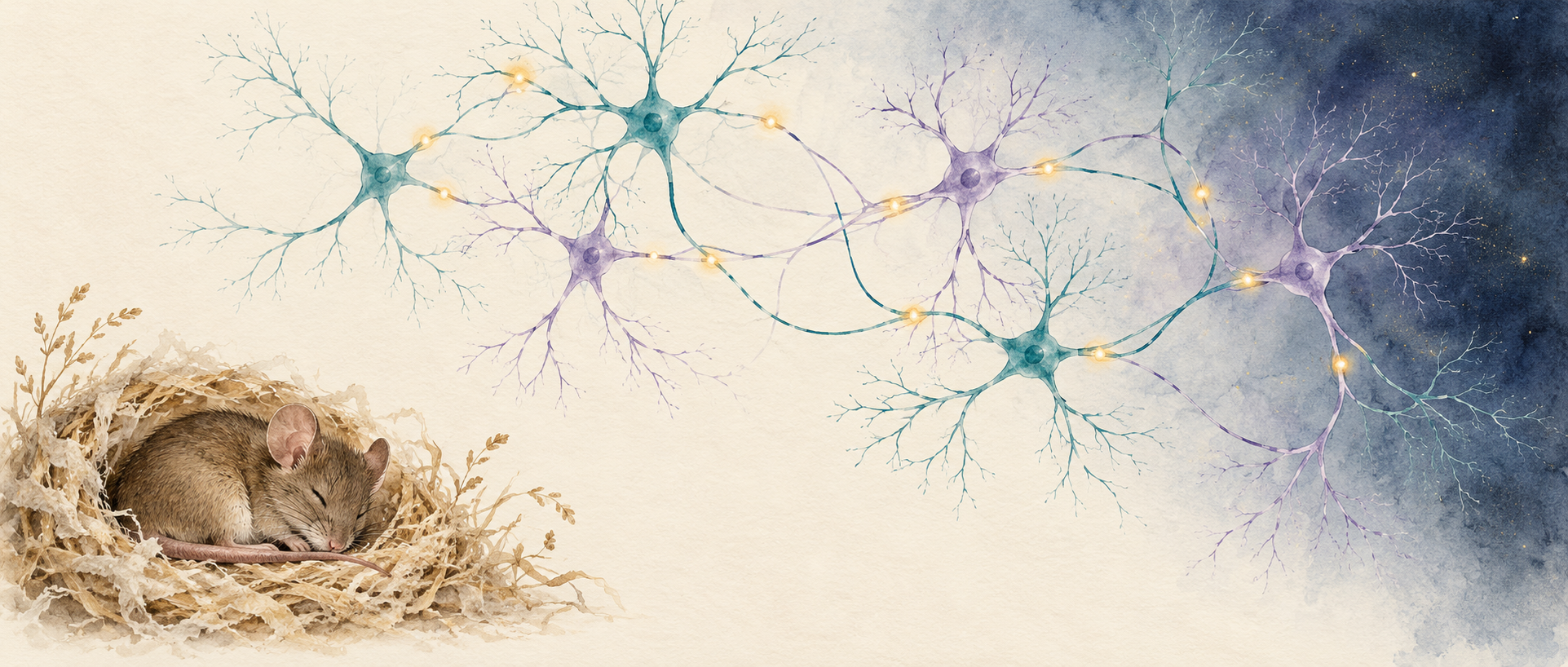
After meeting another mouse and resting for several hours, can a mouse distinguish familiar from unfamiliar? Social memory extends beyond the encounter itself, involving brain activity during sleep. A study in Theranostics follows oxytocin signaling in adult male mice and links a paraventricular hypothalamic (PVN) projection to prelimbic cortex (PrL) during REM sleep with later social discrimination. Coordination within a local inhibitory network provides another part of the account.
Suppressing the PVN–PrL projection during REM sleep reduces later social novelty preference without significant overall sleep-architecture changes. Improved memory after sleep deprivation accompanies increases in local signals including VIP and PV.
Contents · 7 sections
Oxytocin signals vary with sleep state
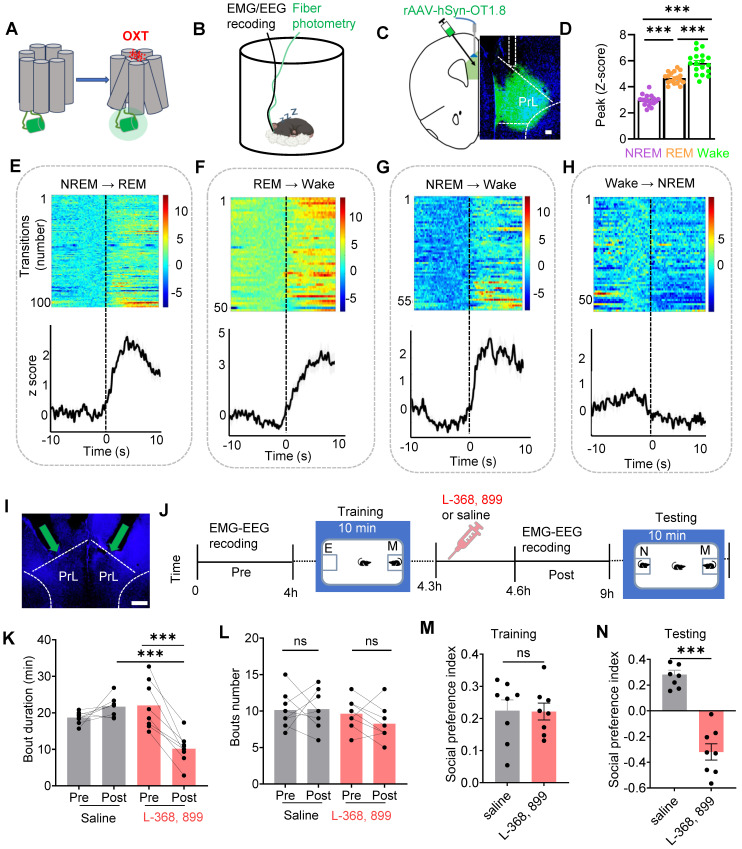
Oxytocin-sensor signals in PrL are higher during REM sleep and waking than NREM. Signals increase around transitions into REM or waking, without the same significant change during waking-to-NREM transitions. Sleep stage supplies a temporal context for this neuropeptide signal.
Mice first encounter a conspecific, rest for several hours, then encounter familiar and unfamiliar mice. Blocking PrL oxytocin receptors after learning reduces REM duration without significantly changing bout count, and lowers novelty preference at testing. Initial social preference is comparable between groups.
Source: Liu YC et al. · Theranostics · CC BY
Original figure and English caption ↗
Reducing upstream release changes both sleep and memory
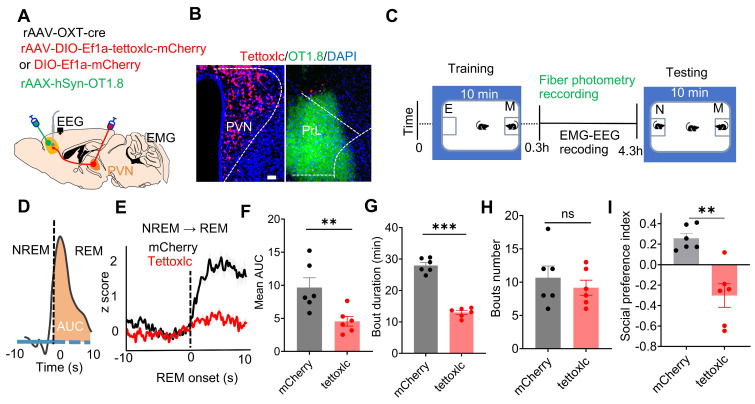
Increasing PVN oxytocin-associated activity raises PrL oxytocin signals. Conversely, reducing synaptic-release function in this population weakens PrL signals during REM and waking, including responses around REM onset. Upstream modulation accompanies changes in cortical signaling.
Following this broader upstream intervention, waking increases, both sleep types shorten, and later social-memory measures decline. Their coexistence motivates a narrower test of one projection within one sleep stage.
Source: Liu YC et al. · Theranostics · CC BY
Original figure and English caption ↗
REM-stage projection activity affects later discrimination
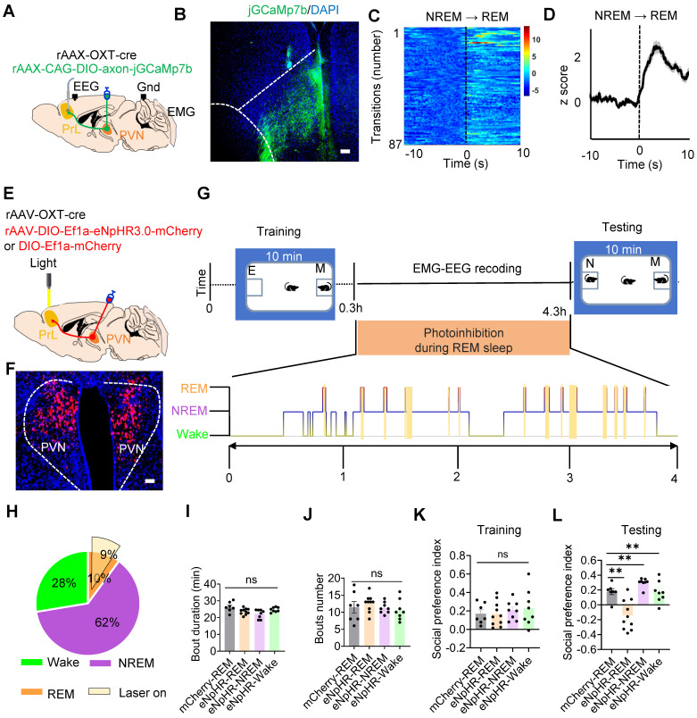
Calcium signals from PVN-derived terminals in PrL rise at REM onset. Suppressing the projection only during post-encounter REM reduces later preference for an unfamiliar conspecific. Comparable-duration suppression during NREM or waking does not produce the same memory change.
Initial social preference is similar across groups, and overall sleep duration and bout counts do not change significantly. The later memory difference therefore occurs against broadly comparable sleep architecture, highlighting the timing of post-learning signals rather than sleep quantity alone.
Source: Liu YC et al. · Theranostics · CC BY
Original figure and English caption ↗
Different inhibitory components converge on pyramidal cells
PrL contains more than excitatory pyramidal cells. PV interneurons mainly constrain somatic activity, while VIP interneurons can reduce dendritic inhibition by suppressing SST cells. Building on known connections, the study proposes a balance model combining somatic inhibition and dendritic disinhibition.
At REM onset, presynaptic calcium in VIP and PV cells and VIP-neuropeptide signals increase. Oxytocin-receptor blockade weakens these signals, increases SST-neuropeptide signals, and raises pyramidal-cell calcium activity. These readouts support oxytocin involvement in local coordination.
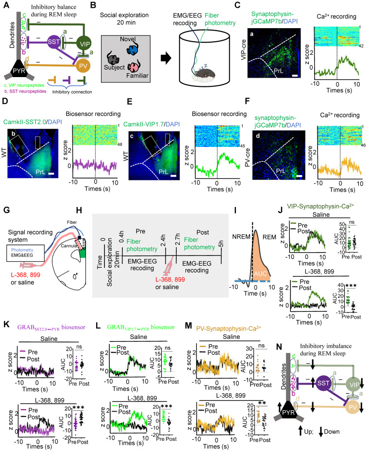
Source: Liu YC et al. · Theranostics · CC BY
Original figure and English caption ↗
Several signals weaken while the SST measure remains similar
After several days of disrupted sleep, social-memory performance declines, REM duration decreases, and PrL oxytocin signals weaken. PVN oxytocin-associated cells also fire less spontaneously. Changes at both ends connect upstream cells and local signaling within the same sleep-deprivation context.
VIP and PV presynaptic calcium and VIP-neuropeptide signals decrease, while pyramidal-cell calcium increases. SST-neuropeptide signals do not differ significantly. Sleep deprivation and receptor blockade have similarities, but differ in SST signaling. The local network therefore does not respond identically to the two conditions.
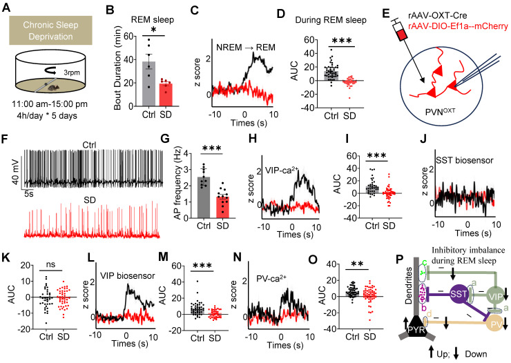
Source: Liu YC et al. · Theranostics · CC BY
Original figure and English caption ↗
Increasing REM-stage signaling improves testing preference
In sleep-deprived mice, increasing PVN–PrL activity during REM improves later social-memory measures while initial social preference remains comparable. Sleep-architecture effects are limited: NREM bout count increases slightly, while several other measures do not change significantly.
In another group, intranasal oxytocin also improves testing preference. VIP and PV presynaptic calcium responses rise; VIP remains different from normal controls, whereas the corresponding PV comparison is nonsignificant. SST-neuropeptide signals remain similar. Behavioral improvement accompanies recovery in some network responses.
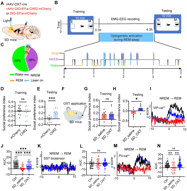
Source: Liu YC et al. · Theranostics · CC BY
Original figure and English caption ↗
Consolidation involves more than increased activity
The study in adult male mice connects REM-stage neuropeptide input and local inhibitory coordination with later social discrimination. Stronger pyramidal-cell activity does not automatically accompany better memory. Signal timing and its distribution across somatic and dendritic regulation also matter.
Sleep deprivation affects multiple stages, and cellular labeling has limitations in population coverage. The study nevertheless brings a specific relationship into view: after a social encounter, sleep continues organizing activity for memory, with oxytocin contributing to cortical network coordination.