Release off the beat: high-frequency stimulation improves movement in mice
In dopamine-depleted male mice, high-frequency stimulation favors asynchronous GABA release from pallidal PV terminals and changes subthalamic firing. Altering release mode also changes the motor effect of low-frequency stimulation.

Why can changing the frequency of a pulse train change movement? Subthalamic-nucleus deep brain stimulation may work through when signals arrive as well as how much neurons fire. A Nature Communications study examines GABA release timing in dopamine-depleted male mice. Stimulation at 130 Hz improves movement, while 20 Hz lacks the same effect. Following inhibitory input from external globus pallidus, or GPe, to STN reveals release that becomes less tightly locked to individual pulses and neuronal activity that becomes less synchronized.
GPe PV terminals provide an important input for high-frequency STN stimulation to improve model movement. Reducing release with an asynchronous component weakens improvement; Syt1 knockdown increases low-frequency asynchronous-release proportion and enables a 20-Hz optogenetic motor benefit.
Contents · 7 sections
Equal pulse counts still produce different movement
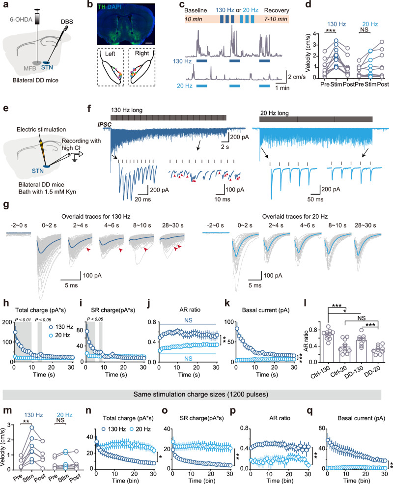
Electrical STN stimulation at 130 Hz raises velocity and reduces immobility in the model; 20 Hz does not significantly improve movement. Equalizing both trains to 1,200 pulses retains the difference, extending the explanation beyond pulse number alone.
Bilateral dopamine depletion provides the motor-deficit context. Linking behavior with STN synaptic recordings allows the study to examine how frequency changes local transmission.
Source: Xu Z et al. · Nature communications · CC BY-NC-ND
Original figure and English caption ↗
At high frequency, release extends beyond each pulse
Synchronous release responds promptly to each pulse. Asynchronous events are less tightly locked, occur between pulses and can outlast stimulation, changing temporal structure as well as the amount of signaling.
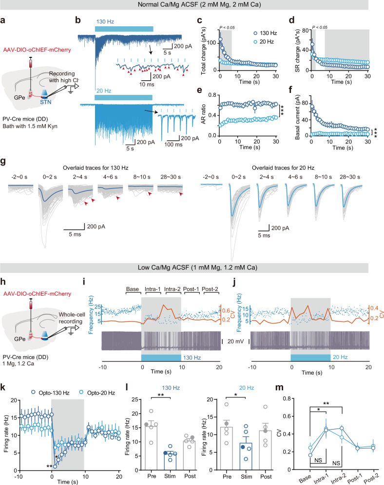
In STN slices, synchronous components weaken during the first seconds of sustained 130-Hz stimulation as asynchronous events become prominent. Around four seconds, AR is a dominant transmission mode; 20 Hz retains more pulse-locked release. High-frequency photostimulation of GPe PV terminals similarly produces stronger asynchronous GABA signaling.
At 45 Hz, the AR proportion lies between 20 and 130 Hz. Walking time increases, while overall motor improvement is limited. This intermediate condition connects graded release composition with behavioral responses.
Source: Xu Z et al. · Nature communications · CC BY-NC-ND
Original figure and English caption ↗
Pallidal cell types contribute differently
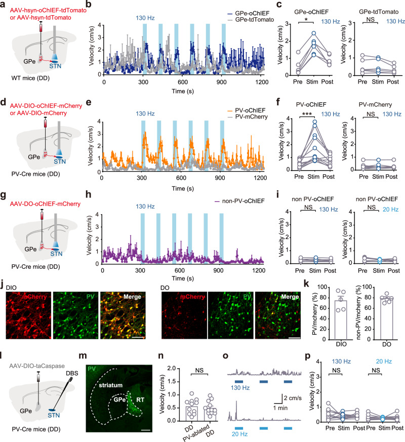
GPe provides substantial STN GABA input, but cell types differ. High-frequency activation of PV terminals increases model velocity, whereas comparable non-PV-terminal activation has no clear benefit.
Blocking STN GABA receptors removes the high-frequency electrical motor benefit. Ablating GPe PV cells or locally inhibiting their STN terminals also removes or markedly diminishes improvement, identifying an important inhibitory input.
Source: Xu Z et al. · Nature communications · CC BY-NC-ND
Original figure and English caption ↗
Changing firing rates and timing relationships
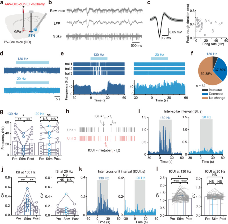
In awake recordings, high-frequency PV-terminal activation does not uniformly silence STN. About one third of 32 units reduce firing, most show no clear change and a few increase, revealing heterogeneous responses.
Inhibited units show more variable spike intervals, paired-unit timing becomes more dispersed and low-beta activity decreases. Optical 20-Hz slice stimulation can also lower mean firing without significantly increasing temporal variability, separating average rate from timing structure.
Some timing changes persist into the 30-second observation window after high-frequency stimulation. Terminal release and neuronal responses each have temporal dynamics, extending the circuit picture beyond the stimulation train.
Source: Xu Z et al. · Nature communications · CC BY-NC-ND
Original figure and English caption ↗
Reduced asynchronous signaling weakens movement improvement
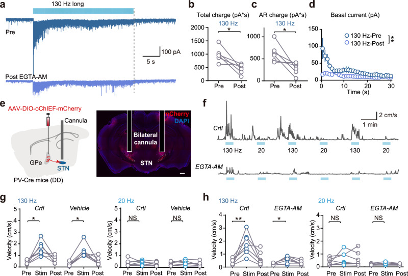
Calcium buffering reduces asynchronous current induced by high frequency and also affects synchronous release. Applied at STN, it substantially weakens the high-frequency PV-terminal velocity benefit, although a significant increase remains; 20 Hz still lacks a clear motor effect.
Source: Xu Z et al. · Nature communications · CC BY-NC-ND
Original figure and English caption ↗
A different release balance enables a low-frequency benefit
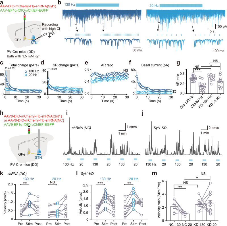
Reducing synaptotagmin-1, or Syt1, in GPe PV cells weakens synchronous events and raises the AR proportion during 20-Hz photostimulation toward high-frequency levels, changing the temporal structure of inhibitory input.
The previously ineffective 20-Hz optical stimulation now raises velocity and reduces immobility. Frequency stays the same while terminal release changes, linking release mode with the motor effect.
Source: Xu Z et al. · Nature communications · CC BY-NC-ND
Original figure and English caption ↗
Effective stimulation also depends on when signals arrive
The study provides a synaptic explanation for high-frequency stimulation: GPe PV inhibition changes firing rate in some STN cells as well as temporal organization. In this male dopamine-depletion model, altering release mode changes low-frequency motor effects. Following these timing changes through larger networks can clarify how stimulation effects depend on parameters and circuit state.