Memory-network rhythms under near-infrared light
In mice, intermediate near-infrared illumination accompanies improved spatial memory and changes in dentate-gyrus activity, rhythmic coupling and cellular structure.
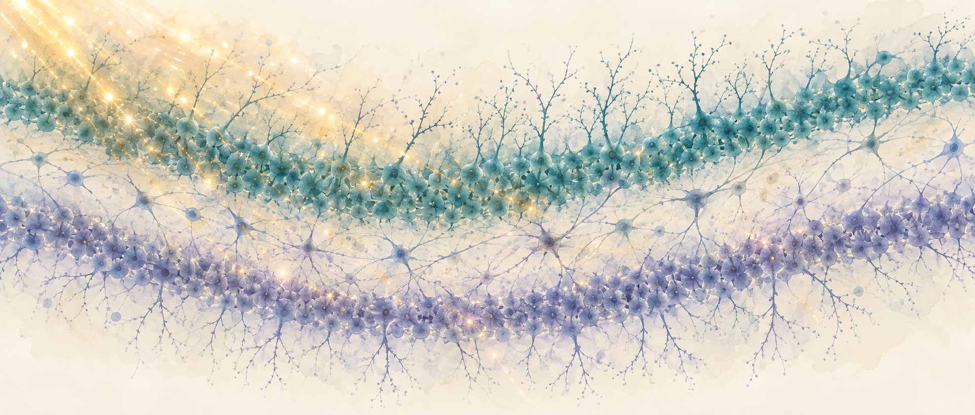
A mouse searching a circular platform for a familiar escape hole relies on spatial cues and memory. What happens if near-infrared light passes through the skull toward the hippocampus during learning? A study published in Advanced Science in 2025 brings photobiomodulation into this setting. Following behavior, neuronal activity and interactions between light and cellular density, the team finds that the same illumination can produce different responses across tissue structures.
At an intermediate tested intensity, 808-nm near-infrared light improves short-term spatial-memory performance in adult male mice and enhances selected dentate-gyrus rhythms and calcium responses in both excitatory and inhibitory neurons. Optical modeling, transcriptional findings and morphology provide mechanistic clues.
Contents · 7 sections
More light does not mean greater benefit
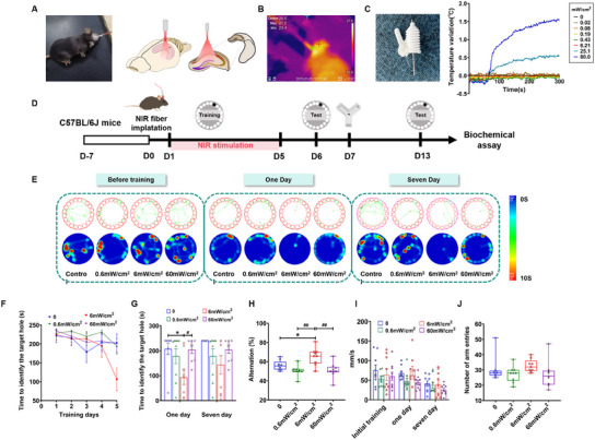
The team compares three illumination intensities in adult male mice without a disease model. At the intermediate intensity, mice find the target hole faster one day after training and show a higher alternation proportion in the Y maze. Lower and higher intensities do not produce the same improvement, suggesting distinct response ranges.
The advantage is transient: supplementary observations indicate an effect lasting roughly two days, and the hole-finding difference is no longer significant seven days after training. Platform movement speed and total Y-maze arm entries remain similar, without increased overall activity accompanying the memory changes.
Source: Pan WT et al. · Advanced science (Weinheim, Baden-Wurttemberg, Germany) · CC BY
Original figure and English caption ↗
The dentate gyrus responds more strongly
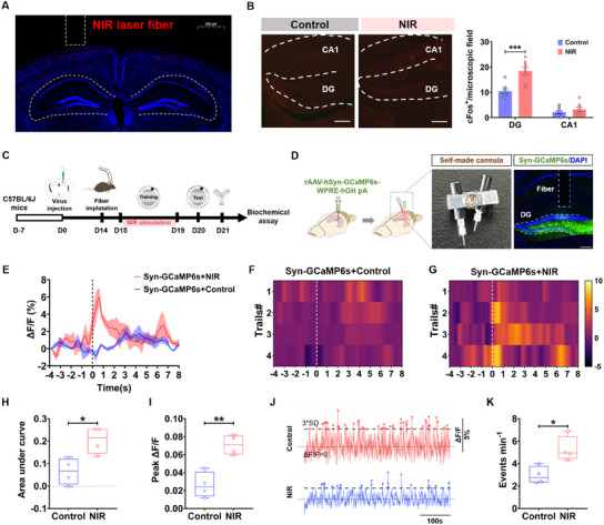
Illumination covers the hippocampus, but activity changes are particularly prominent in the dentate gyrus. Its c-Fos-positive cells increase, while the corresponding CA1 change is not significant. As an important entry point for spatial information into hippocampal memory networks, the dentate gyrus shifts attention from whether light has an effect to which cells respond.
When mice enter the correct hole, dentate-gyrus population calcium responses are stronger in the illuminated group; calcium events are also more frequent during Y-maze exploration. These recordings align cellular responses with important behavioral moments, bringing the observations closer to the memory task than post-task activity labeling alone.
Source: Pan WT et al. · Advanced science (Weinheim, Baden-Wurttemberg, Germany) · CC BY
Original figure and English caption ↗
Both excitatory and inhibitory cells respond
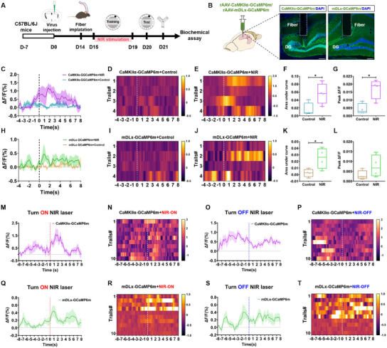
Examining excitatory and inhibitory populations separately reveals increased integrated calcium responses in both upon target-hole entry. Excitatory-cell peaks also rise, whereas the inhibitory-cell peak change is not significant. The measurements describe related but nonidentical responses.
During free movement, calcium signals in both populations rise when illumination begins and fall when it ends. Near-infrared light therefore acts beyond excitatory cells, recruiting excitatory and inhibitory components together and raising the question of how network rhythms change.
Source: Pan WT et al. · Advanced science (Weinheim, Baden-Wurttemberg, Germany) · CC BY
Original figure and English caption ↗
Slow and fast rhythms become more coupled
Local field potentials show increased dentate-gyrus theta, alpha and high-gamma power at the intermediate intensity. Beta and low- and mid-gamma power do not increase significantly relative to controls. The change does not span every band, and CA1 responses are much less widespread.
Theta phase and gamma amplitude also become more strongly coupled: slower timing is more closely coordinated with faster activity. This organization changes alongside memory performance, describing the effect on information processing more specifically than an overall increase in activity.
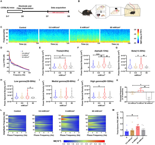
Source: Pan WT et al. · Advanced science (Weinheim, Baden-Wurttemberg, Germany) · CC BY
Original figure and English caption ↗
Cell arrangement influences light distribution
Why is the dentate-gyrus response more prominent? The team turns to its dense granule-cell layers. Scattering, refraction and absorption influence light propagation, including differences between nuclei and their surroundings. Cellular arrangement may therefore give this region a different local light-energy distribution.
Optical analogues using particle suspensions and tissue-image-based simulations indicate greater local energy accumulation in dense cellular layers. Their relationship to the distribution of activated cells suggests that tissue microstructure participates in photomodulation rather than simply receiving uniform illumination.
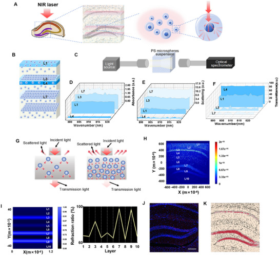
Source: Pan WT et al. · Advanced science (Weinheim, Baden-Wurttemberg, Germany) · CC BY
Original figure and English caption ↗
Energy-related genes accompany structural changes
Single-cell transcriptional analyses highlight gene sets related to neurite formation and oxidative phosphorylation in both neuronal populations, alongside increased cytochrome-c-oxidase-related transcripts. These findings connect light responses with mitochondrial energy processes and provide molecular clues to network changes.
Morphology reveals longer dendrites, more branches and denser dendritic spines. Structures involved in receiving information also change, though concurrent transcriptional and morphological changes do not establish which step is essential. They form multiple levels of evidence consistent with the behavioral and activity findings.
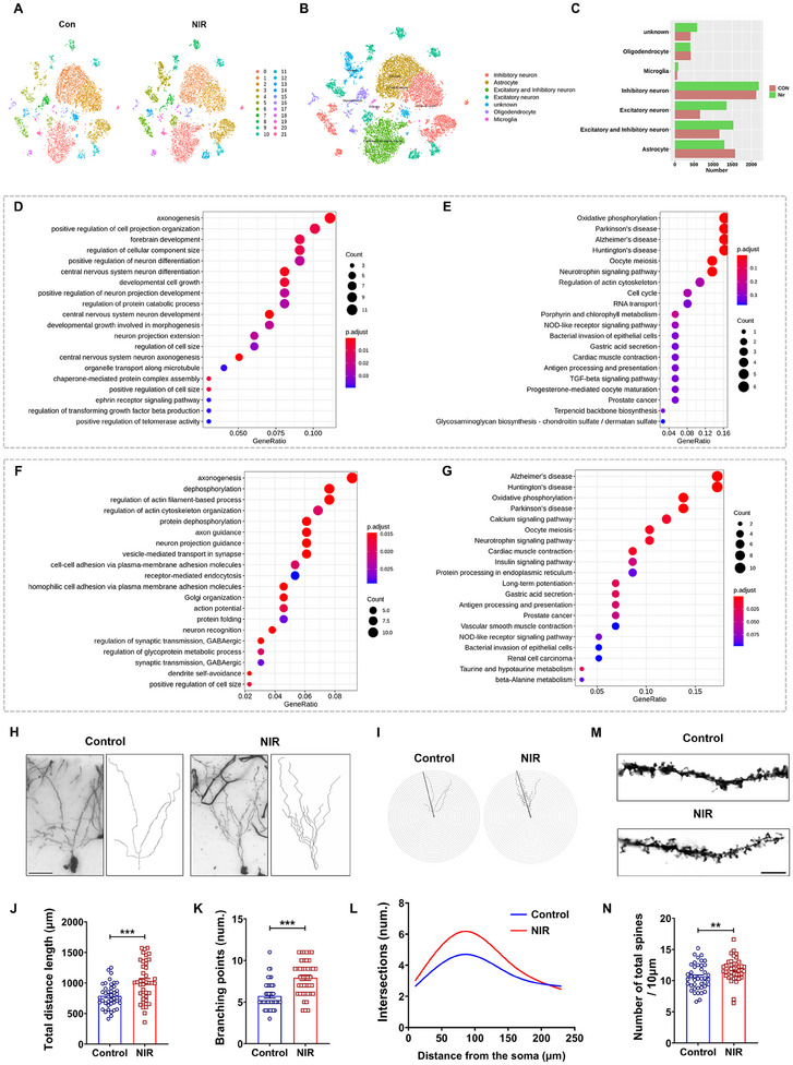
Source: Pan WT et al. · Advanced science (Weinheim, Baden-Wurttemberg, Germany) · CC BY
Original figure and English caption ↗
Postoperative performance accompanies rhythmic changes
In an anesthesia-and-surgery-related cognitive-impairment model, illumination during training improves recent hole finding and alternation, alongside increased dentate-gyrus activity labeling, selected rhythms and cross-frequency coupling. The seven-day hole-finding advantage remains nonsignificant. Behavioral improvement also occurs in an inflammatory-challenge model, but measured TNF-α does not significantly decrease in either model.
This adult male mouse study connects illumination with memory tasks, network rhythms and cellular-layer structure. The responses are shaped jointly by tissue organization and cellular activity. Extending these short-term model effects to human cognitive intervention requires accounting for differences in brain structure and light propagation.
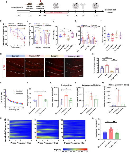
Source: Pan WT et al. · Advanced science (Weinheim, Baden-Wurttemberg, Germany) · CC BY
Original figure and English caption ↗