CA3 neurons help memory adapt when an exit changes location
After IL-1β challenge, some male mice learn the original location but struggle when the exit moves. Dorsal-CA3 activity relates to updating, and activation improves measured performance.
Finding a familiar exit and realizing that it has moved are different demands. Cognitive flexibility requires retaining experience while adapting choices to change. A study published in CNS Neuroscience & Therapeutics in 2025 has mice learn a circular-maze exit during IL-1β challenge and then moves it to the opposite side. Some males keep searching the old location, while others adjust more readily. The team follows this difference to dorsal hippocampal CA3, dCA3, and spatial-information updating.
IL-1β affects spatial reversal in males while initial learning and original-location retrieval remain largely intact. Activating dCA3 excitatory cells improves adaptation to the relocated exit, whereas suppression impairs reversal in a separate experiment.
Contents · 6 sections
Learning the original location does not ensure updating
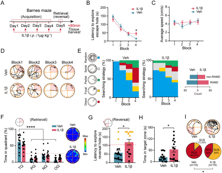
Following repeated inflammatory-signal challenge, initial exit-finding time and average speed remain similar to controls. Both groups prefer the original target region during retrieval. The main difficulty appears after relocation: challenged males take longer to find the new exit and spend longer in the former target zone.
Not every male shows the difficulty. Classification by reversal performance separates less adaptable mice from those retaining adaptability, with the former more prevalent after challenge. Females under the same conditions do not show significant reversal impairment, leading later cellular experiments to focus on males.
Among fifteen challenged males, nine fall in the poorer-adaptability category and six retain adaptability, compared with two less adaptable controls. Different outcomes under the same inflammatory signal motivate the search for neural regulation.
Open-field distance and center time do not change significantly after single or repeated challenge. This places updating difficulty alongside largely preserved overall movement and distinguishes initial learning from reversal.
Source: Zuo W et al. · CNS neuroscience & therapeutics · CC BY
Original figure and English caption ↗
More adaptable mice show greater dCA3 activity labeling
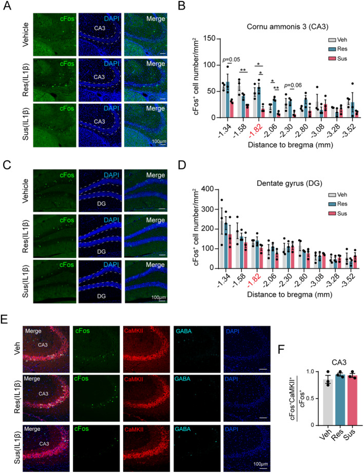
Post-task tissue shows more c-Fos-positive dCA3 cells in more adaptable mice. Dentate gyrus lacks the same group difference, and tested ventral-hippocampal changes are nonsignificant. Activity does not rise or fall uniformly across the hippocampus.
Most dCA3 activity labeling overlaps with CaMKII, pointing to an excitatory population. Overlap proportions remain similar across performance groups, while active-cell density differs more prominently. This leads the investigation to real-time task responses.
Source: Zuo W et al. · CNS neuroscience & therapeutics · CC BY
Original figure and English caption ↗
Activity differs when approaching the old location
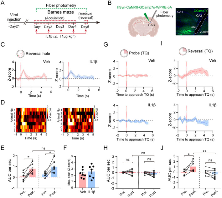
Population calcium recordings specify the difference. When mice find the relocated hole, dCA3 integrated responses and peaks do not differ significantly between controls and IL-1β groups. Original-location retrieval responses are also similar. The difference concentrates in reapproaching the former target zone during reversal.
At that moment, controls increase activity while challenged mice show an attenuated response. The region’s function therefore depends on the event being processed: responding to a familiar location that is no longer useful may relate particularly to updating choices.
Source: Zuo W et al. · CNS neuroscience & therapeutics · CC BY
Original figure and English caption ↗
Activating dCA3 reduces persistence at the old location
Increasing dCA3 CaMKII-marked activity during inflammatory challenge shortens relocated-exit search time, reduces former-target occupancy and lowers the proportion showing poor adaptability. Initial-learning latency and original-target preference during retrieval remain similar.
The intervention also changes exploration in another setting: open-field exploration increases without a significant center-time change, whereas mean maze speed remains similar. Better relocation performance thus accompanies both higher neural activity and context-dependent exploration changes.
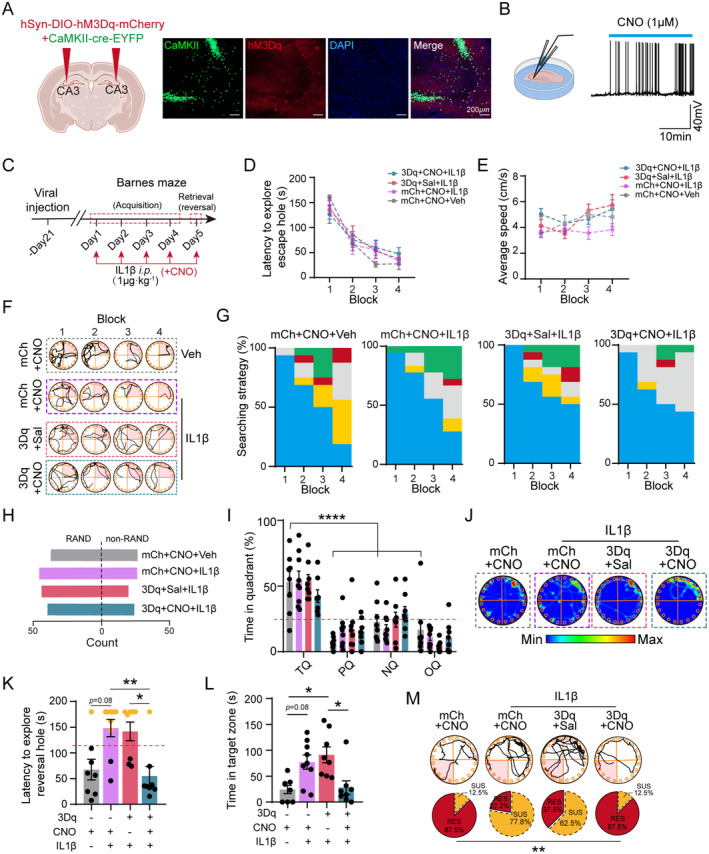
Source: Zuo W et al. · CNS neuroscience & therapeutics · CC BY
Original figure and English caption ↗
Suppressing dCA3 makes relocation harder
A separate experiment directly suppresses dCA3 excitatory cells without added IL-1β. Mice still learn the original exit and prefer its region, but search more slowly after relocation and remain longer in the former zone. The proportion with poorer adaptability increases, complementing the activation findings.
Suppressing CaMKII-marked dentate-gyrus activity mainly delays initial learning, without significantly changing the tested retrieval or reversal outcomes. The two hippocampal regions therefore affect different components of the same task sequence.
Dentate gyrus is an important CA3 input, but connected regions need not serve identical functions. Slower initial learning and difficulty updating a previous rule dissociate in these experiments, illustrating cooperation between hippocampal components for establishing and changing spatial experience.
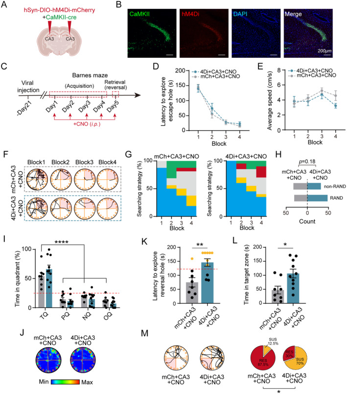
Source: Zuo W et al. · CNS neuroscience & therapeutics · CC BY
Original figure and English caption ↗
Updating spatial experience involves a wider network
Tracing identifies dCA3 outputs to ipsilateral and contralateral hippocampal regions and basal-forebrain areas including septum and vertical diagonal band. These connections situate local activity within a broader network, although individual outputs are not manipulated to identify which carries the relocation benefit.
The study separates retaining a location from adapting when it changes. Inflammatory challenge can leave original-location memory intact while making updating harder for some males. Recruiting dCA3 cells relates to meeting that changing demand, linking cognitive adaptability with a specific behavioral transition and hippocampal node.
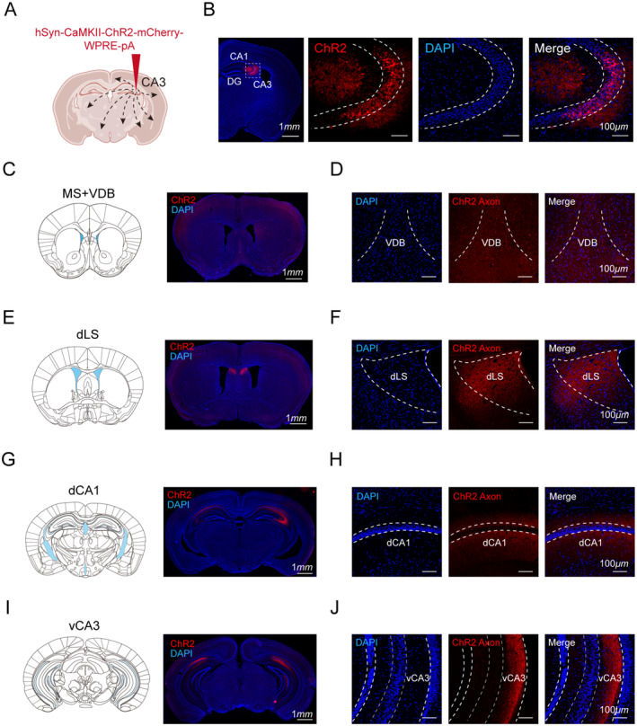
Source: Zuo W et al. · CNS neuroscience & therapeutics · CC BY
Original figure and English caption ↗