More visual activity, weaker memory: a neural turning point in blue-light effects
A male-mouse study connects spatial-memory changes during prolonged blue-light exposure with visual input engaging local prefrontal inhibition.
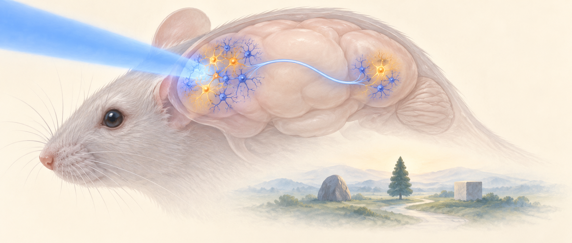
Light entering the eye does more than make things visible: it can change brain state, influencing rhythms, mood and cognition. A Communications Biology study asks why spatial memory worsens under a specific prolonged blue-light exposure. Its answer takes an apparently paradoxical turn: activity rises in visual-related regions while some prefrontal cells become quieter. A connection between them links external light with memory performance.
Excitatory input from V2M can increase inhibitory-population signals in PrL while reducing local excitatory-population signals. Reducing this input improves spatial-memory readouts under blue-light conditions.
Contents · 6 sections
How light arrives matters
Blue light has more than one relationship with cognition. Earlier studies report benefits to some cognitive measures after brief daytime exposure, while other conditions can produce different outcomes. This study focuses on male mice receiving sustained daytime exposure, comparing wavelength, intensity and duration.
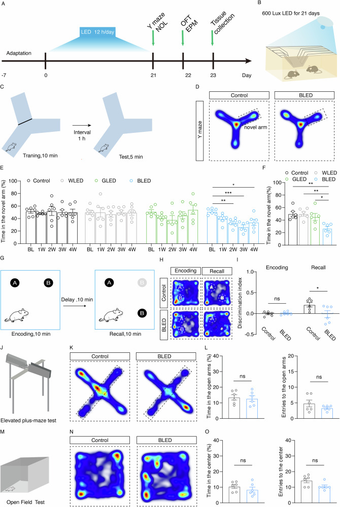
The main condition is 600 lux for twelve hours daily over twenty-one days. White and green light at the same intensity provide wavelength comparisons, alongside normal housing illumination. The results concern animals within a defined environment, where duration and biological state shape the effect of light.
Changed locations become harder to distinguish
Blue-light mice spend a smaller proportion of time in the novel maze arm and show a lower discrimination index for a relocated object. Object-location encoding shows no significant group difference; the change appears during later recall. The main maze measure is also lower than under white or green light at the same intensity.
Shorter daily exposures of four or eight hours do not produce the same significant spatial-memory decline. Open-field center exploration and elevated-maze open-arm measures also show no clear changes. The difference between spatial-task and anxiety-related readouts directs attention toward changes within the memory-related network.
Source: Zhang Y et al. · Communications biology · CC BY-NC-ND
Original figure and English caption ↗
Visual and prefrontal activity do not rise together
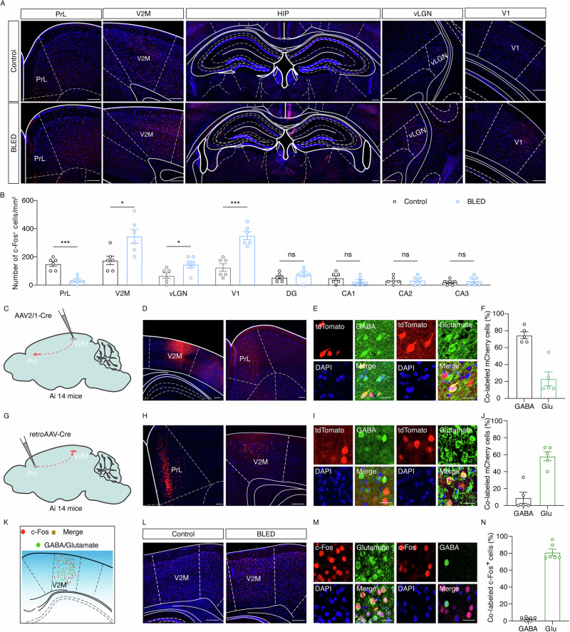
Activity-related labeling increases in primary visual cortex, medial secondary visual cortex (V2M) and ventrolateral geniculate nucleus, but decreases in prelimbic cortex (PrL). Tested hippocampal subregions show no significant labeling change. The effect therefore differs across regions rather than uniformly raising or lowering activity throughout the memory-related network.
Tracing supports a V2M–PrL connection, and the blue-light-associated V2M increase occurs predominantly in excitatory cells. PrL contains both excitatory and inhibitory populations. This raises a possibility: stronger visual input may engage a local brake before altering the population involved in cognition.
Source: Zhang Y et al. · Communications biology · CC BY-NC-ND
Original figure and English caption ↗
Changing the input changes memory readouts
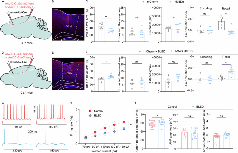
Reducing PrL excitatory cells worsens spatial-memory performance, whereas reducing the relevant V2M population projecting to PrL attenuates task changes under blue light. Changes at different locations yield different behavioral directions: the label “excitatory” alone does not describe a cell’s role within the network.
Increasing activity in the V2M–PrL projection-associated population accompanies poorer spatial-task performance, while decreasing it improves readouts under blue light. Total distance, maze entry counts and object-location encoding do not show similarly significant changes. Lower firing in PrL excitatory cells in the blue-light group supplies a physiological counterpart.
Source: Zhang Y et al. · Communications biology · CC BY-NC-ND
Original figure and English caption ↗
How can excitatory input produce inhibition?
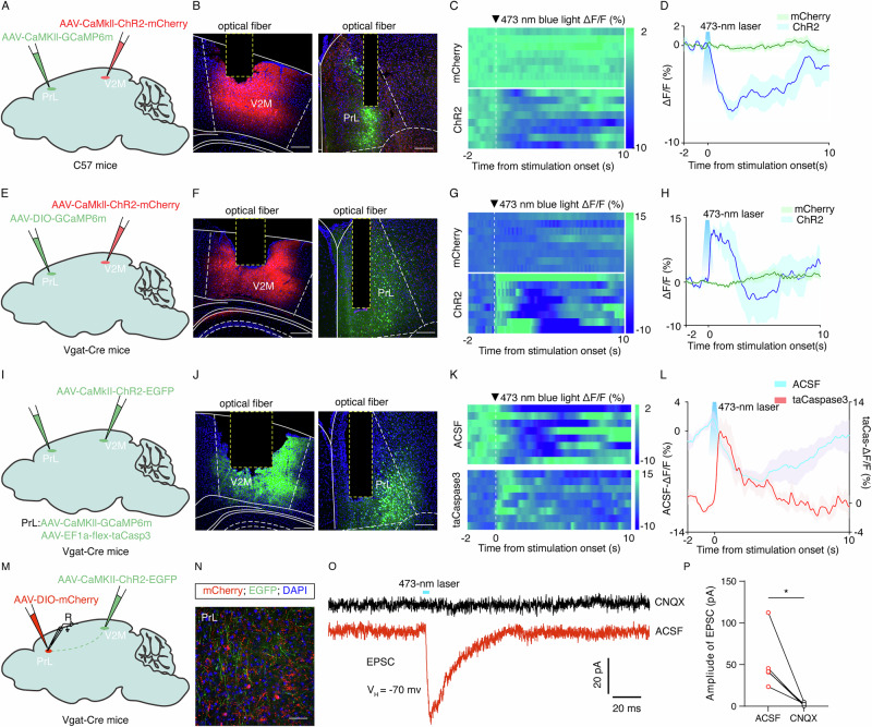
Raising activity in the V2M excitatory population rapidly increases calcium signals in PrL GABAergic cells while reducing signals in local excitatory cells. After reducing the local inhibitory population, the same source activation instead raises excitatory signals. This reversal places the inhibitory population at the center of the explanation.
Electrophysiology also shows that V2M input can evoke excitatory synaptic currents in PrL. Excitation between regions can therefore coexist with inhibition within the recipient region. Like applying more force to a brake pedal, stronger input need not strengthen the final output: it may quiet another population.
Source: Zhang Y et al. · Communications biology · CC BY-NC-ND
Original figure and English caption ↗
A cellular relay lies between light and memory
The study connects the external lighting environment with local cellular relationships. Beyond finding lower memory measures under blue light, it explains how greater visual-region activity can accompany reduced prefrontal excitatory output. Local cellular relays can turn the direction of an effect between regional input and final output.
The results come from young male mice under a specific sustained daytime exposure. Sleep involvement and the upstream retinal route to V2M are not fully resolved here. The new insight is that understanding light’s cognitive effects requires knowing who receives the input and how it is relayed, beyond whether activity increases in a particular region.