Learning without retaining: clues in a septum–hippocampus circuit
In mice, tau accumulation impairs selected cholinergic cells and septum–hippocampus theta coordination. Activating relevant inputs during early consolidation improves later spatial memory.
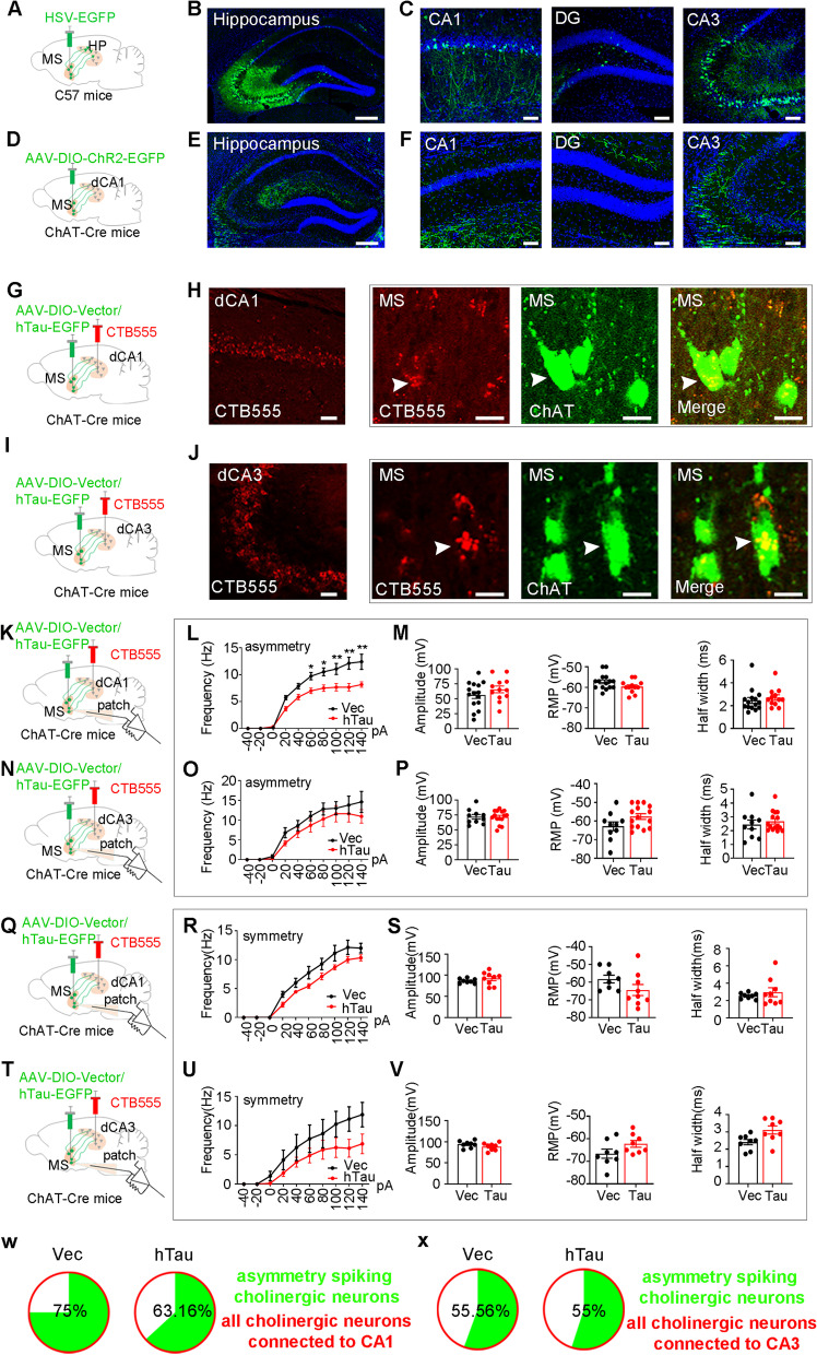
Mice gradually learn an exit’s location during training but later fail to retain it: something may go wrong after acquisition. A Molecular Neurodegeneration study published on April 14, 2023 accumulates human tau in the mouse medial septum. Spatial memory declines despite comparable training performance. The investigation points to cholinergic cells connected to hippocampal CA1 and to rhythmic coordination after learning. Helping the circuit depends on both when its activity is increased and the rhythm used.
Tau accumulation preferentially affects asymmetric-firing cholinergic cells connected to CA1 and reduces septum–CA1 theta coherence. Activating cholinergic input during early consolidation improves memory performance; local acetylcholine-receptor blockade removes this benefit.
Contents · 7 sections
Changes emerge over time, separating learning from retention
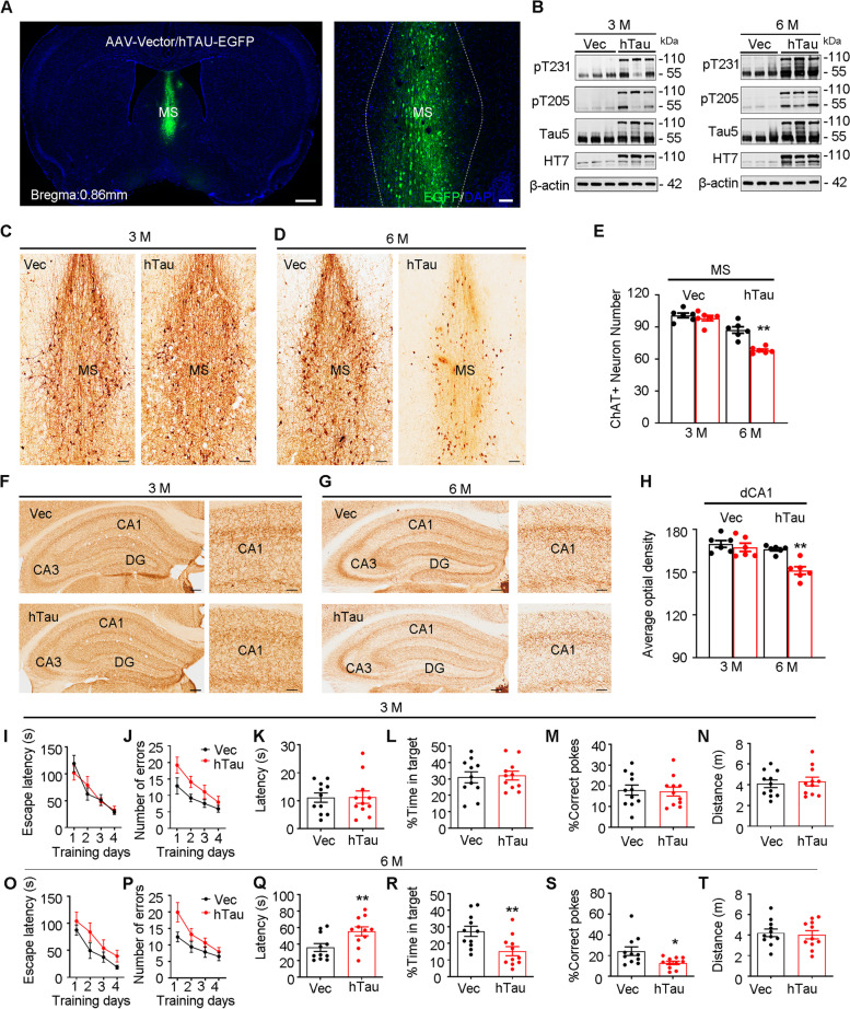
The medial septum, part of the basal forebrain, supplies important cholinergic input to the hippocampus. Abnormal tau accumulation and cholinergic changes both occur in Alzheimer’s disease, motivating an examination of their relationship within this region over time.
With broader regional tau expression, ChAT-positive counts, hippocampal cholinergic fibers and task performance show no significant changes at three months. At six months, marker-positive counts and fiber intensity decline. Training performance remains comparable, but later testing shows slower target localization, fewer correct pokes and less target occupancy, without significantly different distance traveled.
Source: Wu D et al. · Molecular neurodegeneration · CC BY
Original figure and English caption ↗
Focusing on cholinergic cells
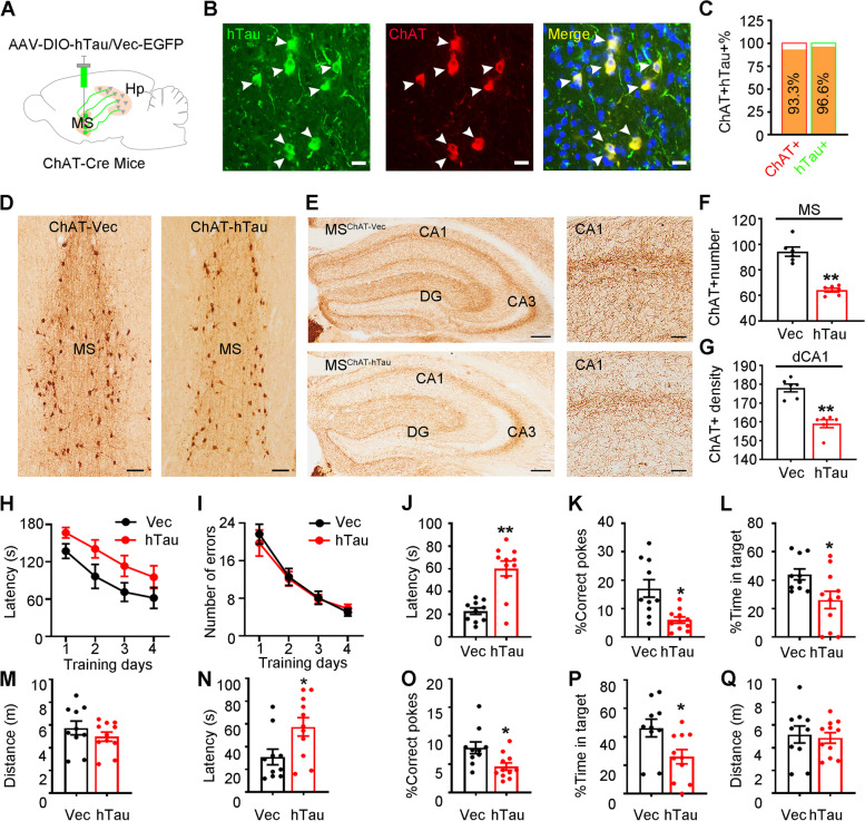
When tau accumulation is concentrated in ChAT-associated cholinergic cells, reduced marker-positive counts and weaker CA1 cholinergic fibers appear by three months. Changes emerge earlier than at the corresponding regional-expression time point, suggesting that the targeted cellular population matters.
Tau accumulation in this population again leaves training performance without a significant group difference, while spatial memory tests one day and one week later worsen. Initial acquisition and subsequent retention become dissociated in these mice.
Source: Wu D et al. · Molecular neurodegeneration · CC BY
Original figure and English caption ↗
Cholinergic cells have different electrical profiles
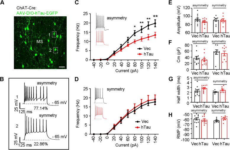
Recorded cholinergic cells fall into symmetric- and asymmetric-firing profiles, with the latter more common. Tau accumulation reduces input-driven firing in asymmetric cells, without significantly changing excitability in symmetric cells. A population often considered as one group contains different vulnerabilities.
The difference chiefly concerns how many spikes cells generate in response to input. Action-potential amplitude, half-width, capacitance and resting potential do not change significantly, indicating selective electrical alterations rather than deterioration of every measured property.
Source: Wu D et al. · Molecular neurodegeneration · CC BY
Original figure and English caption ↗
The CA1-connected branch is more vulnerable
The septum connects with multiple hippocampal regions. Projection tracing reveals that the asymmetric-cell excitability change appears in the CA1-connected population, whereas corresponding CA3-connected cells show no significant change. Symmetric-firing cells in either tested pathway likewise lack this alteration.
Combining molecular identity, firing profile and projection destination reveals the vulnerable branch. The investigation moves from a brain region toward a connected population with a particular activity profile.
Source: Wu D et al. · Molecular neurodegeneration · CC BY
Original figure and English caption ↗
Rhythms remain, but coordination weakens
Two regions can retain activity while coordinating less effectively. Simultaneous field recordings reveal reduced power in parts of the septal theta range, without significant corresponding CA1 power differences. Theta coherence between the regions falls before, during and after the task.
Coherence describes rhythmic relationships between signals rather than acetylcholine release. Local CA1 rhythms remain while their relationship with septal activity weakens, directing attention toward consolidation after learning and the possible importance of coordinated communication.
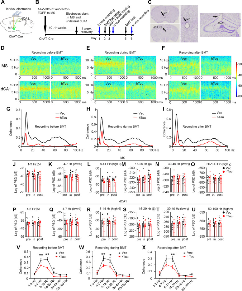
Source: Wu D et al. · Molecular neurodegeneration · CC BY
Original figure and English caption ↗
Helping retention depends on stage and rhythm
The initially tested activation condition during retrieval does not improve performance. Moving activation to immediately after new learning, under a theta-rhythmic condition, improves later target localization, correct pokes and target occupancy. Two faster rhythms do not significantly improve performance. Delaying the same theta condition until three hours after learning also yields no significant benefit.
These comparisons link the benefit to early consolidation. They demonstrate sensitivity to the particular tested timings and rhythms rather than defining a universal memory clock for every task.
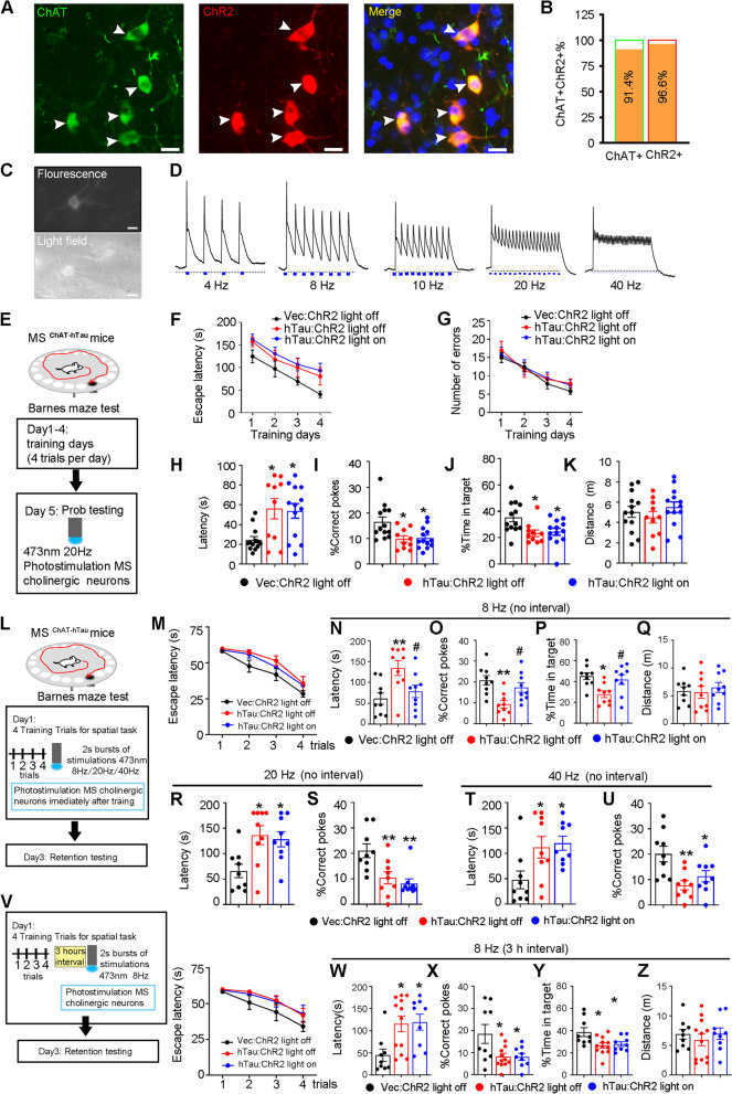
Source: Wu D et al. · Molecular neurodegeneration · CC BY
Original figure and English caption ↗
Localizing the benefit to hippocampal input
Increasing septal cholinergic input within CA1 during early consolidation also improves later spatial memory. Local blockade of the tested acetylcholine receptors removes this benefit, while distance traveled remains without a significant difference. Cholinergic signaling therefore has experimental support as part of the circuit effect.
Local human-tau accumulation in mice does not encompass the full human disease, and reduced labeling does not resolve every cell’s fate. The study locates memory vulnerability at the intersection of firing profile, projection destination and post-learning stage—three levels that may all matter when supporting an affected network.
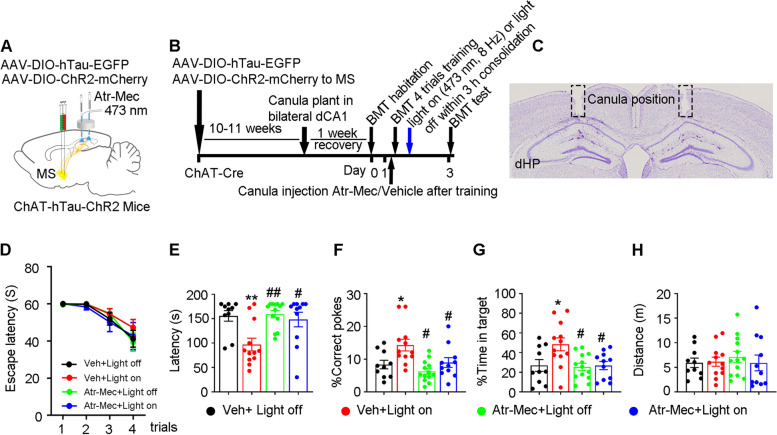
Source: Wu D et al. · Molecular neurodegeneration · CC BY
Original figure and English caption ↗