How does the heart enter post-seizure risk? Clues linking the locus coeruleus and cardiac receptors
In DBA/1 mice, a study connects seizure-induced respiratory arrest with locus coeruleus noradrenergic signaling, cardiac tyrosine hydroxylase activity and myocardial β1-adrenergic receptors across ECG and calcium recordings.
Research on sudden unexpected death in epilepsy (SUDEP) often centers on seizure-induced respiratory arrest (S-IRA), yet respiratory collapse frequently coincides with cardiac rhythm disturbances. Published in iScience on February 27, 2023, a study in DBA/1 mice extended the focus from the locus coeruleus (LC)—the brain’s primary noradrenergic nucleus—to the heart. The protective effect of atomoxetine in lowering S-IRA incidence and alleviating post-seizure atrioventricular block was weakened either after LC noradrenergic perturbation or after β1-adrenergic receptor blockade with esmolol, alongside reduced cardiac tyrosine hydroxylase activity, elevated phosphorylated β1-receptor levels and altered bilateral LC calcium signals.
In induced-seizure DBA/1 mouse models, locus coeruleus noradrenergic signaling and β1-receptor pharmacology are functionally linked to seizure-induced respiratory arrest and cardiac arrhythmias; these findings do not establish a direct anatomical LC-to-heart projection or clinical SUDEP prevention in humans.
Contents · 6 sections
Post-seizure vulnerability involves both breathing and cardiac rhythm
Seizure-induced respiratory arrest (S-IRA) is a primary readout in experimental models of epilepsy-related mortality, yet the autonomic nervous system regulates both respiration and circulation. Earlier observations showed that respiratory suppression in DBA/1 mice coincides with cardiac depression, while the norepinephrine reuptake inhibitor atomoxetine lowers S-IRA incidence.
If noradrenergic signaling acts across both central and peripheral sympathetic pathways, the brainstem locus coeruleus (LC) and cardiac adrenergic receptors could work in concert. When the authors first perturbed LC noradrenergic terminals or local LC neurons, atomoxetine’s reduction of S-IRA incidence was weakened, motivating a closer look at peripheral cardiac markers and receptor responses.
Cardiac tyrosine hydroxylase content rises while enzymatic activity falls after S-IRA
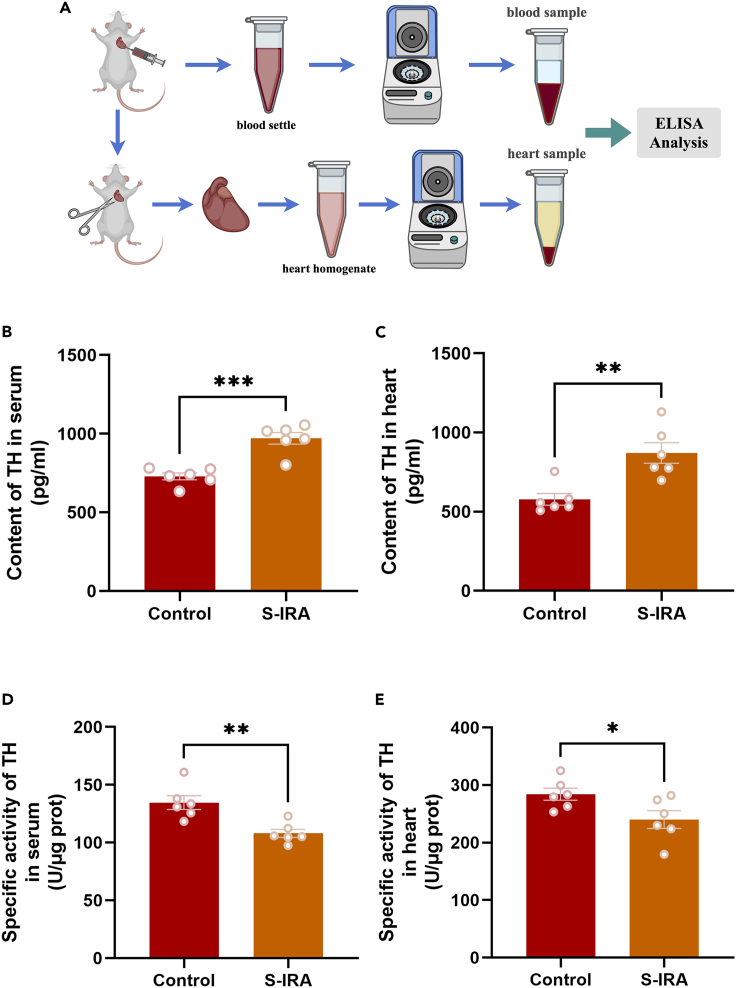
Tyrosine hydroxylase (TH) is the rate-limiting enzyme in norepinephrine synthesis. To assess peripheral noradrenergic status following chemically induced S-IRA, the team measured both TH protein content and specific enzymatic activity in left-ventricular serum and whole-heart tissue from DBA/1 mice.
The two readouts moved in opposite directions: compared with controls, the S-IRA group showed significantly higher TH content in both left-ventricular serum and whole-heart tissue, whereas specific TH enzymatic activity was significantly reduced in both sample types. Higher enzyme abundance alongside lower catalytic activity indicates that protein level alone does not reflect functional output during post-seizure sympathetic stress.
Source: Lian X et al. · iScience · CC BY-NC-ND
Original figure and English caption ↗
Phosphorylated β1-receptors and their ratio increase across cardiac chambers
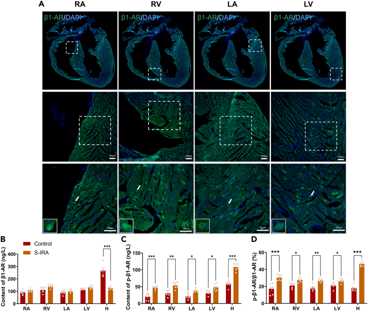
Norepinephrine released by cardiac sympathetic fibers acts primarily on cardiomyocyte β1-adrenergic receptors (β1-AR). The authors sampled the right atrium, right ventricle, left atrium, left ventricle and whole-heart tissue to compare total β1-AR and phosphorylated β1-AR (p-β1-AR) levels.
Across the four individual chambers, total β1-AR content did not differ significantly between control and S-IRA groups, whereas whole-heart total β1-AR was higher in controls. In contrast, both phosphorylated β1-AR content and the p-β1-AR/β1-AR ratio were significantly elevated across the right atrium, right ventricle, left atrium, left ventricle and whole heart in the S-IRA group, pointing to a widespread shift in receptor phosphorylation state.
Source: Lian X et al. · iScience · CC BY-NC-ND
Original figure and English caption ↗
Atomoxetine alleviates post-seizure atrioventricular block, and esmolol reverses the protection
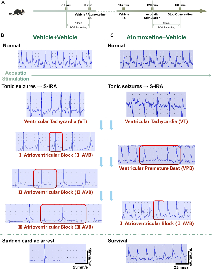
To track cardiac electrophysiology directly, the authors recorded surface electrocardiograms (ECG) in both acoustic and chemically induced seizure models. Control mice rapidly developed ventricular tachycardia, ventricular premature beats and second- to third-degree atrioventricular block following S-IRA, culminating in cardiac arrest; mice pre-treated with atomoxetine still showed transient rhythm disturbances but experienced markedly fewer lethal high-grade blocks and survived.
When the selective β1-AR antagonist esmolol was co-administered with atomoxetine, both the reduction in S-IRA incidence and the alleviation of high-grade atrioventricular block were reversed. Administering esmolol alone produced only transient bradycardia without inducing atrioventricular block or altering baseline mortality, supporting a requirement for β1-AR signaling in atomoxetine’s protective effect.
Source: Lian X et al. · iScience · CC BY-NC-ND
Original figure and English caption ↗
Seizure-phase LC calcium surges are dampened by atomoxetine and restored with esmolol
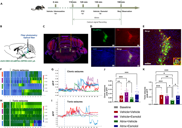
On the brainstem side, the authors expressed GCaMP6m under the DBH promoter in bilateral LC noradrenergic neurons and used fiber photometry to record population calcium activity during chemically induced clonic and tonic seizure phases. Seizure episodes were accompanied by pronounced bilateral LC calcium surges above baseline.
Compared with vehicle or esmolol-treated groups, mice pre-treated with atomoxetine exhibited a significantly lower LC calcium peak during tonic seizures, whereas adding esmolol restored a higher tonic-phase peak. The authors suggest that presynaptic norepinephrine accumulation after transporter blockade may restrain further calcium influx through feedback regulation while reflecting functional coupling across the brain–heart axis.
Source: Lian X et al. · iScience · CC BY-NC-ND
Original figure and English caption ↗
Functional coupling is evident, while anatomical projections and cell-specific control remain open
Taken together, pharmacological perturbations, cardiac enzyme and receptor assays, surface ECG recordings and bilateral LC calcium photometry indicate that post-seizure vulnerability spans more than a single organ: central LC noradrenergic signaling and β1-receptor-dependent cardiac rhythm stability show clear functional interplay.
At the same time, the authors explicitly note several limitations: the study relied on pharmacological and chemical perturbations alongside limb-lead ECG rather than projection-specific optogenetic or chemogenetic control, did not map the anatomical multisynaptic pathway from the LC to cardiac sympathetic preganglionic neurons, and did not measure broader hemodynamic parameters such as ejection fraction. Addressing these questions will be essential to define the exact circuit architecture linking brainstem noradrenergic activity to cardiac survival.