Hippocampal learning responses weaken after BPA exposure
In developmentally exposed male mice, spatial-task performance, hippocampal input measures and learning-related calcium responses change. Increasing CA1 activity improves performance, while activation attempts in other subregions produce severe seizure-like responses.

When a mouse searches for a remembered location, its hippocampal network processes environmental cues. How does exposure to bisphenol A alter this process? A study in Environment & Health examines developmentally exposed male mice across behavior, cellular branching, synaptic input and real-time activity. Hippocampal responses during learning weaken, with cellular changes differing across subregions.
BPA-exposed mice show poorer spatial-task performance, lower synaptic event frequency across hippocampal regions and smaller learning-related calcium peaks with altered timing. Increasing CA1 excitatory-cell activity improves associated behavior.
Contents · 6 sections
Finding a remembered location takes longer
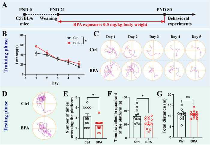
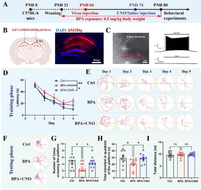
The study examines sustained exposure from after weaning into early adulthood, with behavioral tests after exposure ends. Exposed mice take longer to find the platform during water-maze learning. During a later test without the platform, they cross its former location fewer times and spend less time in the target quadrant. Changes occur during both learning and later spatial-preference measurements.
Total movement distance during the test does not differ significantly. The result centers on location-search performance rather than a reduction in total distance. The team then examines the hippocampal network supporting the task, asking how its inputs, cellular structure and responses change.
Source: Bi N et al. · Environment & health (Washington, D.C.) · CC BY-NC-ND
Original figure and English caption ↗
Input measures fall within and into the hippocampus
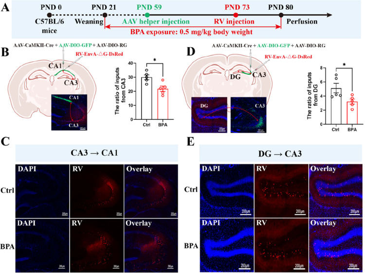
Excitatory connections link dentate gyrus, CA3 and CA1, while entorhinal cortex also supplies hippocampal inputs. Trans-synaptic tracing shows lower dentate-gyrus–CA3 and CA3–CA1 input ratios in exposed mice, together with lower corresponding entorhinal inputs to dentate gyrus and CA1.
The measure compares labeled upstream cells with labeled starter cells and describes relative changes in network input. It links behavioral differences with several connected elements and motivates a cellular question: do neurons in these networks retain comparable input-receiving structures and electrical responses?
The hippocampus contains interconnected internal stages and receives direct cortical inputs. Changes across these connections bring network state, beyond any one region, into focus.
Source: Bi N et al. · Environment & health (Washington, D.C.) · CC BY-NC-ND
Original figure and English caption ↗
Simpler branches, but uneven excitability changes
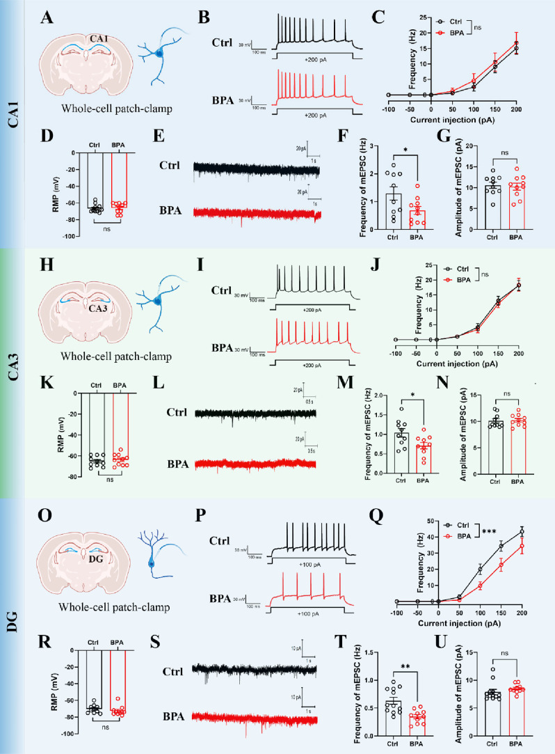
Excitatory neurons in all three subregions have lower branching complexity, indicating altered input-receiving structure. Electrical measurements reveal a more specific pattern: intrinsic excitability and resting potential in CA1 and CA3 do not change clearly, while dentate granule cells have lower intrinsic excitability.
Miniature excitatory synaptic event frequency falls in all three regions, while individual event amplitudes remain comparable. Resting potential does not change significantly, and a presynaptic-associated marker falls while the measured postsynaptic marker is comparable. Different network components therefore show different forms of change.
For CA1 and CA3, comparable responses to direct stimulation do not imply comparable inputs in the intact network. This distinction matters for the subsequent activity measurements: intrinsic cellular properties and the degree of recruitment during a task describe different aspects of neuronal function.
Source: Bi N et al. · Environment & health (Washington, D.C.) · CC BY-NC-ND
Original figure and English caption ↗
Lower peaks with altered response timing
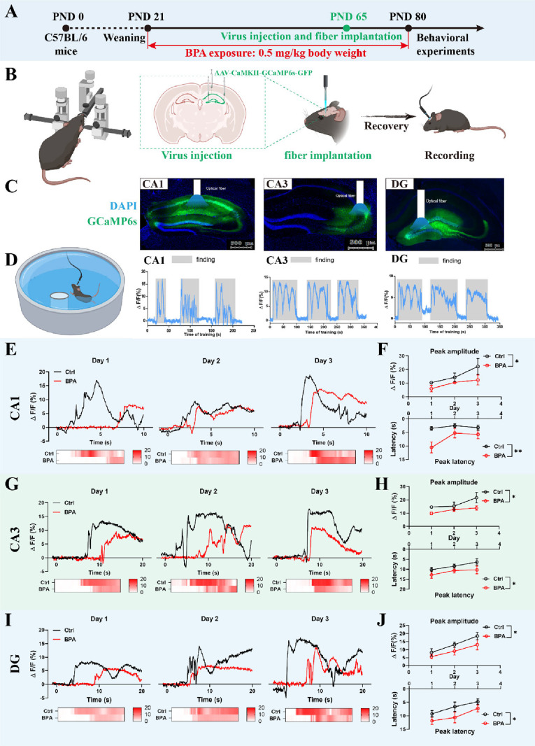
Task-related calcium activity provides a dynamic readout. Hippocampal responses are greater during platform search than while resting on it and vary over learning. In exposed mice, CA1, CA3 and dentate responses have lower peaks with altered peak timing.
CA1 and CA3 retain comparable intrinsic excitability but show weaker learning-related responses, alongside fewer input events. Combining cellular measurements with task activity reveals a network-level difference: cells can retain some intrinsic electrical properties while responding differently during behavior.
Source: Bi N et al. · Environment & health (Washington, D.C.) · CC BY-NC-ND
Original figure and English caption ↗
Performance improves after increasing CA1 activity
Increasing dorsal CA1 excitatory-neuron activity improves platform-search performance in exposed mice. Later former-platform crossings and target-quadrant time increase, while total distance remains comparable. The intervention links altered CA1 activity with improved spatial-task behavior.
Attempts in other subregions differ: increasing activity in CA3 or dentate-associated cells produces severe seizure-like responses and even death, preventing the same spatial behavioral comparison. Different nodes respond very differently to increased activity.
Source: Bi N et al. · Environment & health (Washington, D.C.) · CC BY-NC-ND
Original figure and English caption ↗
Network state shapes learning-related responses
The study connects behavior with branching structure, input events and dynamic activity. Poorer spatial performance does not correspond to an identical loss of function everywhere: some cells change intrinsically, others show altered inputs and task responses, and changing CA1 activity can affect performance.
The findings come from a defined developmental exposure condition in male mice, assessed after exposure ends. They connect an environmental factor with changes across neural levels and show why understanding memory requires attention to both cells and communication between them. Learning-related responses emerge from network participation.