How do hippocampal inputs cooperate when learning a route? Two CA1 inputs and joint potentiation
Spatial-learning and ex vivo hippocampal observations in male mice examine entorhinal and CA3 inputs: single-pathway activity fails to produce the same lasting enhancement seen with coactivation.

Finding a target takes time in an unfamiliar environment, and performance improves with experience. How do hippocampal inputs cooperate? A Communications Biology study examines medial entorhinal and CA3 inputs to CA1, combining spatial-task observations with ex vivo synaptic responses. Coactivation produces lasting enhancement of CA3-derived responses, whereas either pathway alone does not produce the same result. Spatial learning and synaptic cooperation meet along the same connections.
MEC-related inputs influence CA1-projecting CA3 population activity and task performance; in ex vivo CA1, coactivation supports heterosynaptic long-term potentiation.
Contents · 6 sections
CA1 receives entorhinal and intrahippocampal signals
Medial entorhinal cortex (MEC) sends spatially relevant information into hippocampus, while CA3 Schaffer collaterals reach CA1. The inputs share a destination but have different sources. Studying them together asks how they interact beyond the strength of either input alone. The work begins by following CA1-projecting CA3 populations during water-maze navigation.
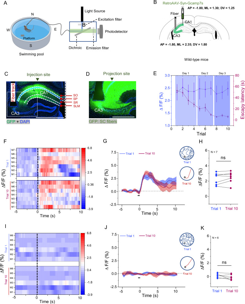
Finding the target becomes faster with training, but CA3 calcium does not simply rise with performance. The short-window average after entering the water does not differ significantly between initial and final trials, and signals closely correlate with swimming speed. Behavior and bulk activity have distinct patterns, prompting tests of how MEC input affects CA3-related responses.
Source: Huang F et al. · Communications biology · CC BY-NC-ND
Original figure and English caption ↗
Increasing MEC activity enhances CA3-related responses
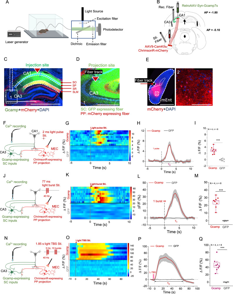
Increasing MEC activity with different tested patterns elicits calcium responses in CA1-projecting CA3 populations, with strength and duration varying by pattern. Control fluorescence does not show the same changes. The regions are functionally linked, demonstrating that external input can influence an intrahippocampal population response.
The influence can pass through several network steps, and population recording captures their combined effect. It does not identify every transmitting cell or synapse, but moves the two inputs from parallel anatomical names to a functional interaction. Behavioral tasks then test the consequences of reducing MEC-related input.
Source: Huang F et al. · Communications biology · CC BY-NC-ND
Original figure and English caption ↗
Reducing MEC input affects activity and performance
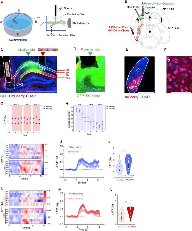
Reducing MEC-related hippocampal input lowers CA3-related calcium in both initial and trained stages and prolongs escape latency. Ex vivo recordings show weaker transmission in the associated input, providing an additional functional observation. Concurrent activity and task changes make the MEC contribution more concrete.
The intervention alters a group of inputs, leaving several possible cell and relay contributions. Behavioral results support their role in normal performance without assigning every effect to one isolated synapse type. The next question focuses on CA1: can converging inputs leave a lasting change in synaptic responses?
Source: Huang F et al. · Communications biology · CC BY-NC-ND
Original figure and English caption ↗
Coactivation leaves a stronger lasting response
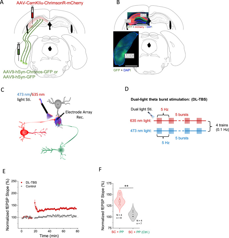
In ex vivo hippocampus, coactivation produces lasting enhancement of CA3-derived synaptic responses, unlike controls. This is described as heterosynaptic long-term potentiation: another input contributes to strengthening the measured pathway. Cooperation becomes a persistent observable effect, with interactions changing subsequent responses.
With NMDA receptors or voltage-gated calcium channels blocked in supplemental observations, the same significant potentiation is absent. Receptor and calcium-related processes enter the explanatory model. Ex vivo plasticity and task results provide evidence at different levels, without establishing a direct causal relay between this plasticity and behavior.
Source: Huang F et al. · Communications biology · CC BY-NC-ND
Original figure and English caption ↗
Each input responds, but neither alone shows the same potentiation
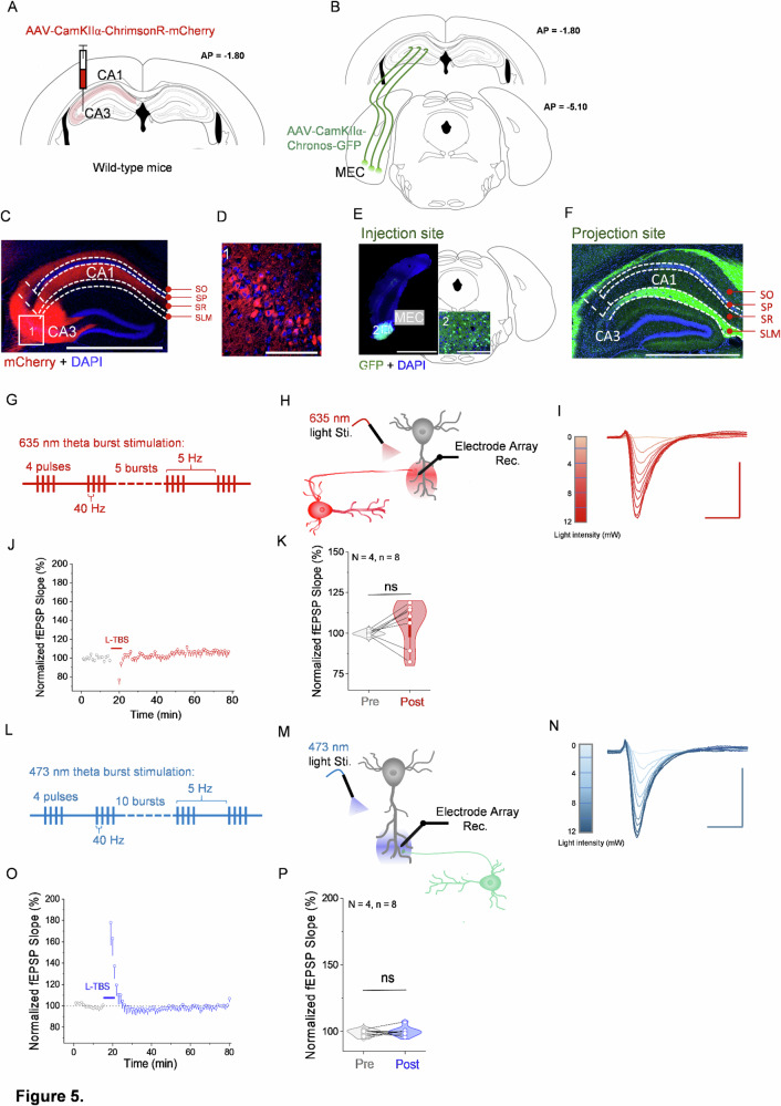
Either CA3–CA1 or MEC–CA1 activity elicits synaptic responses but fails to produce significant lasting potentiation under the tested conditions. The null results clarify the coactivation finding: transmission and persistent change are different questions. An input can respond without independently producing the same plasticity outcome.
Comparing single and joint activity focuses attention on relationships among sources. It is cooperation under the present conditions rather than a universal rule for every activity pattern. For CA1, which inputs arrive matters, and their joint engagement can influence how long an altered response persists.
Source: Huang F et al. · Communications biology · CC BY-NC-ND
Original figure and English caption ↗
Different populations participate with different activity patterns
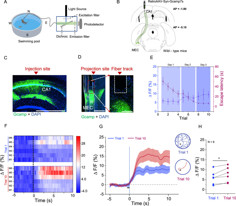
The CA1-projecting MEC population shows higher plotted calcium responses in trained than initial trials, alongside faster escape. This differs from the nonsignificant initial–final short-window comparison in CA3. Inputs associated with the same task need not share one activity curve; source and recording window shape the observed changes.
The male-mouse study combines input contributions during spatial tasks with ex vivo cooperation in CA1. Its clearest insight is that hippocampal processing depends on how sources interact as well as whether one input is active. Performance, population activity and potentiation tell complementary parts of the learning-network story.
Source: Huang F et al. · Communications biology · CC BY-NC-ND
Original figure and English caption ↗