How can a nanoparticle engage a memory-related signaling relay?
Polydopamine improves memory-related performance in an inflammatory mouse model, while hippocampal transmitter changes and septal cholinergic activity suggest a route from material to neural network.

Does supplying one signaling molecule affect only that molecule? A Theranostics study explores the question using polydopamine nanoparticles, whose dopamine-related structure can release dopamine in acidic environments. In male mice, the material improves memory-task performance under inflammatory conditions. Over time, the prominent hippocampal transmitter changes shift from catecholamines toward acetylcholine, suggesting a relay between material chemistry and neural circuitry.
Behavioral improvement accompanies time-dependent transmitter changes, and D2 receptor blockade weakens associated signaling and memory performance. The septum–hippocampus connection offers a clue to this relay.
Contents · 6 sections
The material itself can be a signal source
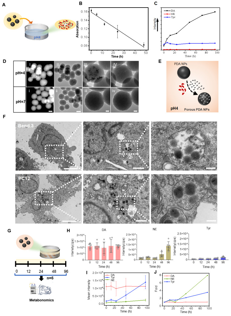
Dopamine contributes to attention, motivation and learning, but does not readily enter the brain from the periphery. Polydopamine offers another idea: rather than simply carrying a separate cargo, the material contains dopamine-related components that can be released. The spherical particles are about 250 nanometers across, and the study first asks how their surroundings change their behavior.
In acidic conditions, small granules and pores appear as dopamine is released, while the overall sphere remains intact. In cell cultures, uptake accompanies an increase in norepinephrine. The seemingly inert dark sphere therefore has environment-dependent chemical activity, with release and cellular metabolism potentially shaping subsequent signaling.
Source: Liu PM et al. · Theranostics · CC BY
Original figure and English caption ↗
Brain evidence sits within a wider distribution
The blood–brain barrier is the first obstacle. Fluorescent surrogate particles with a polydopamine coating produce detectable brain signals, while separate tissue observations show dark particles in brains after polydopamine treatment. These complementary observations support entry into the brain.
The surrogate is not identical to pure polydopamine, and particles also occur in liver, spleen and kidney. The evidence concerns a material capable of reaching the brain, with a broader bodily distribution. The next question is whether that distribution accompanies changes in memory-related behavior and brain chemistry.
Memory-task performance improves without increased movement
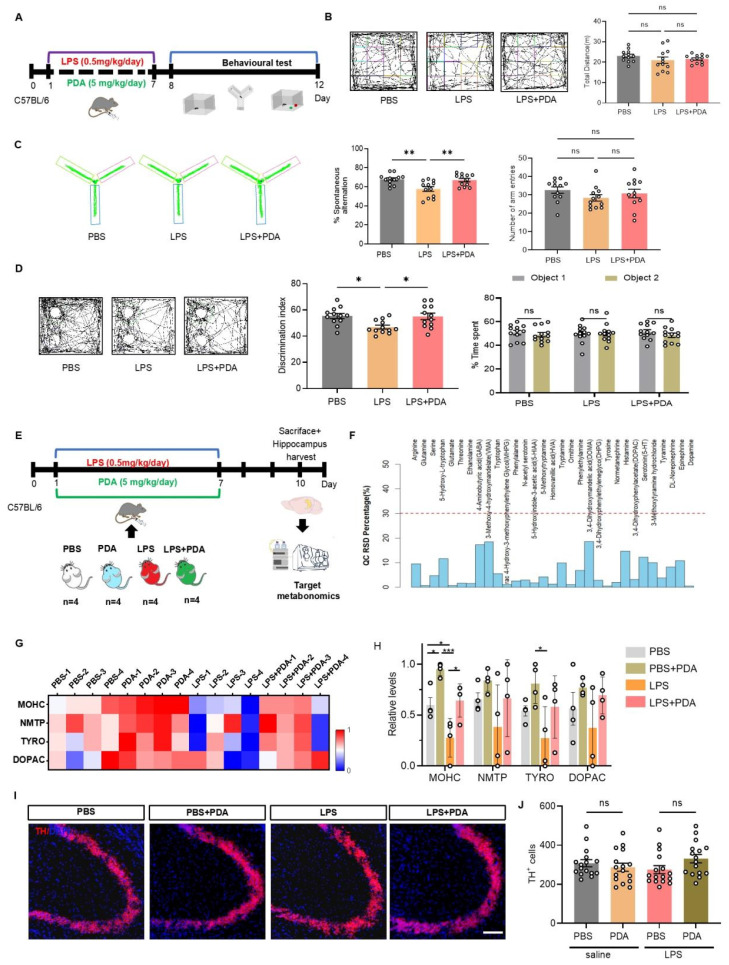
In a lipopolysaccharide-induced inflammatory model, mice perform worse in maze alternation and novel-object discrimination. Both measures improve after polydopamine, approaching controls in the main experiment. Total movement distance and body-weight recovery show no significant change, so the memory-task improvement does not reflect uniform recovery across measures.
Hippocampal analysis also detects changes in several catecholamine-related metabolites, while counts of tyrosine-hydroxylase-positive cells show no clear difference. Behavioral and chemical observations move the story from whether performance changes to which signals accompany it, within this inflammation-related impairment model.
Source: Liu PM et al. · Theranostics · CC BY
Original figure and English caption ↗
Earlier dopamine changes meet later acetylcholine changes
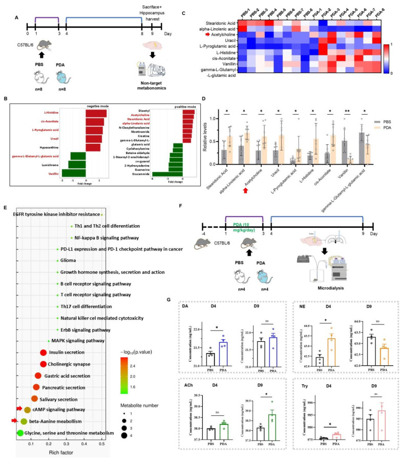
Another transmitter analysis follows the time course. On day four, dopamine, norepinephrine and tyrosine rise, while acetylcholine shows only an upward trend. By day nine, acetylcholine is significantly higher, but dopamine and tyrosine are no longer significantly elevated. The day-nine norepinephrine comparison is also nonsignificant, with a lower numerical value.
This sequence suggests an effect that can propagate through a network rather than remain dependent on one persistently elevated transmitter. In separate fluorescent-sensor observations, acetylcholine-associated signals increase at several time points. Distinct readouts provide different windows, bringing cholinergic signaling into focus.
Source: Liu PM et al. · Theranostics · CC BY
Original figure and English caption ↗
Septum and hippocampus connect the signaling systems
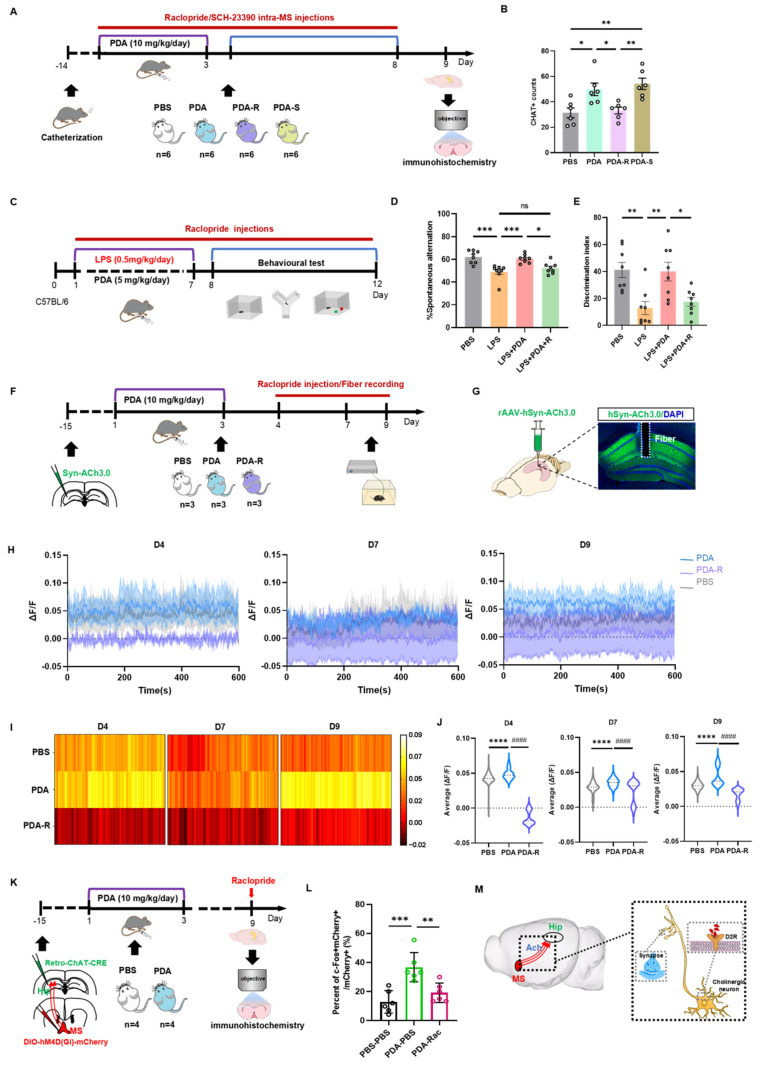
An important cholinergic input to hippocampus originates in the medial septum. After polydopamine, septal ChAT-positive labeling increases and activity-related labeling changes in septal cholinergic cells projecting to hippocampus. Reducing activity in this population lowers the associated activity marker, supporting its involvement in the network response.
D2 receptors provide another clue. Their blockade weakens associated septal labeling, hippocampal acetylcholine fluorescence and behavioral improvement in the inflammatory model. Receptor, transmitter and behavior form an interconnected evidence chain, while the behavioral blockade acts systemically rather than only within the septum.
Source: Liu PM et al. · Theranostics · CC BY
Original figure and English caption ↗
A material can act across more than one level
The study joins material changes, brain transmitters and memory-related circuitry within one story. Polydopamine can release signaling molecules and may also act through antioxidant and anti-inflammatory properties; these contributions are not fully separated here. Its central insight is that a material’s biological effects can extend from its chemical components into another transmitter system.
The transmitter observations span days. Major-organ histology shows no obvious damage, while high concentrations reduce viability in some cell cultures. Biological context and exposure therefore matter. The male-mouse findings pose an engaging mechanistic question: how can changing the source of one signal recruit a circuit and pass the next part of the memory-related effect to another signal?