How can a metabolic receptor shape drug memory? Adiponectin signaling in an amygdala–prefrontal pathway
A mouse study shows that adiponectin receptor 1 in basolateral amygdala neurons projecting to prelimbic cortex dampens excitability and weakens methamphetamine-associated memory.
Persistent drug-associated memories are a major driver of cue-induced relapse in methamphetamine (MA) use disorder. Although the metabolic hormone adiponectin and its receptor AdipoR1 modulate central synaptic plasticity, the specific amygdalocortical projection and downstream ion channel mechanism through which AdipoR1 gates MA-associated reward memory have remained unclear. Published in Cell Reports on December 10, 2024, a study led by Prof. Hao Hong, Prof. Hao Wang, and Assoc. Prof. Yan Long at China Pharmaceutical University ("Adiponectin receptor 1-mediated basolateral amygdala-prelimbic cortex circuit regulates methamphetamine-associated memory") combined pharmacology, viral knockdown and overexpression, Caspase-3 ablation, pathway-specific chemogenetics, GCaMP6s fiber photometry, slice electrophysiology, and physical exercise training in male C57BL/6J mice to show that AdipoR1 in prelimbic cortex (PrL)-projecting basolateral amygdala excitatory pyramidal neurons (BLA CaMKIIα+→PrL) suppresses MA-induced conditioned place preference (CPP) by activating AMPK and downregulating the L-type calcium channel Cav1.3.
Methamphetamine reduces adiponectin and AdipoR1 expression in BLA CaMKIIα+ pyramidal neurons; activating AdipoR1 in BLA CaMKIIα+→PrL projection neurons—or restoring this pathway via long-term physical exercise—activates AMPK to downregulate Cav1.3 L-type calcium channels, reducing neuronal excitability and terminal Ca2+ activity to suppress MA-associated memory formation.
Contents · 6 sections
Systemic or Intracerebroventricular AdipoRon Suppresses MA CPP Formation, and MA Downregulates AdipoR1 in BLA Pyramidal Neurons
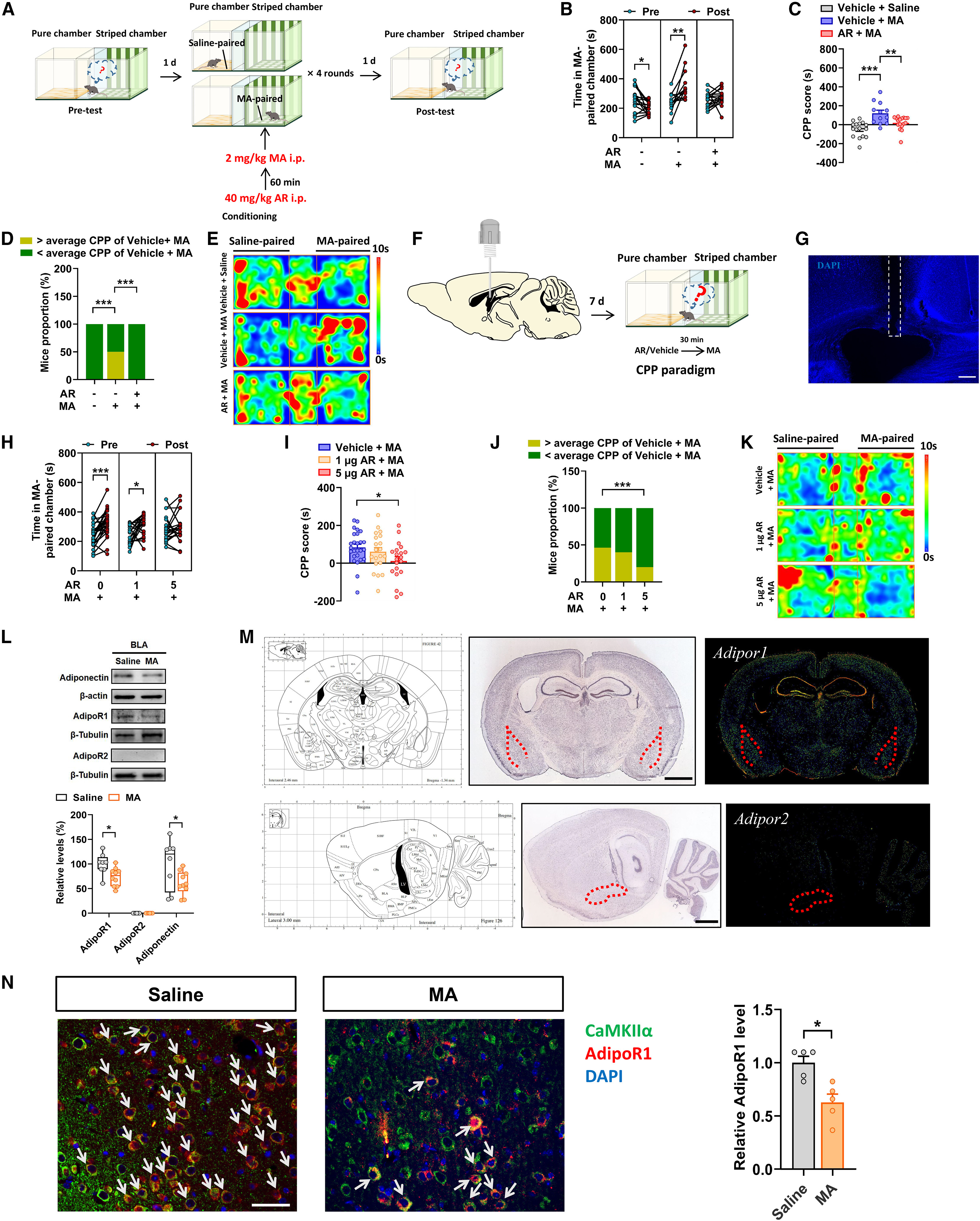
Using a methamphetamine (MA, 2 mg/kg, i.p.) conditioned place preference (CPP) paradigm in male C57BL/6J mice, the authors found that systemic administration of the adiponectin receptor agonist AdipoRon (40 mg/kg, i.p., 60 min prior to MA) or bilateral intracerebroventricular (i.c.v.) infusion of AdipoRon (1 or 5 μg/side) during conditioning significantly suppressed MA-induced CPP formation without altering locomotor velocity, whereas administration before CPP expression or 0.5 mg/kg MA-primed reinstatement had no effect (Figure S1).
Molecular profiling revealed that 2 mg/kg MA conditioning reduced adiponectin levels in the lateral hypothalamus (LH) and significantly decreased both adiponectin and AdipoR1 protein levels in the basolateral amygdala (BLA), with no changes in the VTA or NAc and undetectable AdipoR2 in the BLA. Immunofluorescence colocalization demonstrated that MA selectively reduced AdipoR1 intensity in BLA CaMKIIα+ excitatory pyramidal neurons (PyNs) rather than GAD67+ interneurons.
Source: Zhigang Chen et al. · Cell Reports · © Elsevier Inc.
Original figure and English caption ↗
Overexpressing AdipoR1 in BLA CaMKIIα+ Neurons Suppresses 2 mg/kg MA CPP, Whereas Knockdown Facilitates Suboptimal 0.25 mg/kg MA Reward
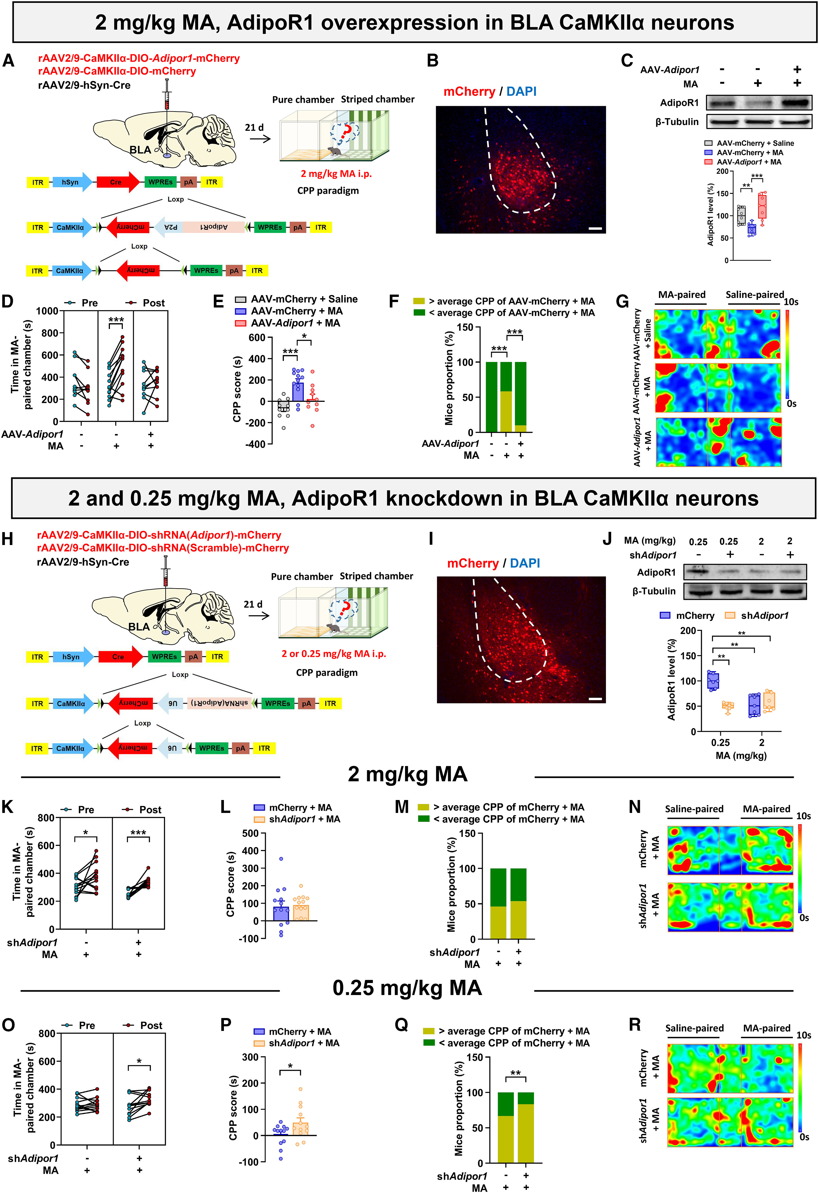
To test the causal role of BLA excitatory neuronal AdipoR1, the authors overexpressed AdipoR1 selectively in BLA CaMKIIα+ neurons using rAAV2/9-CaMKIIα-Adipor1, which robustly suppressed 2 mg/kg MA-induced CPP. When AdipoR1 was knocked down in BLA CaMKIIα+ neurons (rAAV2/9-CaMKIIα-DIO-shRNA(Adipor1)-mCherry + rAAV2/9-hSyn-Cre), it did not further lower AdipoR1 protein or increase CPP under the 2 mg/kg MA dose due to a ceiling effect from MA-induced endogenous AdipoR1 downregulation.
Crucially, under a suboptimal MA conditioning dose (0.25 mg/kg) that did not downregulate endogenous AdipoR1 in controls, knocking down AdipoR1 in BLA CaMKIIα+ neurons markedly enhanced 0.25 mg/kg MA-induced CPP (and promoted intravenous MA self-administration; Figure S4A–C), demonstrating that loss of BLA AdipoR1 heightens susceptibility to MA reward memory.
Source: Zhigang Chen et al. · Cell Reports · © Elsevier Inc.
Original figure and English caption ↗
Intra-BLA AdipoRon Selectively Reduces Prelimbic Cortex c-Fos, and BLA CaMKIIα+→PrL Activity Bidirectionally Gates MA CPP
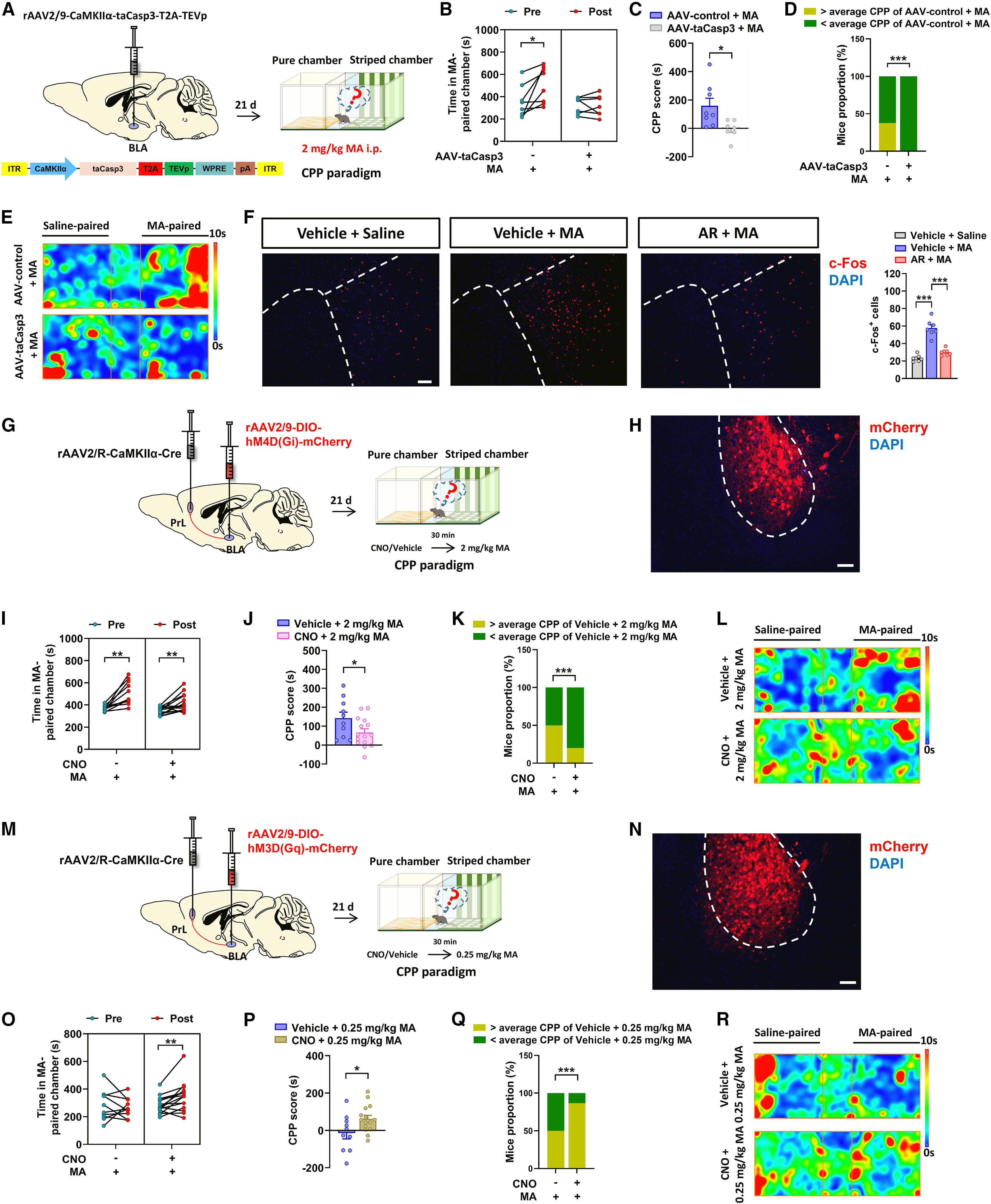
Selective Caspase-3 ablation (taCasp3) of BLA CaMKIIα+ neurons attenuated 2 mg/kg MA-induced CPP, confirming their necessity. Mapping downstream c-Fos expression after intra-BLA AdipoRon microinfusion revealed a selective reduction of c-Fos+ neurons in the prelimbic cortex (PrL), with no significant change in the NAc, lateral septum (LS), anterior cingulate cortex (ACC), or infralimbic cortex (IL) (Figure S4D).
Pathway-specific chemogenetic manipulation confirmed the functional role of the BLA CaMKIIα+→PrL projection: chemogenetic inhibition (hM4Di) of BLA CaMKIIα+→PrL neurons suppressed 2 mg/kg MA-induced CPP, whereas chemogenetic excitation (hM3Dq) of BLA CaMKIIα+→PrL neurons facilitated CPP under the suboptimal 0.25 mg/kg MA dose.
Source: Zhigang Chen et al. · Cell Reports · © Elsevier Inc.
Original figure and English caption ↗
AdipoR1 Overexpression in BLA CaMKIIα+→PrL Neurons Reduces Excitability, and Intra-BLA AdipoRon Suppresses Terminal Ca2+ Activity
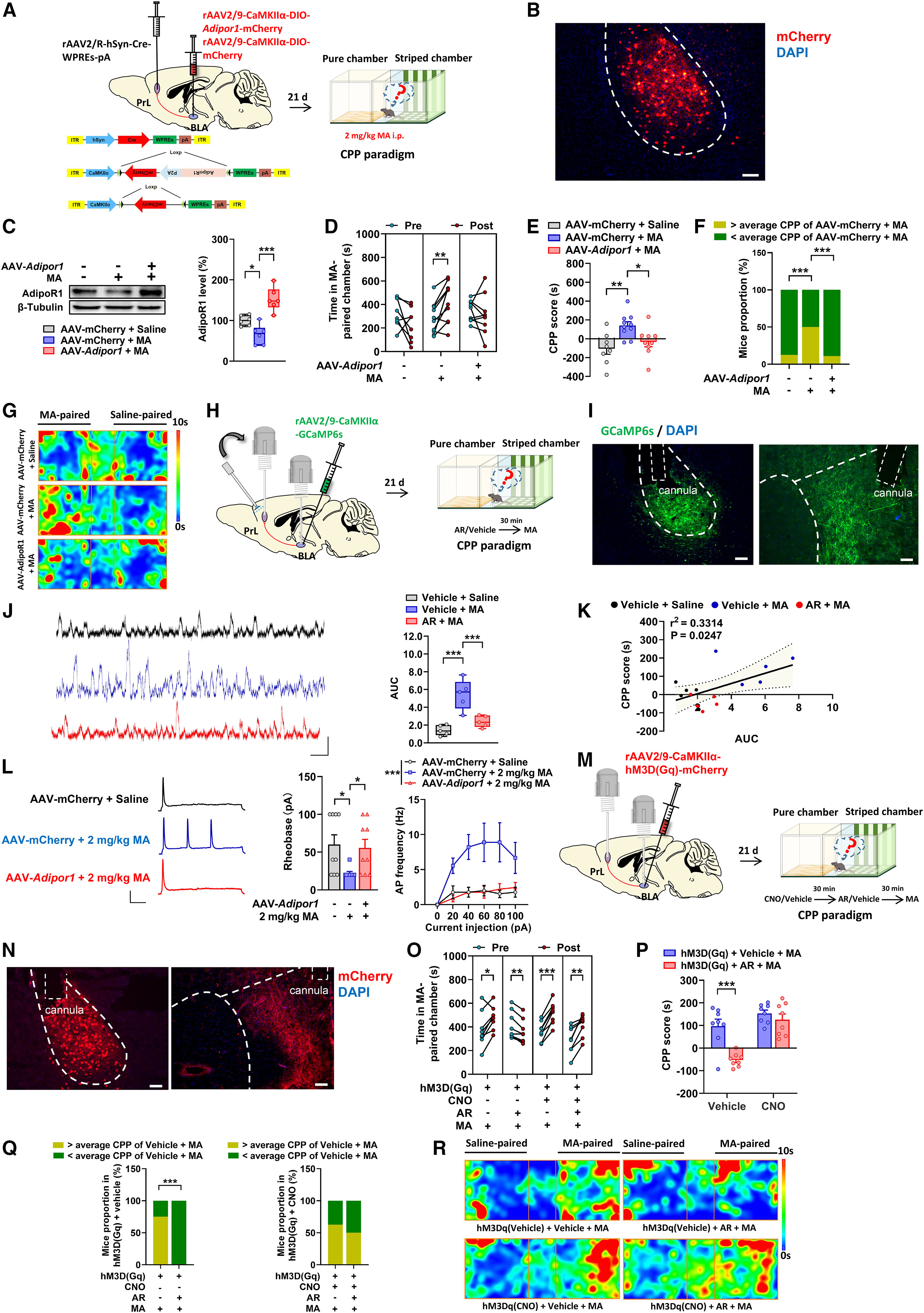
Pathway-specific overexpression of AdipoR1 in BLA CaMKIIα+→PrL neurons suppressed 2 mg/kg MA-induced CPP, elevated the action potential rheobase, and decreased depolarizing current (40 pA)-evoked spike firing in whole-cell patch-clamp recordings. Conversely, pathway-specific AdipoR1 knockdown in BLA CaMKIIα+→PrL neurons lowered rheobase, increased firing rates, and enhanced suboptimal 0.25 mg/kg MA-induced CPP and terminal Ca2+ signals in the PrL (Figure 5).
In vivo GCaMP6s fiber photometry of BLA CaMKIIα+→PrL axon terminals during the fourth conditioning session showed that entry into the MA-paired chamber evoked robust Ca2+ elevations that correlated positively with CPP scores. Intra-BLA AdipoRon infusion suppressed these terminal Ca2+ transients and attenuated MA CPP, whereas concurrent chemogenetic activation (hM3Dq) of BLA CaMKIIα+→PrL neurons reversed the inhibitory effect of intra-BLA AdipoRon on MA CPP.
Source: Zhigang Chen et al. · Cell Reports · © Elsevier Inc.
Original figure and English caption ↗
AdipoR1 Activates AMPK to Downregulate L-Type Calcium Channel Cav1.3 and Suppress BLA CaMKIIα+→PrL Activity
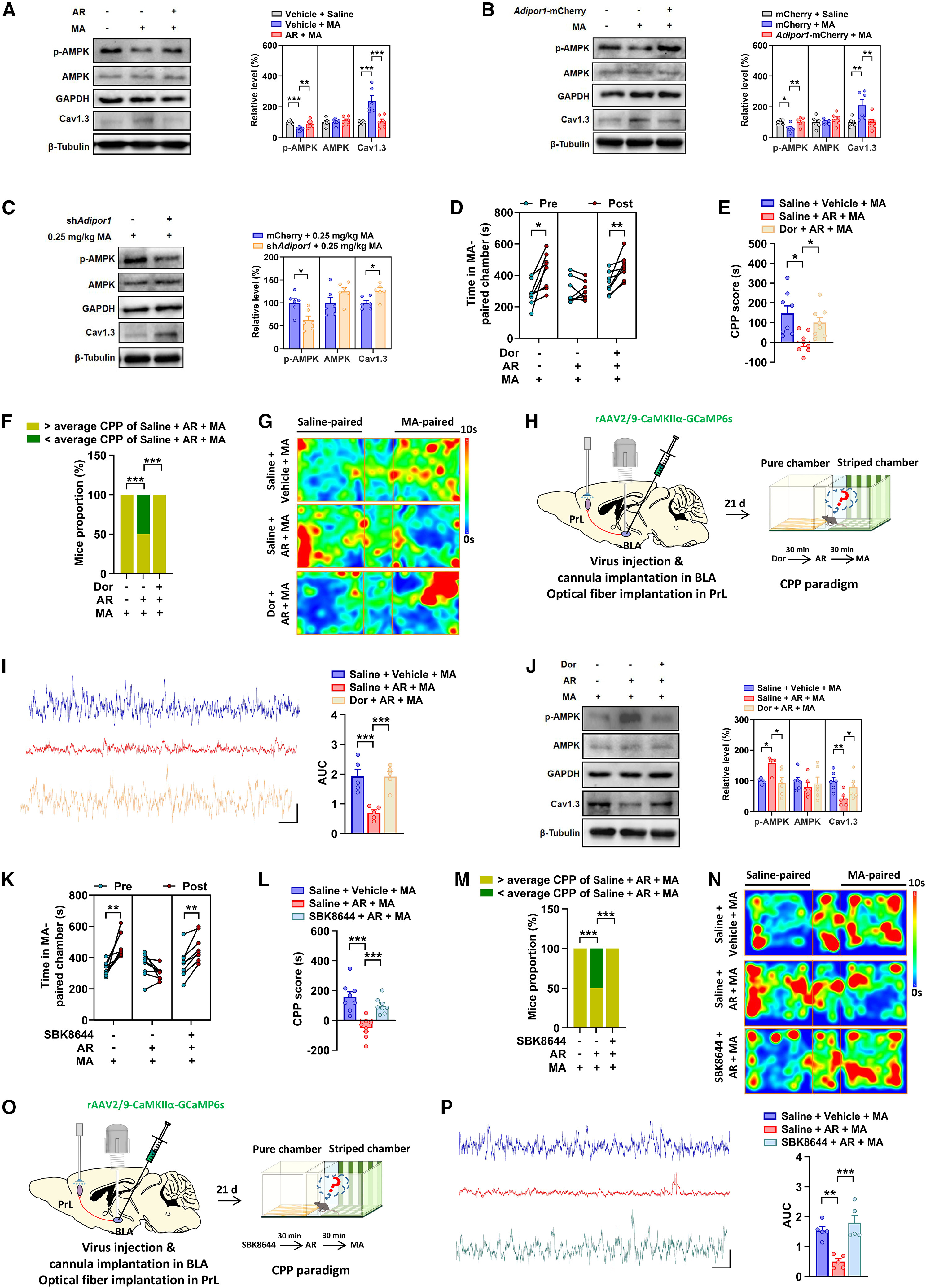
To determine how AdipoR1 regulates neuronal excitability and Ca2+ influx, the authors examined downstream signaling and ion channels. While BKCa (Kcnma1) mRNA was unchanged (Figure S6A), 2 mg/kg MA significantly reduced phosphorylated AMPK (p-AMPK) and upregulated mRNA and protein levels of the L-type voltage-gated calcium channel Cav1.3 (Cacna1d) in the BLA. AdipoRon treatment or BLA AdipoR1 overexpression restored p-AMPK and downregulated Cav1.3, whereas AdipoR1 knockdown under 0.25 mg/kg MA reduced p-AMPK and elevated Cav1.3.
Intra-BLA microinfusion of the AMPK inhibitor dorsomorphin (Dor) blocked AdipoRon-induced Cav1.3 downregulation and reversed the suppression of 2 mg/kg MA CPP and BLA CaMKIIα+→PrL terminal Ca2+ transients by AdipoRon. Similarly, intra-BLA infusion of the L-type calcium channel agonist (S)-(-)-Bay-K-8644 (SBK8644) abolished the inhibitory effects of AdipoRon on both BLA CaMKIIα+→PrL terminal Ca2+ signals and MA-induced CPP.
Source: Zhigang Chen et al. · Cell Reports · © Elsevier Inc.
Original figure and English caption ↗
Long-Term Physical Exercise Attenuates MA-Induced CPP by Restoring BLA AdipoR1–AMPK–Cav1.3 Signaling and Restraining the BLA CaMKIIα+→PrL Circuit
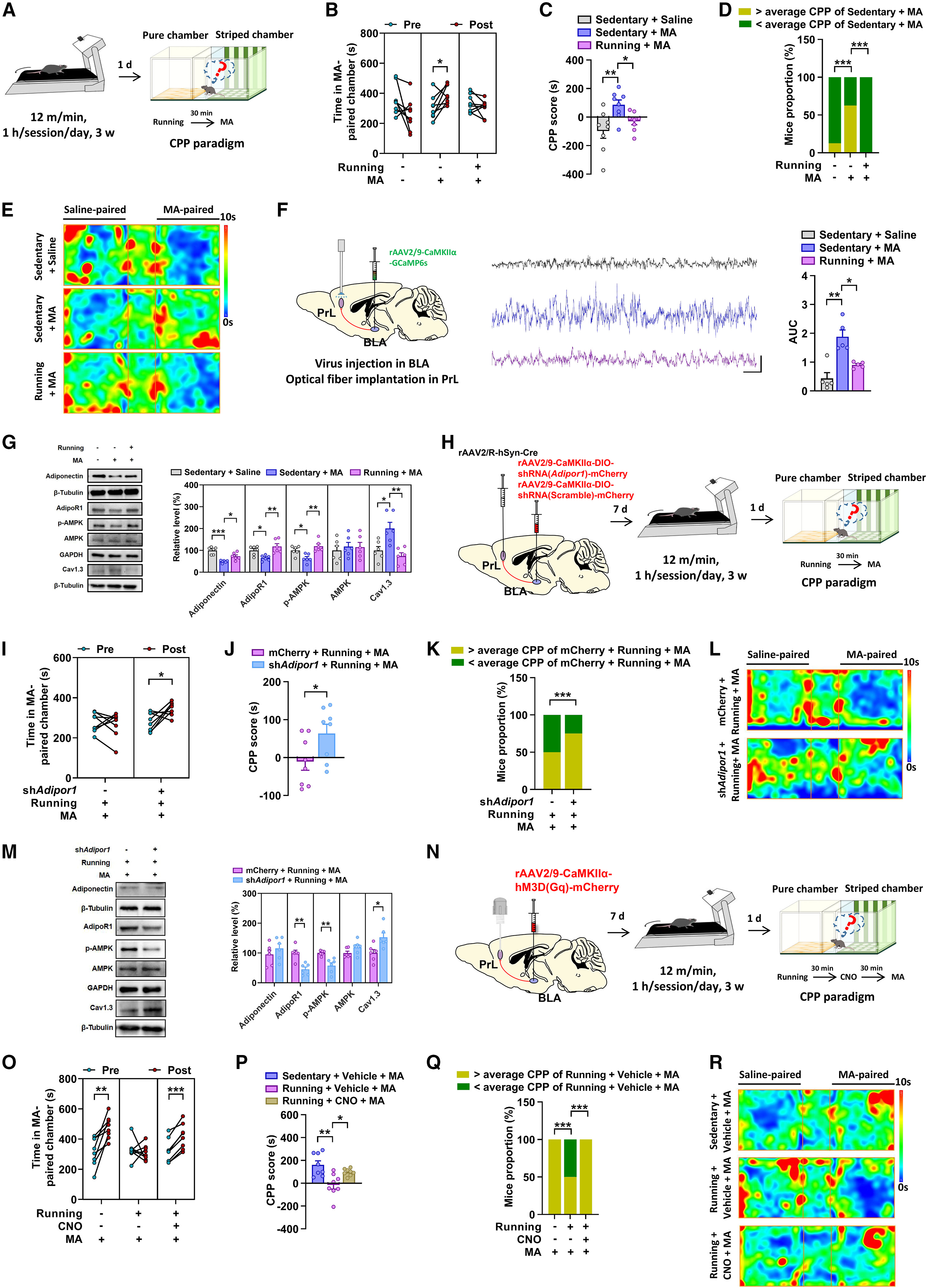
Long-term physical exercise (3 weeks of treadmill running prior to conditioning plus exercise before each conditioning session) significantly suppressed 2 mg/kg MA-induced CPP (as well as MA self-administration; Figure S7G–J), attenuated MA-evoked Ca2+ transients in BLA CaMKIIα+→PrL terminals, and restored BLA adiponectin, AdipoR1, and p-AMPK levels while downregulating Cav1.3 expression.
Crucially, either pathway-specific knockdown of AdipoR1 or chemogenetic activation (hM3Dq) of BLA CaMKIIα+→PrL neurons partially reversed the protective effect of long-term exercise against 2 mg/kg MA-induced CPP. Together, these findings establish the AdipoR1–AMPK–Cav1.3 cascade within the BLA CaMKIIα+→PrL circuit as a critical brake on methamphetamine-associated reward memory and a key mediator of exercise-induced protection.
Source: Zhigang Chen et al. · Cell Reports · © Elsevier Inc.
Original figure and English caption ↗