Histamine-tuned subicular cells contribute to faster alert escape
In mice, TMN histamine input to subiculum promotes faster movement at baseline and contributes to maintaining alert-escape performance. Recipient-cell and receptor interventions reveal distinct roles.
Accelerating during free exploration and rapidly escaping an alerting cue need not depend on identical regulation. A 2024 Nature Communications study follows histaminergic cells in the tuberomammillary nucleus, TMN, and their input to hippocampal subiculum, SUB. Histamine is often linked to arousal and attention; this study asks a more specific question about changing speed: in which contexts does this signal help mice move from slower to faster locomotion? Subicular cells, histamine receptors and downstream outputs provide different levels of evidence.
Stimulating TMN-to-SUB terminals promotes baseline movement, whereas suppression has no significant baseline effect. During alert escape, stimulation increases and suppression reduces distance and maximum speed. SUB excitatory cells and H2-related actions contribute, with RSG-projecting cells providing an output clue.
Contents · 7 sections
Histamine-cell responses vary with context
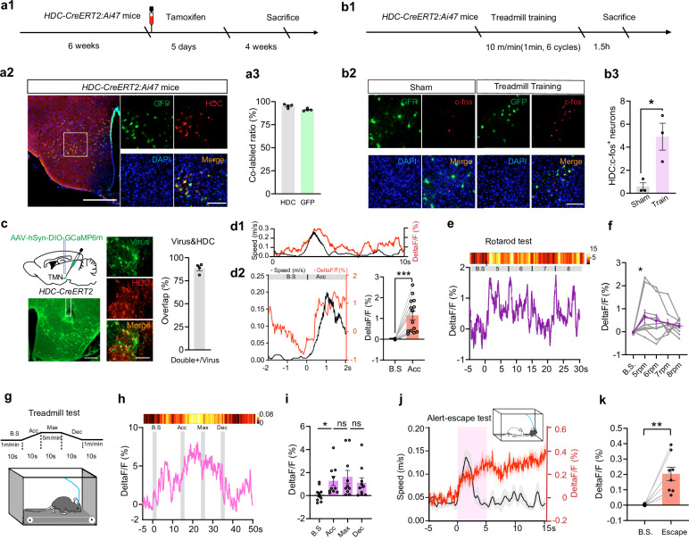
TMN histamine-cell population calcium signals increase during free-movement and treadmill acceleration. On the rotarod, signals rise at movement onset but do not show the same significant increase with further speed changes. They do not simply rise linearly with speed in every task.
During alert escape elicited by social attack, calcium rises rapidly and can remain elevated after speed falls. Increasing or reducing the whole TMN histamine population has no significant baseline free-movement effect, but respectively strengthens or weakens alert escape. Context changes the relationship between activity and action.
Source: Yang L et al. · Nature communications · CC BY-NC-ND
Original figure and English caption ↗
Subiculum and entorhinal cortex differ
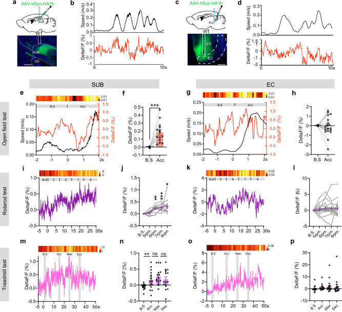
TMN histamine fibers reach SUB and entorhinal cortex, both associated with spatial and movement processing. Local histamine sensing shows SUB increases during free and treadmill acceleration and several alert behaviors. Entorhinal signals are more variable, with nonsignificant tested acceleration comparisons.
On the rotarod, SUB histamine signals increase at later faster stages, differing from the timing of whole-population TMN calcium. Cellular activity and transmitter signals at one recipient region describe different components, directing attention to SUB.
Source: Yang L et al. · Nature communications · CC BY-NC-ND
Original figure and English caption ↗
Promotion at baseline and contribution during alert escape
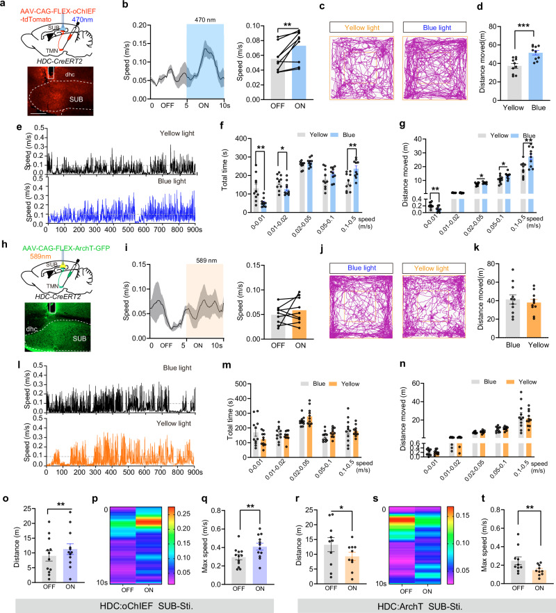
Stimulating TMN terminals in SUB increases transient speed, total distance and faster-movement time during baseline free behavior. Suppressing them does not significantly change baseline speed, distance or speed-bin measures. An ability to increase movement does not make the input indispensable for ordinary locomotion.
During alert escape, the interventions act in opposite directions: stimulation increases escape distance and maximum speed, while suppression reduces both. The same input can supply additional drive or help maintain recruited escape performance depending on context.
Source: Yang L et al. · Nature communications · CC BY-NC-ND
Original figure and English caption ↗
SUB excitatory cells receive the modulation
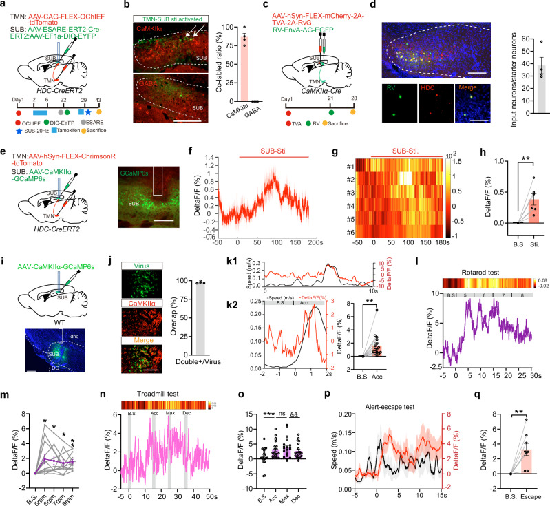
Most SUB cells activity-labeled after histamine-input stimulation carry an excitatory CaMKII marker. Monosynaptic retrograde tracing links excitatory SUB cells to TMN histamine cells, and terminal stimulation increases SUB population calcium, complementing the anatomical connection.
SUB excitatory cells respond at acceleration onset and during alert escape. During treadmill deceleration, their signal decreases relative to maximum-speed movement, whereas corresponding decreases in TMN calcium and SUB histamine sensing are nonsignificant. Different nodes retain distinct dynamics.
Source: Yang L et al. · Nature communications · CC BY-NC-ND
Original figure and English caption ↗
Suppressing recipients weakens movement promotion
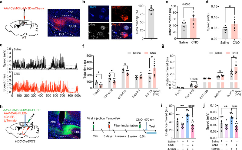
Directly increasing SUB excitatory-cell activity raises average speed and distance in faster speed bins. Combining TMN-terminal stimulation with SUB excitatory-cell suppression markedly weakens the stimulation-induced increases in distance and speed.
This combination links input and recipient activity to the same behavioral measure. Suppression covers the excitatory SUB population rather than only a small histamine-input-tagged subset, supporting a recipient contribution without identifying one exclusive cell subtype.
Source: Yang L et al. · Nature communications · CC BY-NC-ND
Original figure and English caption ↗
Two receptor classes show different intervention outcomes
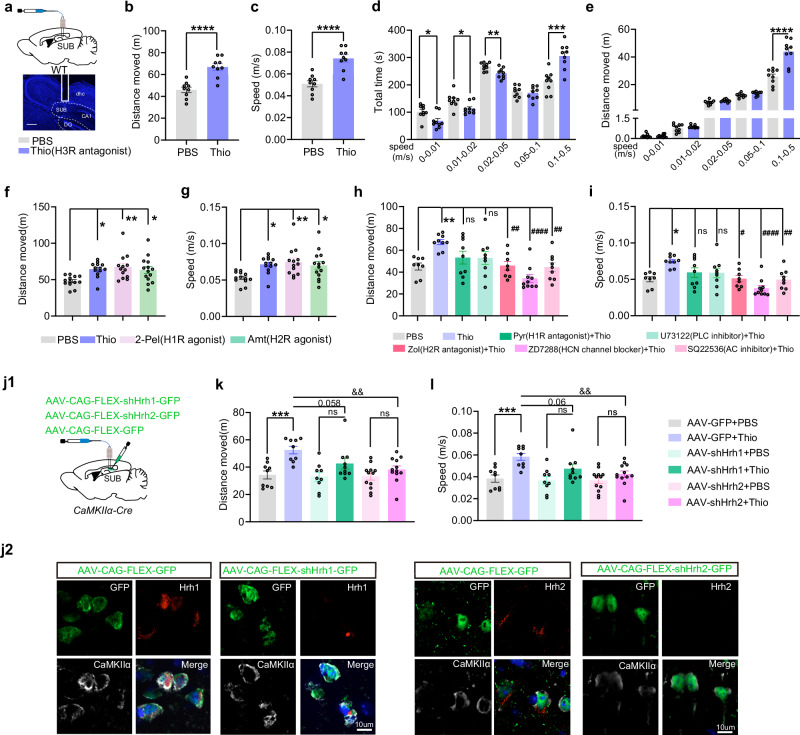
Local H3-receptor blockade promotes faster movement, and this effect disappears after removing most TMN histamine cells. H1- and H2-related activation can each promote movement; blocking H2, adenylyl cyclase or HCN-related actions weakens promotion caused by H3 blockade.
Reducing H2 expression in excitatory SUB cells significantly weakens pharmacological movement promotion relative to controls. Promotion is also nonsignificant after H1 knockdown, but its corresponding difference from controls is nonsignificant. Pharmacological and genetic findings highlight H2-related action while leaving room for receptor cooperation.
Source: Yang L et al. · Nature communications · CC BY-NC-ND
Original figure and English caption ↗
Speed modulation connects to a wider network
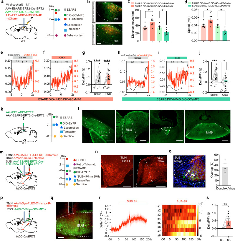
Activating movement-tagged SUB cells promotes locomotion, while suppressing them reduces it. Their axons reach retrosplenial granular cortex, RSG, anteroventral thalamus and medial mammillary nucleus. Some SUB cells recruited by TMN-terminal stimulation project to RSG, and calcium in this projection population rises with terminal stimulation.
RSG provides an output clue, without an isolated terminal intervention establishing behavioral necessity. In adult female and male mice, histamine regulation has a contextual character: rather than uniformly driving acceleration, it changes faster movement according to alerting context and recipient-cell state.
Source: Yang L et al. · Nature communications · CC BY-NC-ND
Original figure and English caption ↗