Drug-context memory recruits a prefrontal–thalamic connection
PVT excitatory cells are recruited when male mice revisit a methamphetamine-paired context. Suppressing prefrontal input weakens established preference; sleep-state measures remain nonsignificantly changed in a separate experiment.

A familiar setting can allow a past drug experience to influence choice again. Remembering the setting and expressing a preference when revisiting it are different stages of the same experience. A 2025 Theranostics study follows the paraventricular thalamic nucleus, PVT, and excitatory input from medial prefrontal cortex, mPFC, in methamphetamine conditioned place preference in male mice. Because PVT also participates in arousal, another question arises: does weakening contextual preference also make animals sleepier? The researchers examine memory stages, cellular connections and sleep recordings.
Suppressing PVT excitatory cells during testing weakens established preference, whereas training-phase suppression has no significant effect. Inhibiting mPFC terminals in PVT also reduces preference. In a separate experiment, suppressing PVT-projecting mPFC cells does not significantly change sleep-state durations over three hours.
Contents · 8 sections
Changes appear after returning to the setting
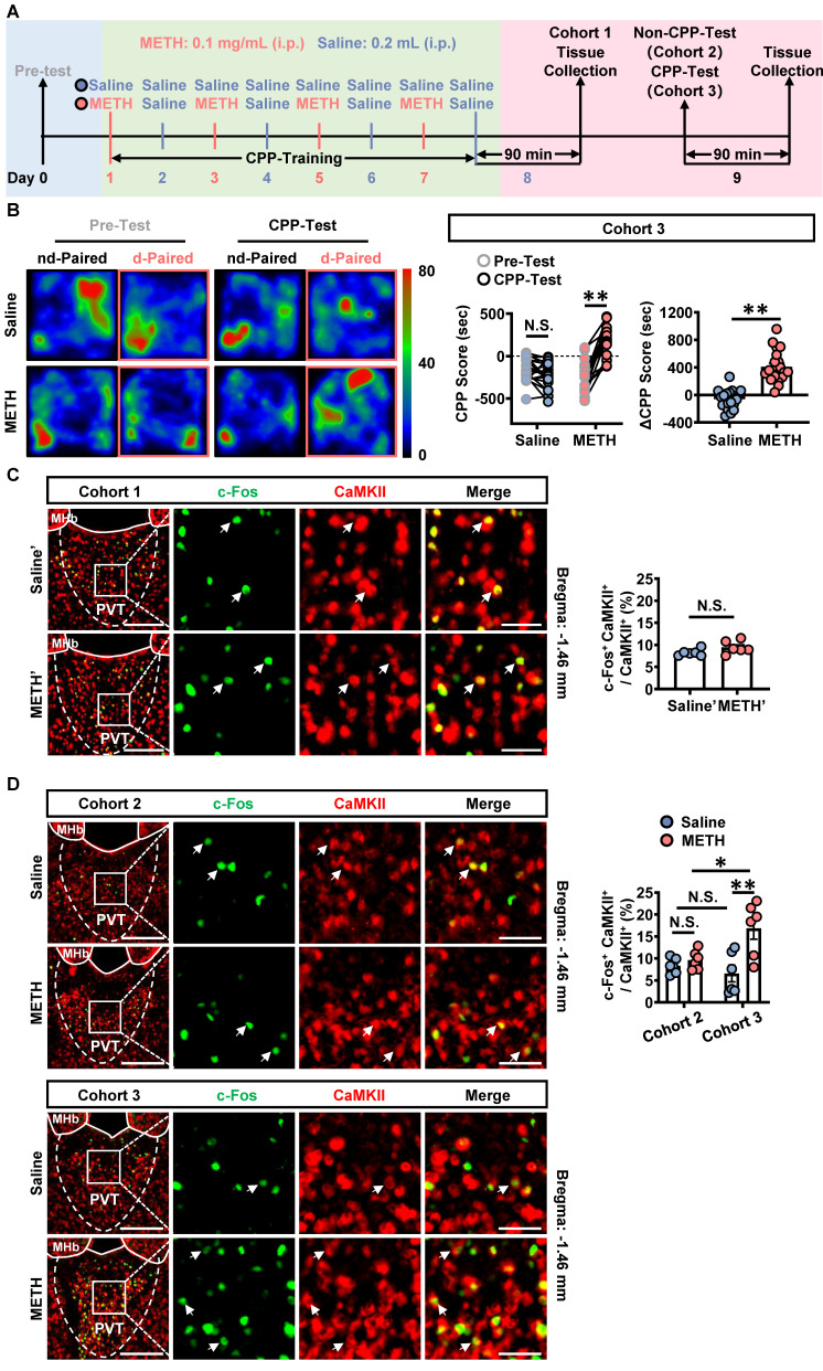
After repeated pairing of one context with the drug, mice prefer that side during a drug-free test, with total travel distance similar to controls. Comparing separate cohorts after training, without testing and after testing, the team finds increased c-Fos activity labeling in CaMKII-marked PVT cells mainly in the drug group that completes the preference test.
The same significant group increase is absent immediately after training. PVT responses stand out when the context returns and the animal makes a choice. This stage difference shifts attention from which cells change after exposure to when the memory-related behavior requires them.
Source: Li X et al. · Theranostics · CC BY
Original figure and English caption ↗
Established preference can still be weakened during testing
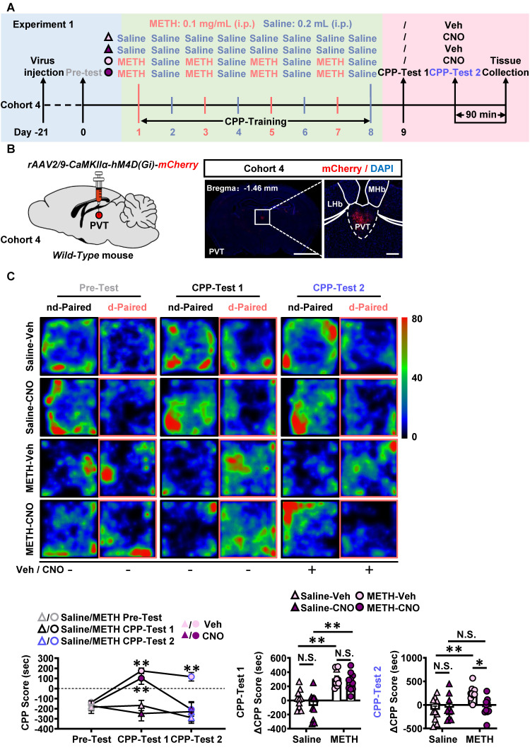
Suppressing PVT excitatory cells during training leaves later preference intact, without a significant difference in preference change from unsuppressed mice. Once preference has developed, however, suppression before testing reduces it in the drug group, without the same effect in controls.
Total travel distance does not decline significantly, so the preference change does not accompany reduced overall movement. The findings point to PVT involvement when an established contextual memory is retrieved and expressed as a choice. Reduced preference in this test does not demonstrate permanent erasure of the memory.
Source: Li X et al. · Theranostics · CC BY
Original figure and English caption ↗
PVT cells fire more readily and excitatory input changes
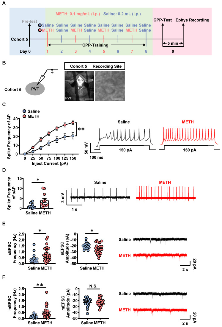
Post-test slice recordings show more current-evoked action potentials and increased spontaneous firing in drug-group PVT cells. Spontaneous excitatory-current frequency and amplitude increase; miniature-current frequency increases while amplitude does not change significantly. Cellular excitability and input measures describe changes at different levels.
Receptor screening finds that blocking GluN2A-containing NMDA receptors or AMPA receptors reduces current-evoked firing in the drug group. D1- or GluN2B-related blockade has no comparable significant effect. The team therefore follows glutamatergic input to identify a source driving PVT responses.
Source: Li X et al. · Theranostics · CC BY
Original figure and English caption ↗
Prefrontal cortex directly excites PVT
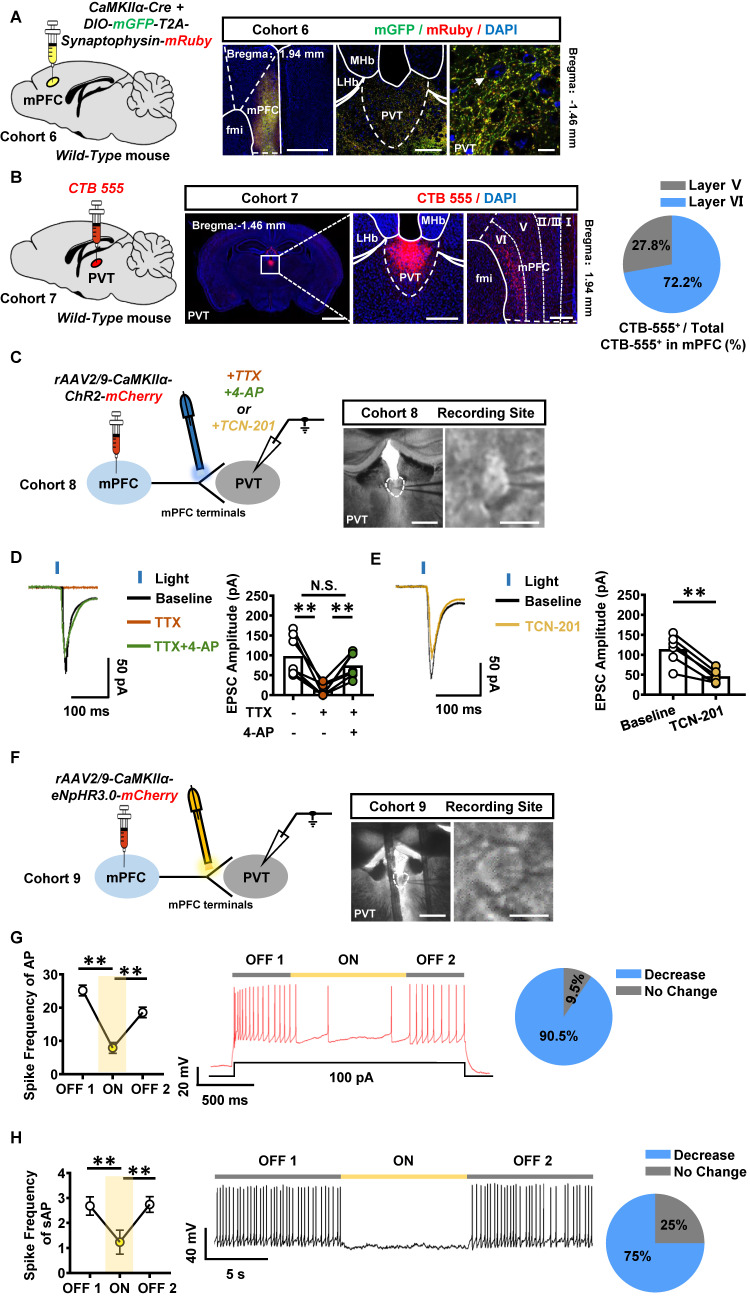
Anterograde and retrograde tracing complement each other: mPFC terminals reach PVT, and PVT-projecting prefrontal cells lie mainly in layers V and VI. Stimulating their terminals evokes excitatory currents in PVT cells; synaptic blocking and recovery experiments support a direct monosynaptic excitatory connection.
GluN2A-related blockade partially reduces these currents. Conversely, suppressing mPFC terminals lowers firing in most sampled PVT cells, with recovery after suppression ends. Prefrontal input is thus linked to immediate PVT activity, motivating an intervention directed at this input.
Source: Li X et al. · Theranostics · CC BY
Original figure and English caption ↗
Reducing this input weakens contextual preference
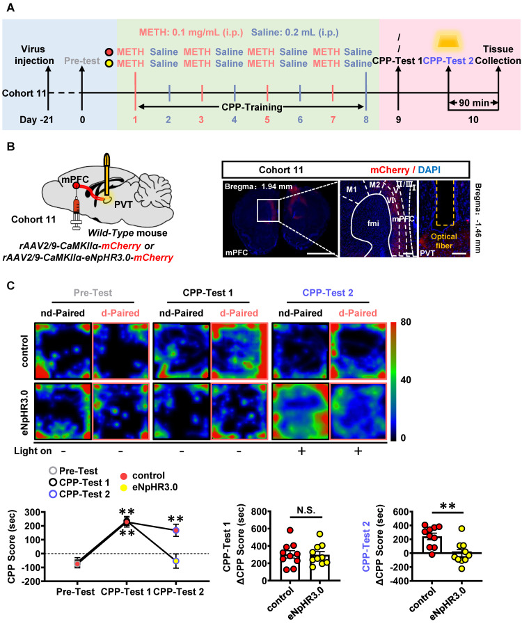
In the same mice, the first test confirms preference for the drug-paired context. During the second test, the team suppresses mPFC excitatory terminals in PVT. Preference declines in the experimental group without the same significant change in viral controls, while total travel distance remains similar.
This intervention acts on an input to PVT rather than broadly suppressing the entire nucleus. It identifies excitatory information arriving from prefrontal cortex as one contributor to expression of an established contextual preference during testing.
Source: Li X et al. · Theranostics · CC BY
Original figure and English caption ↗
Selecting an input differs from broad PVT suppression
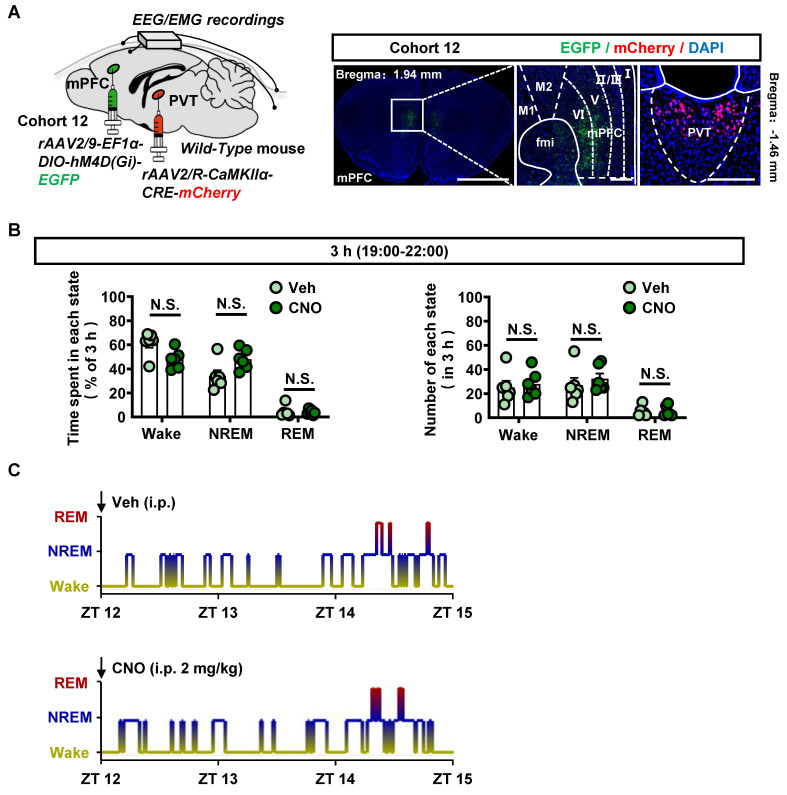
In a separate experiment, broad PVT excitatory-cell suppression reduces wake time and increases non-REM sleep. The team then selects PVT-projecting mPFC cells for suppression in an independent EEG/EMG experiment. Across three hours, durations and episode counts of wake, non-REM and REM sleep do not change significantly.
PVT is not a switch devoted solely to reward. It receives multiple inputs and participates in multiple states. Targeting cells from a particular source can produce different sleep-state outcomes from broader PVT suppression. This observation concerns the short recording period, rather than all arousal functions.
Source: Li X et al. · Theranostics · CC BY
Original figure and English caption ↗
GluN2A and AMPA show different behavioral outcomes
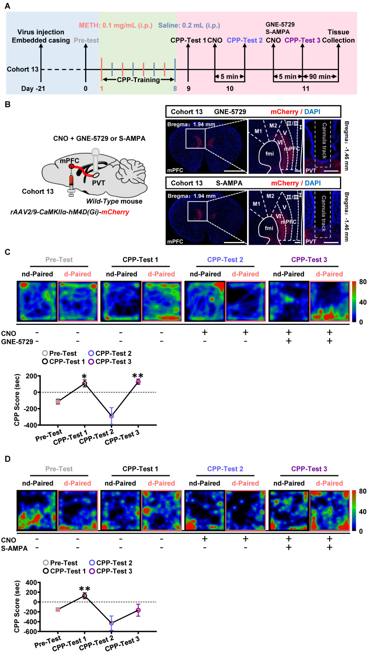
After further suppression of mPFC terminals in PVT, the team adds different receptor-related pharmacological enhancements. With GluN2A-related enhancement, mice again show significant preference relative to pretraining baseline. AMPA-related enhancement does not produce the same significant preference.
Both receptor types contribute to cellular responses in slices, yet their behavioral outcomes differ here. The study connects GluN2A-related action with the circuit intervention. These local PVT pharmacological interventions are not restricted to a single identified synapse.
Source: Li X et al. · Theranostics · CC BY
Original figure and English caption ↗
Retrieving a memory also requires network input
This study in young male mice separates formation of a contextual association from later preference expression, linking the mPFC–PVT excitatory connection to the latter stage. Examining a region’s incoming information more closely helps explain both how a setting influences choice again and why interventions yield different sleep-state outcomes.