Beyond headache: a connection between the mouse body clock and memory
Changing activity in the body-clock center improves memory and activity rhythms in male nitroglycerin-model mice, while mechanical and thermal hypersensitivity remain largely unchanged.
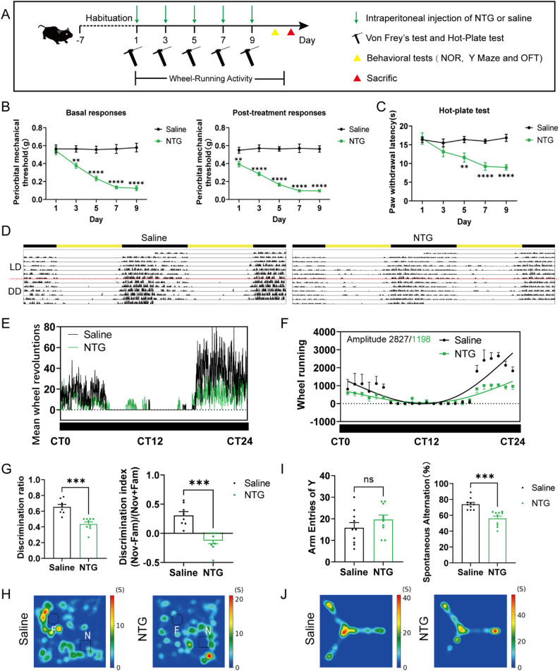
Headache research often starts with pain, but changes in attention, memory and daily rhythms raise a separate question: are they distractions caused by pain, or do they involve distinct neural routes? A study in The Journal of Headache and Pain brings together the suprachiasmatic nucleus, which organizes circadian rhythms, and the dentate gyrus, which processes memory information. In male nitroglycerin-model mice, changing body-clock activity alters memory-task performance without a corresponding change in established hypersensitivity.
Body-clock activity affects memory measures in both directions without parallel changes in hypersensitivity; tracing and reported route stimulation point toward the dentate gyrus.
Contents · 6 sections
Two changes appear in the same model
The suprachiasmatic nucleus (SCN) coordinates daily timing through outputs to other regions. The dentate gyrus (DG), at the hippocampal information gateway, helps distinguish and encode experiences. Timing and memory appear to have separate jobs, yet both depend on recruiting neurons appropriately. The study asks whether these processes interact in a headache-like state.
After nitroglycerin, mechanical and thermal responses become more sensitive, wheel-running declines and rhythms become less organized. Novel-object recognition and Y-maze alternation also decline. Arm entries remain similar; open-field distance increases while center time is unchanged. These measures reveal distinct changes in activity, timing and memory rather than a uniform reduction in movement.
Source: Xie Q et al. · The journal of headache and pain · CC BY-NC-ND
Original figure and English caption ↗
Overall recruitment rises, but populations differ
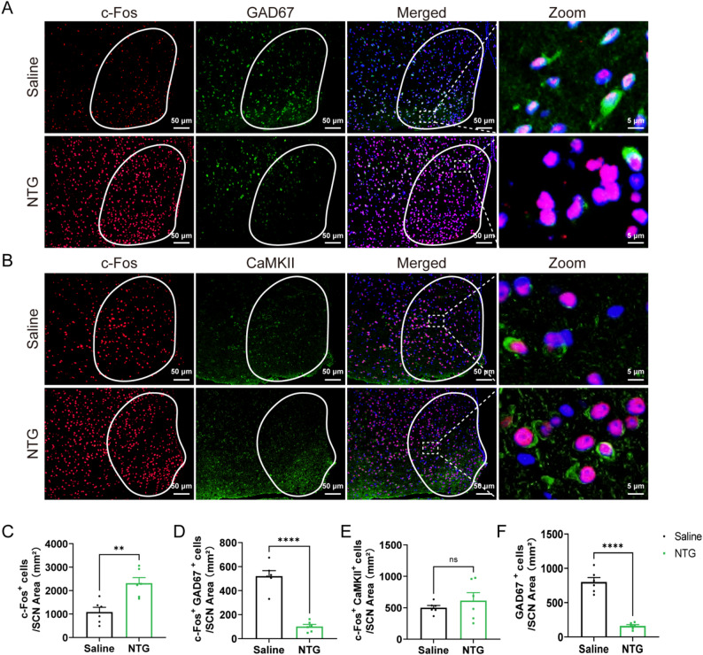
Overall activity-related c-Fos labeling increases in SCN, yet c-Fos/GAD67 co-labeling decreases and c-Fos/CaMKII changes are nonsignificant. Populations within one region move differently. The pattern resembles a redistribution of recruitment rather than a uniform increase or decrease across the clock.
GAD67-label density also falls. This measures detectable molecular labeling at sampling, which can itself change with expression. The question becomes which cells are recruited and how their coordination affects outgoing clock signals. Population differences set the context for the functional observations.
Source: Xie Q et al. · The journal of headache and pain · CC BY-NC-ND
Original figure and English caption ↗
Memory improves while hypersensitivity persists
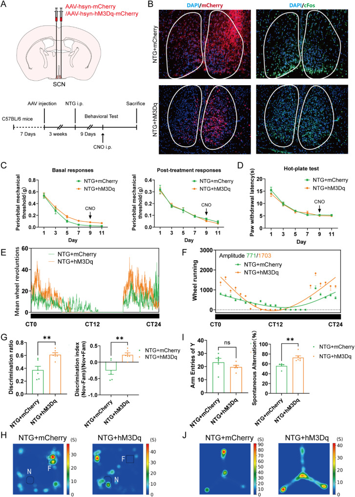
Reducing SCN activity further impairs object recognition and alternation; increasing it improves both measures and organizes wheel-running rhythms. The affected population is predominantly GAD67-labeled and spans the targeted SCN group. Opposite activity changes produce opposite memory outcomes.
Mechanical and thermal hypersensitivity show no significant change with either intervention. Memory can improve while stimulus sensitivity persists. Cognitive changes therefore occupy a distinct place within the same model, able to vary independently from pain-like responses. Their improvement alongside rhythms raises a further question about shared outputs.
Source: Xie Q et al. · The journal of headache and pain · CC BY-NC-ND
Original figure and English caption ↗
Following clock outputs toward the dentate gyrus
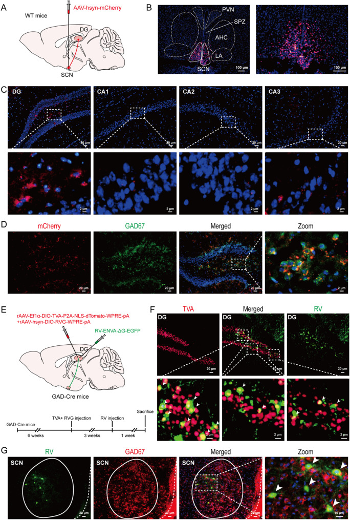
DG recruitment also shifts: overall c-Fos and GAD67 co-labeling increase, while CaMKII co-labeling decreases. Overall c-Fos in the other examined hippocampal subregions is unchanged. Different parts of the hippocampus thus register the state differently, drawing attention to DG.
Converging tracing supports a direct SCN–DG anatomical connection, with some inputs associated with DG inhibitory cells. The clock therefore has a route to a memory gateway. Its local effects depend on recipient cells and synaptic properties; the established observations concern anatomy and altered recruitment in a memory-related region.
Source: Xie Q et al. · The journal of headache and pain · CC BY-NC-ND
Original figure and English caption ↗
Reported route stimulation improves memory in the same direction
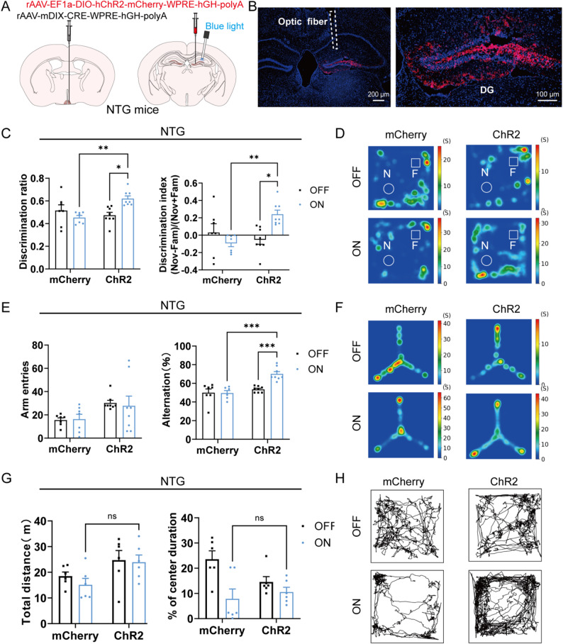
The authors report improved object recognition and alternation after stimulation of their labeled SCN–DG-associated route. Arm entries, open-field distance and center time remain unchanged. The result connects the broader SCN findings with the network near DG and makes a simple increase in activity an incomplete explanation.
Anatomy and behavior complement one another at different levels: images locate connections, while tasks reveal changes accompanying the reported stimulation. How specific cells receive timing signals during natural memory remains unresolved. The route is now functionally associated with improved memory measures in this model.
Source: Xie Q et al. · The journal of headache and pain · CC BY-NC-ND
Original figure and English caption ↗
Memory changes in a headache-like state have their own network clues
The study brings memory changes into the foreground rather than treating them as background to pain-like responses. Clock outputs, dentate recruitment and behavior fit within one framework. The key dissociation remains: memory and rhythms improve without reduced mechanical or thermal hypersensitivity. Sharing a state does not require sharing one driver.
The observations concern male nitroglycerin-model mice and tasks of object recognition and spatial working memory. They suggest that a region organizing time may also help organize conditions for memory processing. The clock–hippocampus link makes activity timing and the handling of new experiences relevant parts of the same research question.