At the moment of recognition, how does subiculum support retrieval?
The extracellular-matrix protein FN1 links subicular firing and BK channels to an entorhinal output supporting object discrimination.
Two objects were encountered yesterday; today, one has been replaced. A mouse prefers to approach the new one, comparing earlier experience with current input. This simple choice requires retrieval and the transfer of information through hippocampal outputs. An Advanced Science study in adult male mice follows the subiculum, an output hub, to an extracellular-matrix protein: fibronectin 1, or FN1. Its relationship with potassium channels, neuronal firing and a selected output route affects the moment of recognition.
Strengthening subiculum–entorhinal output during retrieval improves novelty discrimination, while inhibition reduces it. FN1 knockdown removes novel–familiar signal differentiation and increases BK expression; pathway activation or local BK blockade improves the deficit.
Contents · 7 sections
A population that matters during retrieval
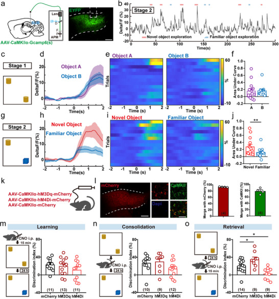
During initial exploration of two identical objects, subicular pyramidal-cell calcium rises similarly for both. One day later, replacing one object produces a stronger signal during novel-object exploration. The population shifts from comparable responses to stronger novelty responses when earlier experience is available.
The behavioral effect depends on intervention timing. Activation or inhibition before learning, or six hours afterward, does not significantly change next-day discrimination. During retrieval, activation improves the index and inhibition reduces it. Object-location inhibition produces only a nonsignificant trend. In these tasks, the prominent contribution occurs during object comparison.
Source: Fei F et al. · Advanced science (Weinheim, Baden-Wurttemberg, Germany) · CC BY
Original figure and English caption ↗
Different subicular exits produce different outcomes
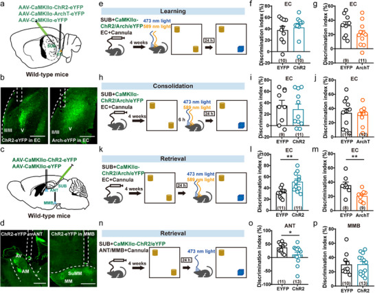
Subiculum sends information to several regions, whose outputs have distinct functions. Its entorhinal terminals show stronger calcium during novel-object exploration, unlike anterior-thalamic and mammillary terminals. Strengthening entorhinal output improves retrieval discrimination, and inhibition reduces it, without significant effects during learning or the tested consolidation time.
Activating anterior-thalamic terminals instead reduces novelty discrimination, while inhibition does not cause the same deficit. Mammillary activation and inhibition, and related retrosplenial inhibition, do not significantly change performance. Object-location manipulations also lack significant effects. Increasing output broadly is therefore not equivalent to strengthening a selected route.
Source: Fei F et al. · Advanced science (Weinheim, Baden-Wurttemberg, Germany) · CC BY
Original figure and English caption ↗
An extracellular protein participates in discrimination
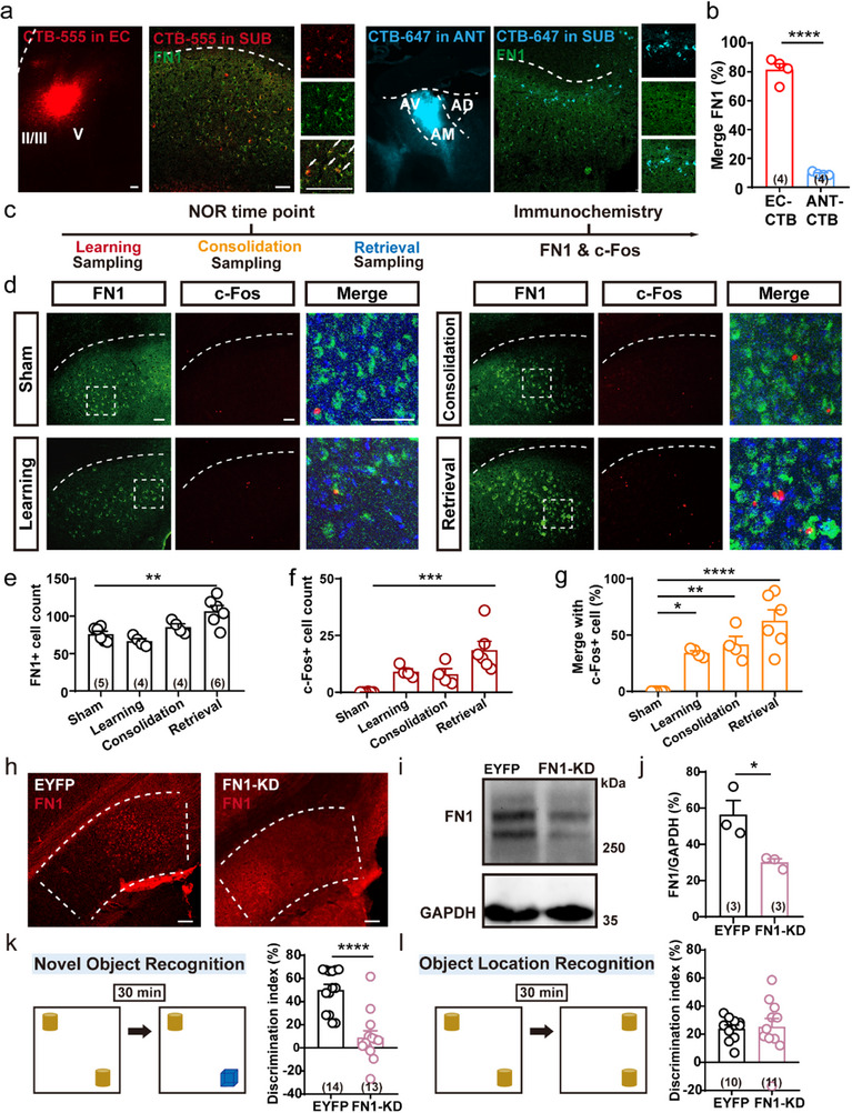
FN1 emerges while comparing output populations. It is found mainly in entorhinal-projecting subicular pyramidal cells, with little expression in anterior-thalamic-projecting cells. FN1 and the activity marker c-Fos increase after retrieval, unlike learning and the tested consolidation condition. An extracellular-matrix protein thus appears in the population contributing during retrieval.
Reducing subicular FN1 impairs novel-object discrimination, without significant corresponding deficits in object location, Y-maze, water maze, conditioned fear or affect-related tests. Suppressing neural activity or c-Fos/AP-1-related activity also lowers FN1. The protein moves from a population marker to a functional participant whose level responds to neural activity.
Source: Fei F et al. · Advanced science (Weinheim, Baden-Wurttemberg, Germany) · CC BY
Original figure and English caption ↗
Stronger output restores discrimination
After FN1 reduction, sampling responses to identical objects remain comparable, but novel–familiar calcium differentiation disappears at retrieval. The deficit does not eliminate every object response; the population loses its previous differentiation. Molecular and retrieval-related activity changes coincide in the behavioral deficit.
Strengthening subiculum–entorhinal output during retrieval improves discrimination after FN1 knockdown. Activation during learning or consolidation does not provide the same compensation, nor does stimulation of the thalamic branch. Both route and timing must align for improvement, linking the deficit to a retrieval circuit whose activity can compensate.
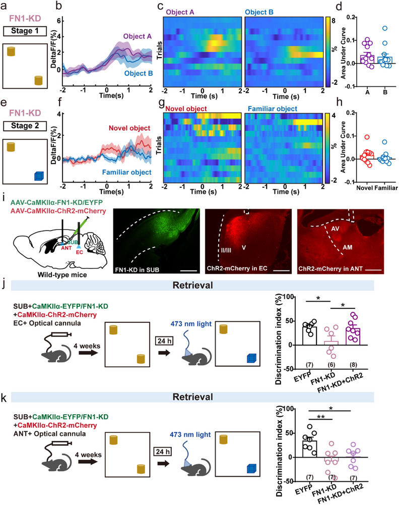
Source: Fei F et al. · Advanced science (Weinheim, Baden-Wurttemberg, Germany) · CC BY
Original figure and English caption ↗
Why does potassium-channel braking increase?
In slices, FN1 knockdown reduces spikes under larger current inputs, lowers spike amplitude, increases fast afterhyperpolarization and reduces the fraction of burst-firing cells. Rheobase and slow afterhyperpolarization do not significantly change; increased low-current firing is also nonsignificant. The alteration concerns responsiveness under stronger input rather than a uniform decrease in activity.
BK potassium channels provide the next clue. They contribute to membrane repolarization after spikes, acting as part of the firing brake. FN1 knockdown increases principal-subunit protein and KCNMA1 transcript, while KCNMB4 does not significantly change. Local paxilline improves discrimination. Channel expression, altered firing and pharmacological rescue support BK involvement.
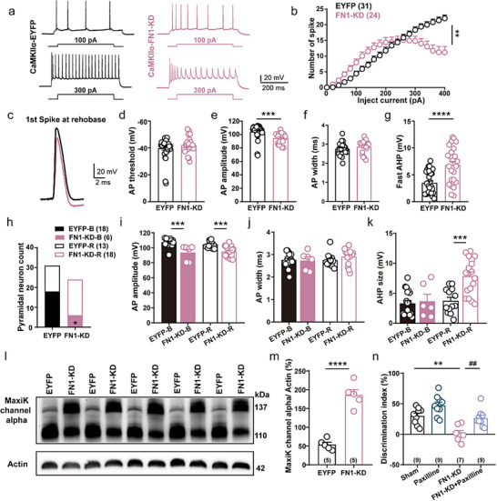
Source: Fei F et al. · Advanced science (Weinheim, Baden-Wurttemberg, Germany) · CC BY
Original figure and English caption ↗
How does hippocampal input reach this route?
Tracing shows stronger CA1 and local inputs to entorhinal-projecting subicular cells, whereas thalamic-projecting cells receive more presubicular input. CA1 inhibition removes novel–familiar calcium differentiation during retrieval. Activity and anatomy align: subiculum does not distribute the same hippocampal information equally across all outputs.
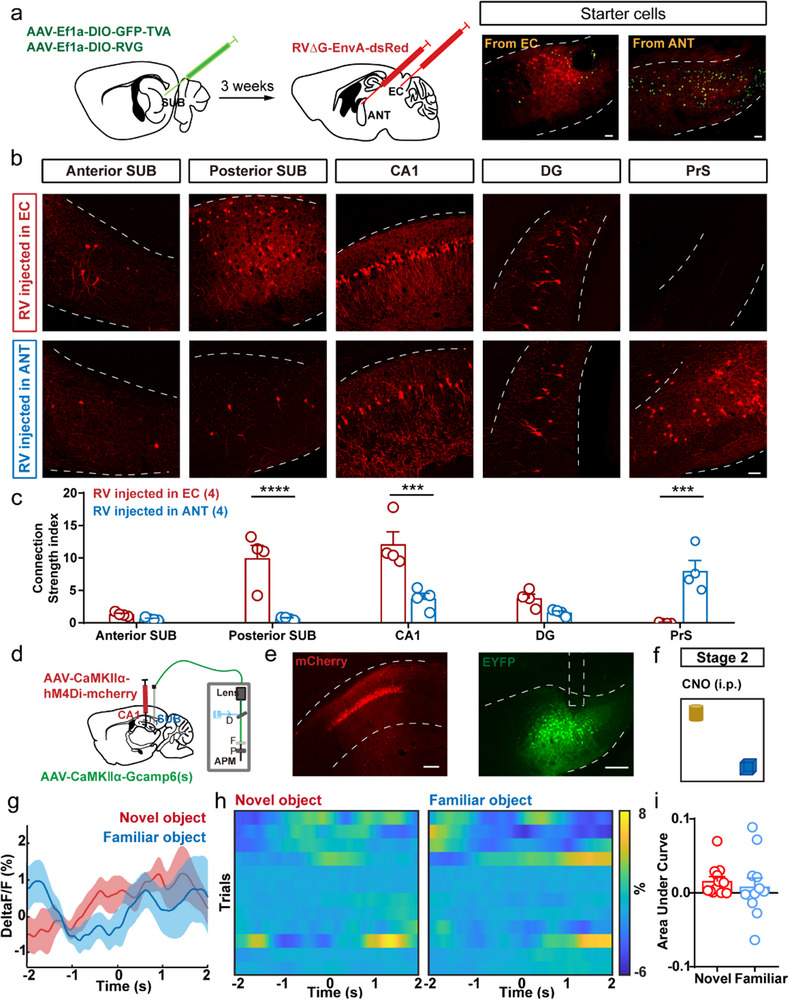
Source: Fei F et al. · Advanced science (Weinheim, Baden-Wurttemberg, Germany) · CC BY
Original figure and English caption ↗
The extracellular environment matters for retrieval
In this adult male-mouse task, FN1, BK-associated excitability and subiculum–entorhinal output influence behavior when memory is retrieved. Extracellular matrix is more than a scaffold around neurons: it relates to whether cells generate adequate output at a crucial moment. Retrieval brings cellular environment, firing state and information routing together.