An ultrasound switch for a prefrontal pathway: what changes after stress?
Sonogenetics makes selected prefrontal cells responsive to ultrasound; dorsal raphe and its serotonergic cells contribute to reduced immobility in stressed mice.
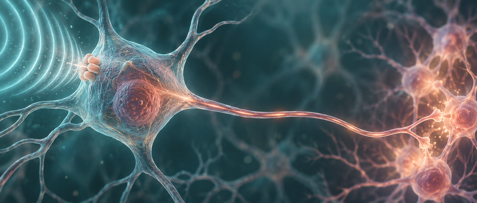
The prefrontal cortex is central to emotion regulation, yet raising activity across the whole region need not produce the expected result. Its many outputs can carry information in different directions. A Molecular Psychiatry study selects one route: the excitatory projection from medial prefrontal cortex to dorsal raphe. Researchers express the mechanosensitive channel MscL-G22S in these cells and trigger activity with ultrasound. In chronically restrained male mice, this combination of cellular targeting and acoustic stimulation changes immobility in two behavioral tests.
MscL expression combined with ultrasound produces clear DRN calcium and serotonin-sensor responses and reduces post-stress immobility; DRN or serotonergic-cell inhibition blocks the behavioral effect.
Contents · 6 sections
How does ultrasound reach a selected neural output?
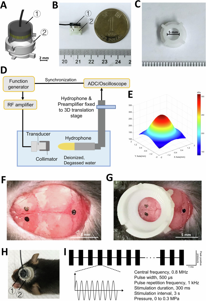
Projection identity alone does not make a neuron selectively responsive to ultrasound. Researchers introduce MscL-G22S into DRN-projecting excitatory mPFC cells to increase mechanical sensitivity. Genetic targeting constrains responsive cells, while acoustic positioning constrains the stimulated region. Together they connect a physical signal to a defined neural output.
Ultrasound does not recognize cellular identity. Adding a mechanosensitive channel to projection-defined cells biases which population responds within the acoustic field, directing influence toward its output network. This allows a comparison: does stimulating broad prefrontal excitatory populations have the same behavioral effect as strengthening the route to raphe?
Source: Lei T et al. · Molecular psychiatry · CC BY-NC-ND
Original figure and English caption ↗
Prefrontal stimulation rapidly recruits raphe
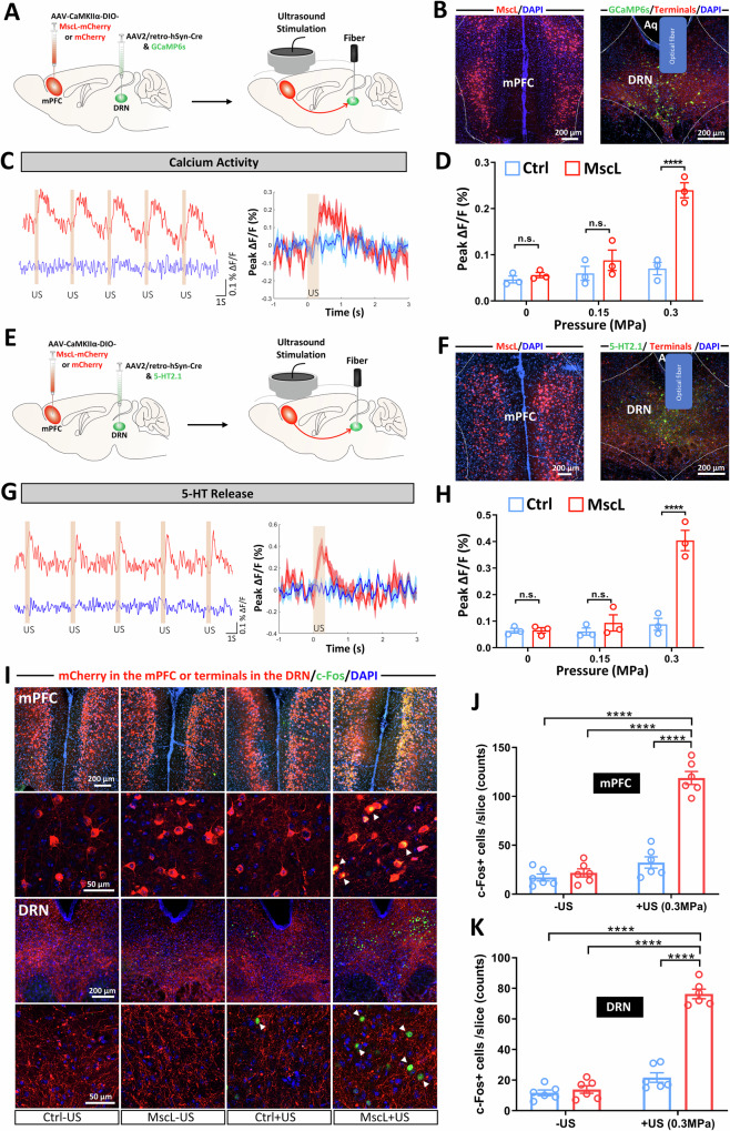
In isoflurane-anesthetized mice, DRN calcium and local serotonin-sensor signals rise within a second of ultrasound, recur with stimulation and recover afterward. Controls without MscL lack similarly clear responses. The selected projection therefore carries effects beyond prefrontal tissue to downstream activity and transmitter signaling.
Longer stimulation increases c-Fos in mPFC and DRN. Recruited DRN cells include serotonergic and other cells. Central amygdala also shows increased activation, while nucleus accumbens, hippocampus, basolateral amygdala and ventral tegmental area do not. Auditory-cortex c-Fos does not differ between groups. A targeted intervention still enters an interconnected network.
Source: Lei T et al. · Molecular psychiatry · CC BY-NC-ND
Original figure and English caption ↗
Less immobility, more struggling
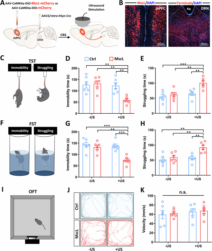
After fourteen days of restraint, male mice show more immobility in tail suspension and forced swimming. Sonogenetic stimulation begins before testing and continues during it. MscL plus ultrasound reduces immobility and increases struggling, unlike channel expression or ultrasound alone. The downstream physiological response is accompanied by an intervention effect in awake behavioral tests.
Open-field velocity does not increase, and center duration does not significantly change. Stimulating a broader mPFC excitatory population also fails to clearly alter the two stress-related behaviors. Raising activity across a region and strengthening one particular output produce different outcomes.
Source: Lei T et al. · Molecular psychiatry · CC BY-NC-ND
Original figure and English caption ↗
Silencing the downstream node removes the change
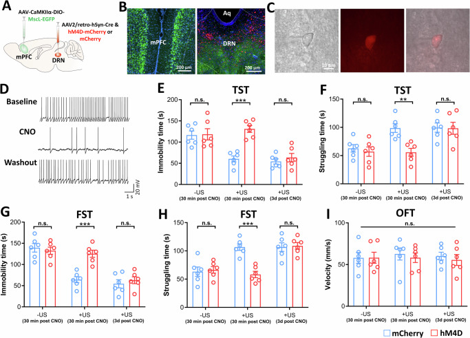
If the mPFC–DRN route contributes to the behavioral effect, blocking its downstream node should alter the outcome. Chemogenetic DRN inhibition removes the sonogenetic reduction of immobility. Without ultrasound, baseline performance does not significantly differ, and renewed stimulation three days later yields comparable behavior. Downstream blockade changes the effect of this intervention.
Restricting inhibition to serotonergic cells also blocks the tail-suspension effect. Their involvement complements the increased serotonin signal: activity in this population contributes to the intervention. Prefrontal output, raphe responses and stress-test behavior form a functionally supported chain.
Source: Lei T et al. · Molecular psychiatry · CC BY-NC-ND
Original figure and English caption ↗
What appears in tissue after stimulation?
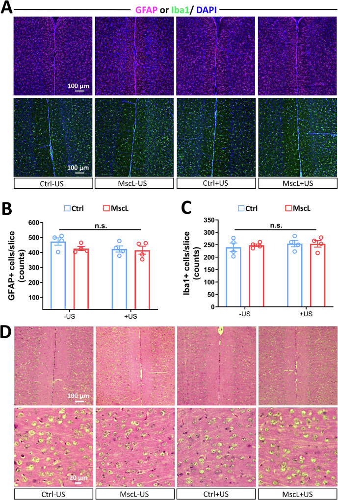
Researchers also examine mPFC tissue after twenty minutes of stimulation. Astrocyte-associated GFAP, microglial Iba1 and routine histology show no overt acute inflammatory activation or tissue damage. The observed behavioral change is accompanied by no clear abnormality in these measured tissue indicators.
Together with cellular, circuit and behavioral results, these tissue observations describe the tested stimulation conditions. They concern a short exposure; repeated stimulation and long-term channel expression involve different timescales. The present strategy requires intracerebral gene delivery and a head apparatus, and its behavioral evidence comes from acute tests in male mice.
Source: Lei T et al. · Molecular psychiatry · CC BY-NC-ND
Original figure and English caption ↗
The route taken by a signal matters
The study turns prefrontal stimulation into a more specific question: which cells respond, and where do they send information? Broad excitatory-population stimulation lacks the same behavioral effect, whereas DRN-projecting cells sensitized with MscL can change the outcome. Sonogenetics serves as both a trigger and a way to reveal functional differences among neural output routes.