Across a brief wait, how do two mammillary populations support working memory?
With shared hippocampal input, Nts and Nos1 cells support different portions of a delay. Grik4 changes activity duration and maze performance.
Can an animal remember which route it just took after a delay of several dozen seconds? Working memory must preserve a temporary cue between its disappearance and the next action. Often described as a relay between hippocampus and thalamus, the mammillary body may help determine how long that cue remains useful. This Research study in male mice identifies distinct temporal contributions from two molecularly defined excitatory populations and links them to a glutamate-receptor gene.
Both populations contribute from delay onset, with Nts involvement over roughly20seconds and Nos1 over40. Grik4 regulates synaptic/activity persistence, also altered in early APP/PS1 mice.
Contents · 7 sections
Distinct populations inside the small mammillary body
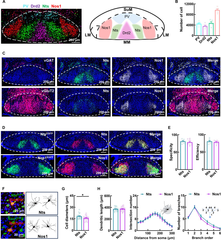
In the Papez memory circuit, Nts and Nos1 cells formed bilaterally symmetric populations, with Nos1 more lateral and both glutamatergic. Distinct cellular organization prompted comparisons of connectivity and activity.
Morphology differed modestly: Nts somata were slightly larger but dendritic length, complexity and branching were comparable. Input processing and activity persistence differentiated the populations more clearly than shape.
Source: Guo Y et al. · Research (Washington, D.C.) · CC BY
Original figure and English caption ↗
Shared input, separated output
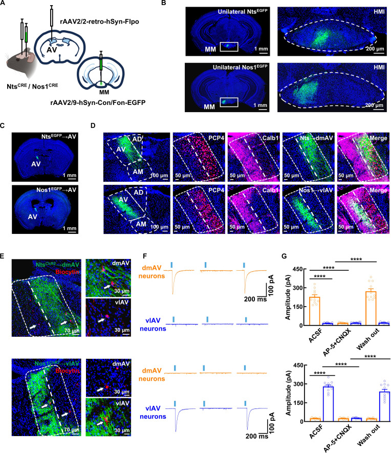
Both populations receive their predominant input from ventral subiculum. Axons originating on one side extend across the mammillary body and provide direct excitatory input to bilateral Nts and Nos1 cells. Output follows a different rule: each mammillary population projects ipsilaterally to anteroventral thalamus, with Nts and Nos1 targeting dorsomedial and ventrolateral subdivisions, respectively.
Stimulation and slice recording supported direct excitatory outputs. Shared subicular input led toward distinct thalamic subdivisions, motivating a test of when each population contributes during the waiting period.
Source: Guo Y et al. · Research (Washington, D.C.) · CC BY
Original figure and English caption ↗
Performance differs when waiting is introduced
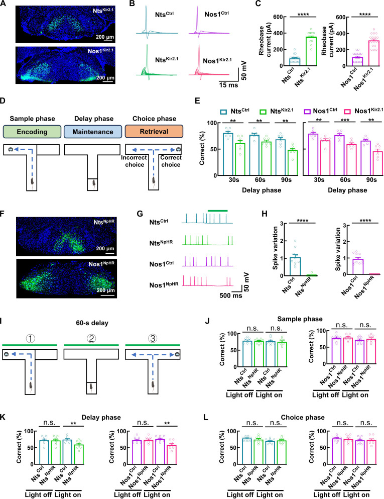
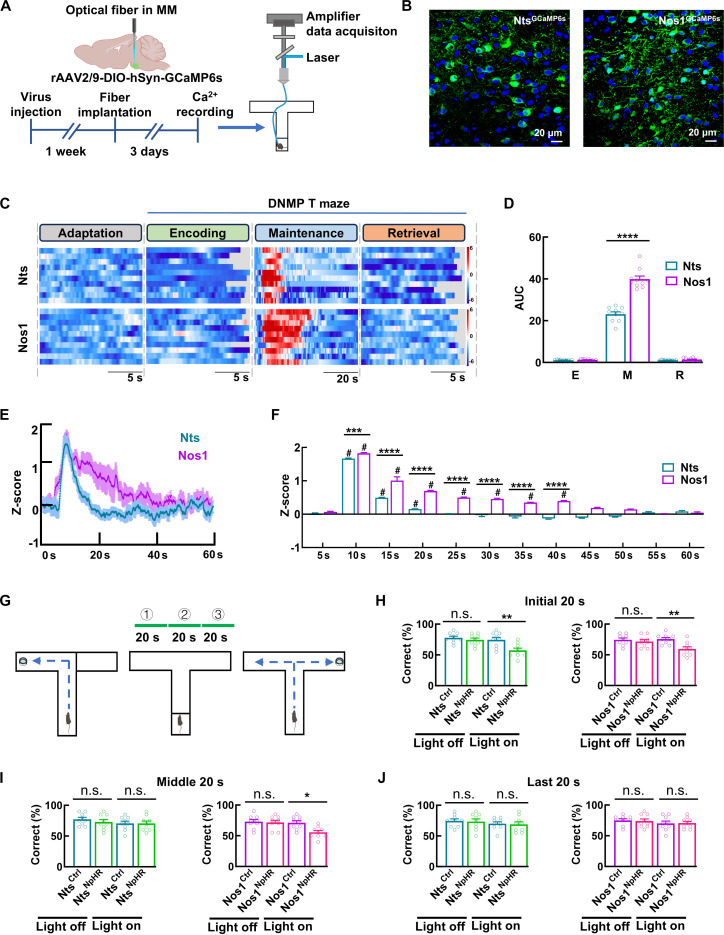
In delayed nonmatching-to-place, a mouse samples one arm and must choose the opposite arm after a delay to obtain reward. Sustained inhibition of either population leaves training and zero-delay choices comparable to controls, but reduces accuracy with 30-, 60- or 90-second delays. General movement, affect-related tests, social memory and water-maze performance do not show corresponding deficits, focusing the interpretation on temporary maintenance.
Phase-specific inhibition affected waiting rather than encoding or retrieval, with matching somatic and output results. The vulnerable period lay between acquiring information and choosing, focusing the next tests on seconds within the delay.
Source: Guo Y et al. · Research (Washington, D.C.) · CC BY
Original figure and English caption ↗
Both respond at the start, but Nos1 contributes longer
Population calcium signals rise at delay onset and then decay. Significant Nts activity lasts approximately 20 seconds, compared with approximately 40 seconds for Nos1, rather than scaling proportionally with total delay length. During a 60-second delay, Nts inhibition affects performance in the initial 20 seconds; Nos1 inhibition is effective in the initial and middle segments, with no comparable effect in the final segment.
The populations contribute together at onset, with a longer Nos1 window rather than sequential handoff. Activity does not scale proportionally with total delay. The authors proposed complementary updating and retention, motivating a search for persistence-related molecules.
Source: Guo Y et al. · Research (Washington, D.C.) · CC BY
Original figure and English caption ↗
Grik4 changes receptors and activity persistence
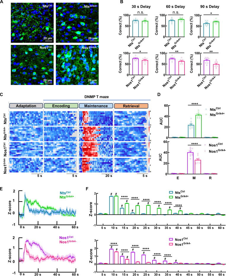
Nos1 cells express more Grik4, encoding the KA1 kainate-receptor subunit. Their kainate currents are larger and decay faster, while AMPA currents and long-term potentiation are stronger. Baseline firing measures, NMDA currents, miniature inhibitory currents and miniature excitatory-current frequency do not show the same differences; the distinction is not a uniform increase in all electrical activity.
Increasing Nts Grik4 or reducing Nos1 Grik4 shifted synaptic features toward the other population and lengthened or shortened activity. Nts gains appeared at90seconds, not30/60; a ceiling effect at shorter delays was proposed, indicating load-dependent expression of the benefit.
Source: Guo Y et al. · Research (Washington, D.C.) · CC BY
Original figure and English caption ↗
Functional change before cell loss
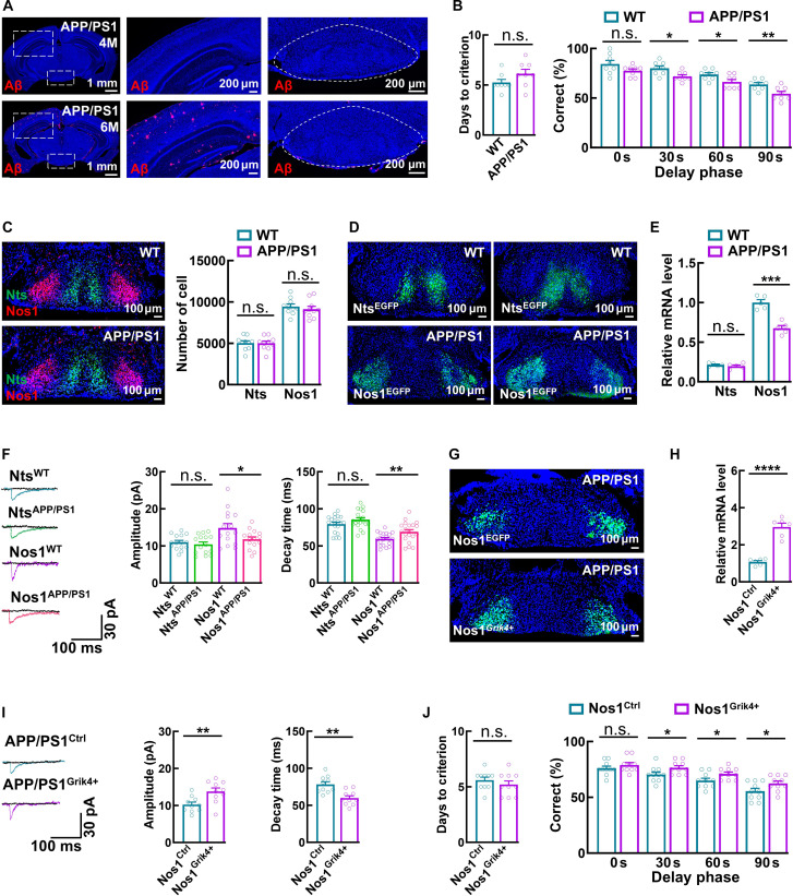
Four-month-old APP/PS1 mice already show working-memory impairment without overt plaque deposition or loss of mammillary Nts and Nos1 cells in this study. Nos1 Grik4 expression and kainate currents change, whereas Nts does not show the same alteration. Targeted Nos1 overexpression improves electrophysiology and maze performance, indicating that functional impairment need not wait for cell loss.
Source: Guo Y et al. · Research (Washington, D.C.) · CC BY
Original figure and English caption ↗
Working-memory timing gains cellular and receptor detail
The male-mouse study gives memory for a recent route a cellular timescale. Shared input, distinct output and receptor-linked persistence reveal temporal organization, while early APP/PS1 changes show functional alteration before cell loss. Populations need not look markedly different to contribute over different portions of one delay. Receptors, synapses and seconds-long activity reveal the mammillary body as a memory node with internal temporal organization.