Why does risk assessment falter under threat? Astrocytic D-serine clues in the basolateral amygdala
In DISC1-N model mice, activating basolateral amygdala astrocytes restores WFS1 neuron excitability via D-serine and NMDA receptors, improving risk-assessment behavior.
Accurate risk assessment in the face of potential threats is essential for survival, whereas dysregulated risk appraisal and maladaptive avoidance are prominent behavioral hallmarks across schizophrenia, bipolar disorder, and related psychiatric conditions. Although Disrupted-in-Schizophrenia 1 (DISC1) is a well-established psychiatric risk gene, how DISC1 dysfunction disrupts central threat evaluation—and specifically how astrocyte-neuron communication contributes to this process—has remained poorly understood. Published in Neuron in April 2024, Jie Tu, Fan Yang, Liping Wang, and colleagues at the Shenzhen Institute of Advanced Technology, Chinese Academy of Sciences, investigated inducible DISC1-N-terminal fragment transgenic (DISC1-N) mice and uncovered a specialized astrocyte-to-neuron signaling axis within the basolateral amygdala (BLA). Combining single-nucleus RNA sequencing, microdialysis-coupled liquid chromatography-mass spectrometry, patch-clamp electrophysiology, and in vivo fiber photometry, the authors show that impaired astrocytic D-serine release in the BLA of DISC1-N mice diminishes the excitability of Wolfram syndrome 1-positive glutamatergic (BLAWFS1) neurons, causing severe risk-assessment deficits. Optogenetic stimulation of BLA astrocytes or local D-serine supplementation restores BLAWFS1 neuronal firing via NMDA receptors and normalizes risk-assessment and avoidance behaviors.
In DISC1-N mice, impaired D-serine synthesis and vesicular release from basolateral amygdala (BLA) astrocytes reduces the excitability of neighboring WFS1-positive glutamatergic (BLAWFS1) neurons, causing risk-assessment deficits that can be rescued by astrocytic activation or exogenous D-serine acting at neuronal NMDA receptors.
Contents · 5 sections
DISC1-N Mice Exhibit Selective Risk-Assessment Deficits and Diminished Threat Avoidance
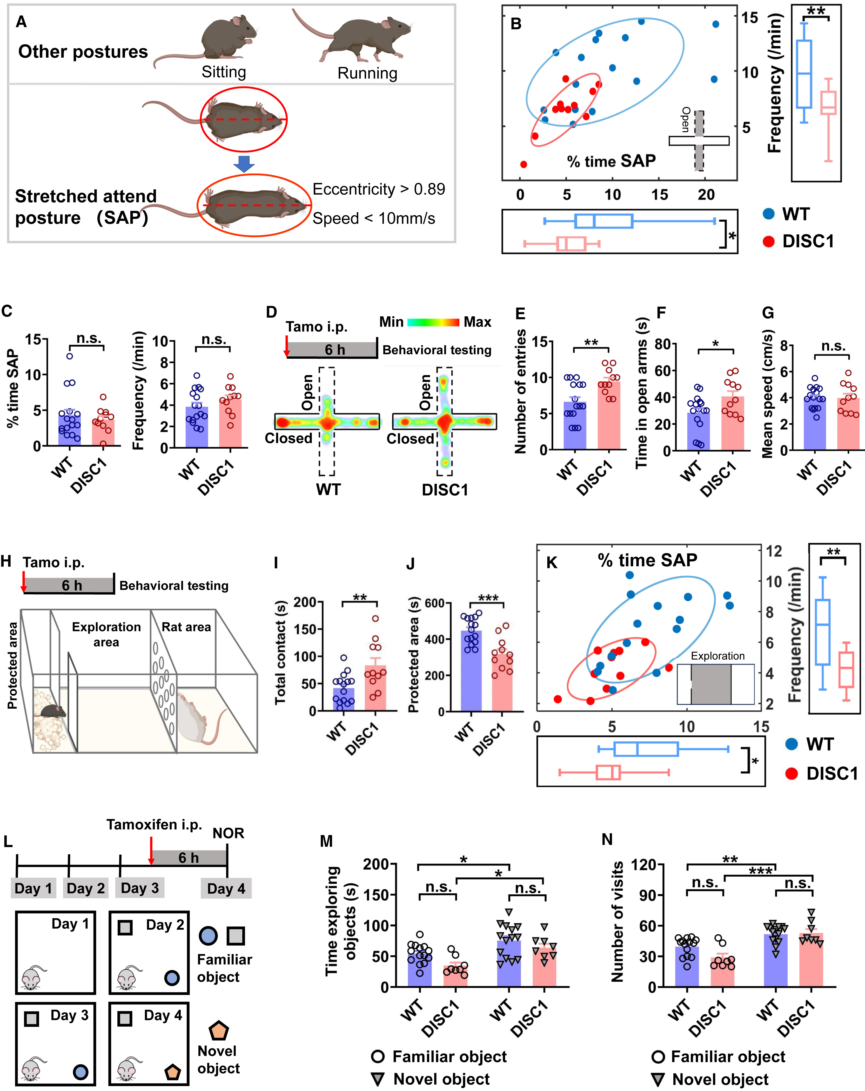
To evaluate risk appraisal and defensive avoidance across uncertain and explicit threat environments, the authors tested inducible DISC1-N transgenic mice and wild-type (WT) littermates 6 hours after intraperitoneal tamoxifen administration across the elevated plus maze (EPM), open-field test (OFT), light-dark box (LDB), rat exposure test (RET), and overhead looming paradigm. Specifically, they quantified stretched attend posture (SAP)—an investigatory posture in which an animal stretches forward and retracts without locomotion—as a behavioral readout of active risk assessment.
In the EPM, tamoxifen-induced DISC1-N mice exhibited significantly reduced SAP duration and frequency on the open arms alongside diminished risk avoidance (increased open-arm entries and time spent) without changes in locomotion speed, whereas corn-oil-treated DISC1-N and WT mice showed no behavioral differences. Consistently, in the RET paradigm—where mice encounter an anesthetized rat across a wire mesh screen—DISC1-N mice spent significantly more time in contact with the high-risk grid wall, less time in the protected chamber, and displayed fewer SAP bouts in the exploration zone. Novel object recognition (NOR) and sucrose preference tests confirmed normal novelty preference and absence of anhedonia, establishing that reduced threat avoidance in DISC1-N mice stems specifically from impaired risk assessment.
Source: et al. · Neuron · Original figure reproduced for academic literature review under fair scholarly commentary; copyright remains with the authors/publisher.
Original figure and English caption ↗
snRNA-seq and Microdialysis LC-MS Reveal Impaired Astrocytic Vesicle Release and Reduced Serine in the BLA
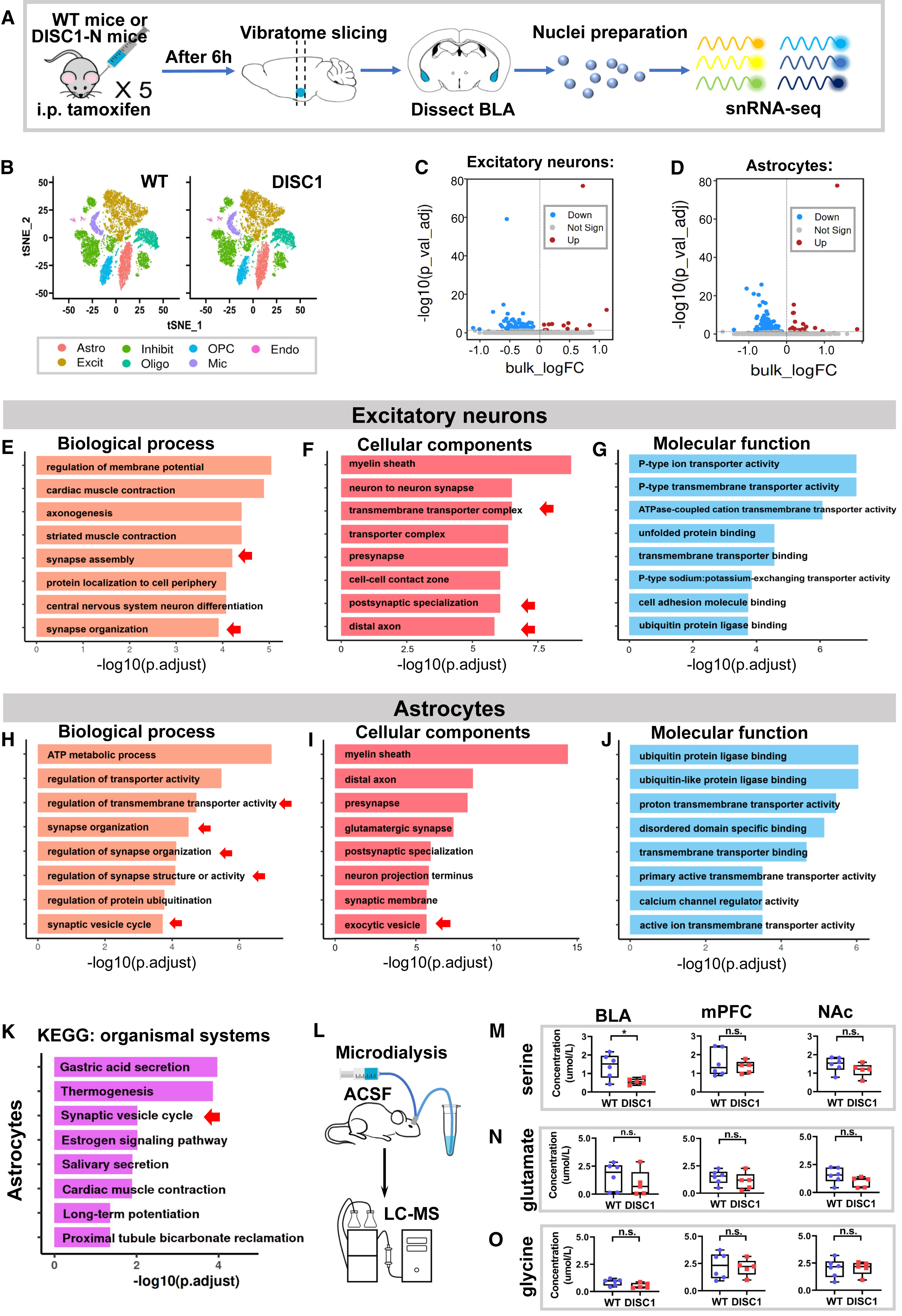
To dissect cell-type-specific molecular alterations in the basolateral amygdala (BLA), the researchers performed 10x Genomics single-nucleus RNA sequencing (snRNA-seq) on 11,122 nuclei from DISC1-N mice and 12,121 nuclei from WT controls. Among seven major cell types, excitatory neurons (CAMK2A+, 111 DEGs) and astrocytes (AQP4+, 103 DEGs) harbored the highest number of differentially expressed genes. Downregulated genes in excitatory neurons were enriched for synapse assembly and glutamatergic synaptic transmission (including reduced Ntrk2 and voltage-gated calcium channel subunits Cacna1a and Cacna1c, accompanied by reduced action potential firing at 130–140 pA in whole-cell patch-clamp recordings), whereas astrocytic DEGs were enriched for synapse organization, exocytic vesicles, and the synaptic vesicle cycle (including reduced Nlgn3, Snap25, Calm1, and Atp6v0c).
To test whether impaired astrocytic vesicular machinery alters extracellular gliotransmitters, the team collected microdialysates from the BLA, medial prefrontal cortex (mPFC), and nucleus accumbens (NAc) for liquid chromatography-mass spectrometry (LC-MS). Extracellular serine concentrations were significantly and selectively reduced in the BLA of DISC1-N mice—with no change in the mPFC or NAc, and unchanged glutamate and glycine levels across all three regions. Quantitative PCR further confirmed decreased BLA mRNA expression of phosphoglycerate dehydrogenase (Phgdh) and the neutral amino acid transporter Slc7a10, which govern astrocytic serine synthesis and D-serine transport.
Source: et al. · Neuron · Original figure reproduced for academic literature review under fair scholarly commentary; copyright remains with the authors/publisher.
Original figure and English caption ↗
Stimulating BLA Astrocytes Boosts Extracellular Serine Release and Rescues Risk-Assessment Deficits in DISC1-N Mice
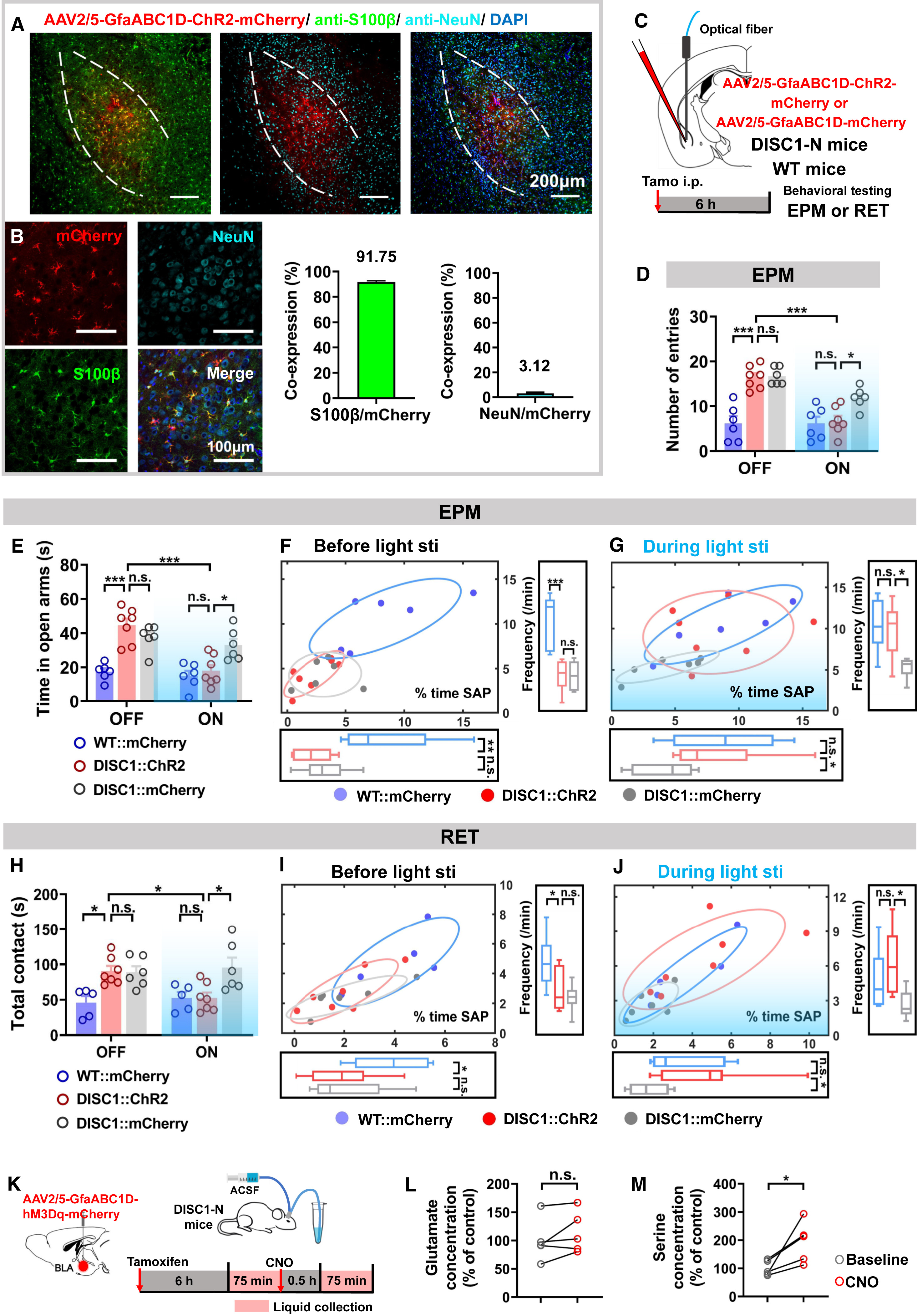
To test whether enhancing BLA astrocyte activity rescues impaired risk assessment, the authors injected astrocyte-specific AAV2/5-GfaABC1D-ChR2-mCherry into the BLA of DISC1-N mice (achieving 91.75% colocalization with S100β and only 3.12% with NeuN) and compared behavior during light-off versus blue-light photostimulation (20 Hz, 25 ms pulses). Whereas DISC1-N mice exhibited excessive open-arm exploration in the EPM, prolonged rat contact in the RET, and diminished SAP frequency/duration during the light-off phase, blue-light stimulation of ChR2-expressing BLA astrocytes immediately normalized open-arm entries and duration, reduced high-risk rat contact time, and restored SAP risk-assessment behaviors to WT levels.
This behavioral rescue was independently replicated in Aldh1l1-CreERT2 × DISC1-N double-transgenic mice injected with Cre-dependent AAV2/5-DIO-ChR2-mCherry in the BLA (89.8% S100β colocalization). Furthermore, chemogenetic activation of BLA astrocytes in DISC1-N mice (via AAV2/5-GfaABC1D-hM3Dq-mCherry plus CNO) coupled with microdialysis and LC-MS significantly increased extracellular serine concentrations in the BLA without altering extracellular glutamate levels.
Source: et al. · Neuron · Original figure reproduced for academic literature review under fair scholarly commentary; copyright remains with the authors/publisher.
Original figure and English caption ↗
BLA Astrocytes Selectively Excite WFS1+ Glutamatergic Neurons via D-Serine Acting at NMDA Receptors
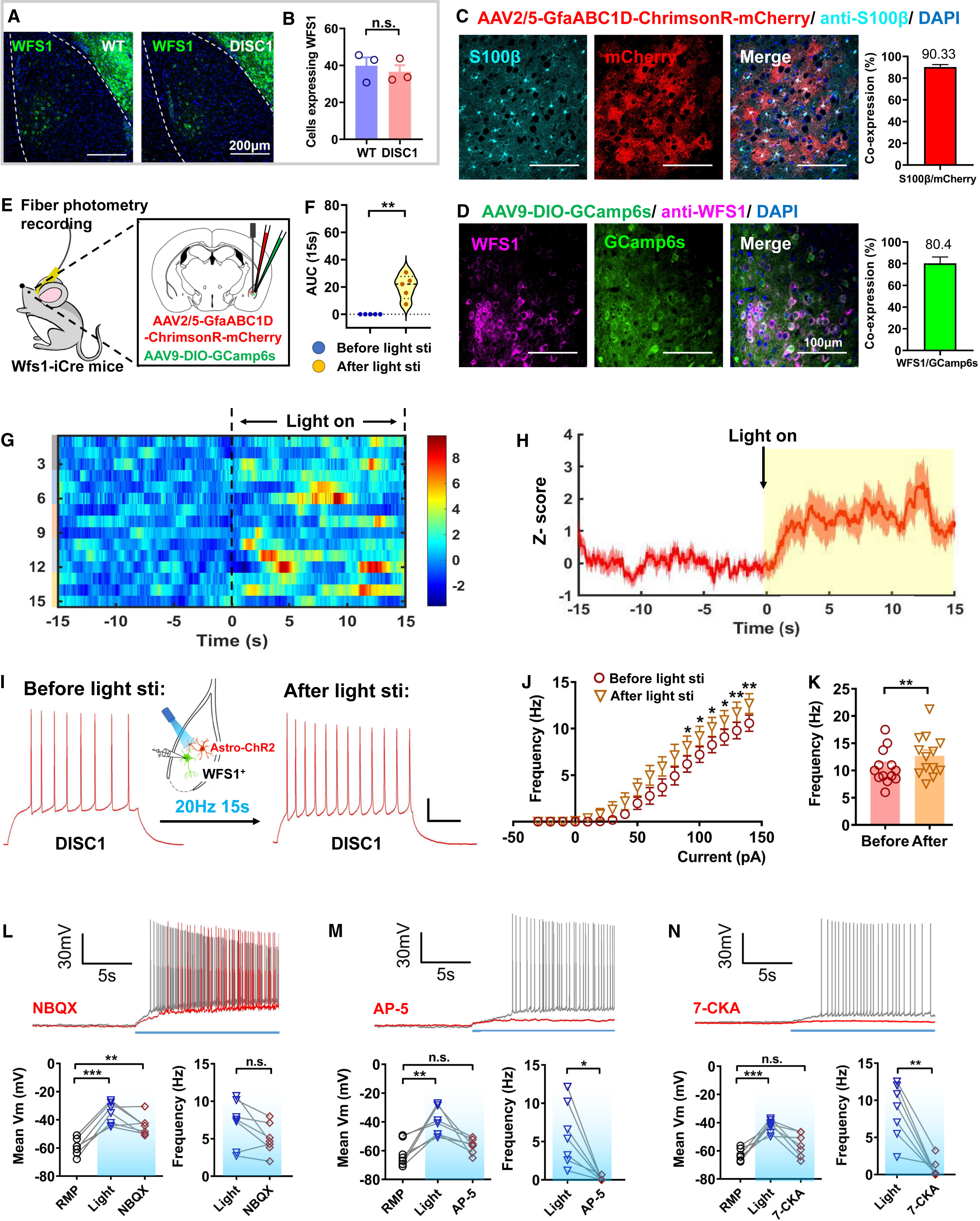
To determine how BLA astrocytes modulate nearby neurons, the authors recorded from neurons within 100 μm of ChR2-expressing astrocytes during 15-second blue-light stimulation in acute brain slices. Recorded BLA neurons segregated into two distinct populations: 'high-response' neurons that depolarized and fired action potentials with an average latency of ~5.6–5.9 seconds (consistent with gliotransmitter signaling), and 'low-response' neurons that exhibited only subthreshold depolarization (~5.16 mV) without firing regardless of stimulation frequency or duration. Notably, high-response neurons in DISC1-N mice exhibited significantly reduced current-evoked firing rates compared with WT high-response neurons. Combining snRNA-seq candidate screening with patch-seq single-cell qPCR of recorded neurons identified Wolfram syndrome 1 (Wfs1)—expressed in ~32% of BLA neurons, >90% of which are vGlut+ excitatory neurons—as a specific molecular marker of high-response neurons.
In Wfs1-iCre mice co-expressing ChrimsonR in BLA astrocytes (AAV2/5-GfaABC1D-ChrimsonR-mCherry) and GCaMP6s in BLAWFS1 neurons (AAV9-DIO-GCamp6s), in vivo fiber photometry coupled with 590 nm yellow-light stimulation demonstrated robust, sustained Ca2+ elevations in BLAWFS1 neurons upon astrocytic activation, and ChR2-astrocyte photostimulation in slices restored current-evoked firing rates of DISC1-N BLAWFS1 neurons. Pharmacological dissection in slices revealed that whereas the AMPA receptor antagonist NBQX (50 μM) did not block astrocyte-evoked action potential firing in BLAWFS1 neurons, both the competitive NMDA receptor antagonist AP-5 (50 μM) and the selective NMDAR glycine-site antagonist 7-chlorokynurenic acid (7-CKA, 20 μM) completely abolished astrocyte-induced firing, proving that astrocytic D-serine acts at the NMDAR glycine-binding site to excite BLAWFS1 neurons.
Source: et al. · Neuron · Original figure reproduced for academic literature review under fair scholarly commentary; copyright remains with the authors/publisher.
Original figure and English caption ↗
Exogenous D-Serine Recapitulates Behavioral Rescue and BLAWFS1 Neuronal Activity Is Required for Astrocyte-Mediated Recovery
To test whether exogenous D-serine directly rescues risk-assessment deficits, the researchers microinfused D-serine into the BLA of DISC1-N mice prior to behavioral testing. Compared with ACSF vehicle controls, intra-BLA D-serine administration significantly reduced open-arm entries and duration while increasing open-arm SAP percentage in the EPM, and reduced high-risk grid contact time while elevating SAP frequency and duration in the RET. Furthermore, in vivo fiber photometry in Wfs1-iCre versus Wfs1-iCre × DISC1-N mice during the RET revealed that whereas BLAWFS1 neurons in control mice exhibited a sharp Ca2+ surge upon entering the high-risk zone, DISC1-N mice displayed a significantly blunted high-risk-zone Ca2+ response (AUC) and reduced Ca2+ event frequency in the exploration area.
Finally, to test whether BLAWFS1 neuronal activity is required for astrocyte-mediated behavioral rescue, the team co-expressed AAV2/5-GfaABC1D-ChR2-eYFP in astrocytes and AAV9-DIO-hM4Di-mCherry in BLAWFS1 neurons of Wfs1-iCre × DISC1-N mice. Chemogenetic inhibition of BLAWFS1 neurons with CNO completely abolished the ability of blue-light astrocyte stimulation to restore risk avoidance and SAP behavior in both the EPM and RET paradigms. Notably, direct chemogenetic activation of BLAWFS1 neurons (via AAV9-DIO-hM3Dq-mCherry) without astrocytic modulation elevated normalized risk-assessment scores (SAP) but paradoxically increased time spent in risky zones in DISC1-N mice, highlighting the unique role of BLA astrocytes in coordinating balanced threat appraisal and defensive avoidance.