Quiet but more vigilant: how serotonin changes brain state
After an alarm signal, zebrafish move less yet respond more readily to threats. The study links synchronized neural activity to serotonin and its receptors, with related vigilance features in male mice.

An animal facing danger may stop rather than run faster, attending to changes around it. A study published in Nature Communications on March 22, 2024 starts with alarm responses in zebrafish to investigate this combination of reduced movement and heightened responsiveness. Serotonergic signaling from the dorsal raphe nucleus changes the rhythm of forebrain activity. In both fish and mice, sustained activation of these neurons can produce less movement alongside greater sensitivity to aversive stimuli.
Sustained raphe serotonin-neuron activation can induce low-frequency synchrony and vigilance-related behavior involving 5-HT7 receptors. Pulsed stimulation in mice does not produce the same EEG transition, highlighting the importance of activation pattern.
Contents · 8 sections
Less movement does not mean less responsiveness
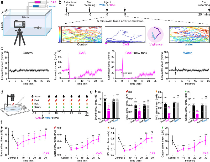
After exposure to conspecific alarm substance, zebrafish briefly swim rapidly, remain near the bottom, then swim more slowly. This state persists even after transfer to fresh water. Movement alone cannot distinguish immobility from heightened monitoring of danger.
Response thresholds to sound and several other aversive stimuli fall and then gradually recover, unlike water controls. Reduced movement together with greater responsiveness provides the behavioral basis for studying vigilance.
Source: Zhao Y et al. · Nature communications · CC BY
Original figure and English caption ↗
Forebrain cells adopt a shared rhythm
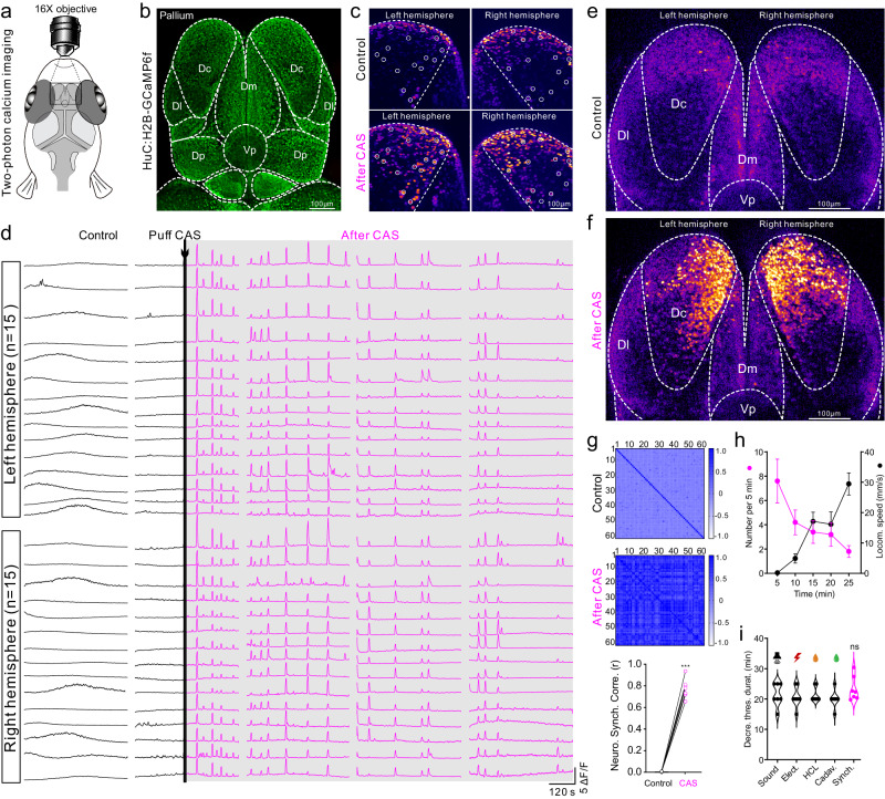
Imaging reveals a change in the dorsal pallium. Previously less coordinated calcium activity becomes low-frequency, high-amplitude synchronized oscillation, concentrated in central zone Dc. Correlations between neurons rise, as if independent pendulums begin keeping time together.
As synchronized events decline, swimming speed rises again. Their duration resembles the period of lowered response thresholds, adding a neural pattern to the behavioral description of vigilance.
Source: Zhao Y et al. · Nature communications · CC BY
Original figure and English caption ↗
Alarm recruits raphe serotonin signaling
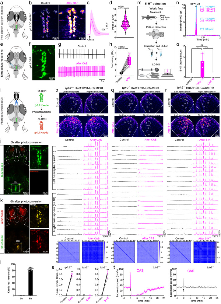
Alarm substance increases calcium activity and sustained firing in dorsal raphe serotonin neurons. Tracing links them to Dc, while dorsal-pallium samples show an early increase in 5-HT that returns near control levels after an hour.
Neuromodulators can change how populations receive inputs and organize activity, linking an external event to a longer-lasting state. That time span is central here: the initial rapid swim has ended while slower behavior and coordinated neural rhythms continue, gradually subsiding as the alarm response fades.
Fish lacking the serotonin-synthesis gene tph2 lack the normal synchrony and movement reduction. Added 5-HT restores synchrony, implicating serotonin in generating the state rather than merely accompanying alarm.
Source: Zhao Y et al. · Nature communications · CC BY
Original figure and English caption ↗
A receptor links the chemical signal to firing rhythm
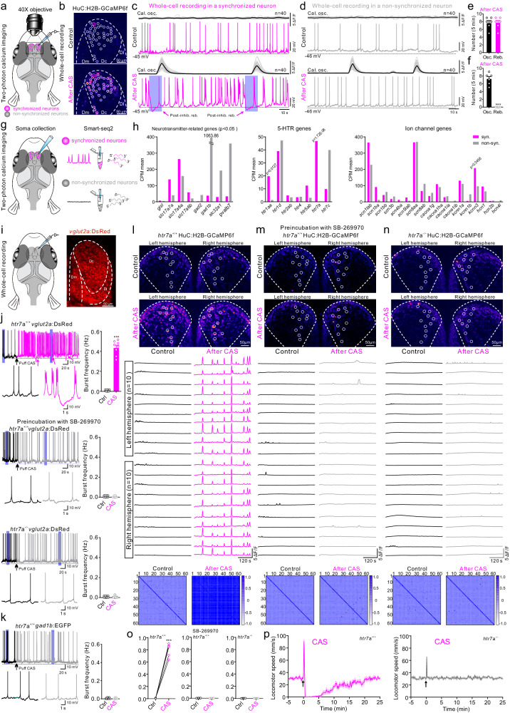
Synchronized cells predominantly show glutamatergic features and enriched htr7a transcripts. Their firing shifts toward bursts, with post-inhibitory rebounds aligned to calcium oscillations. Recorded GABAergic cells do not show the same firing transition.
Blocking 5-HT7 receptors or losing htr7a prevents alarm-induced bursting and synchrony. Mutant fish also lack the normal sustained movement reduction, connecting receptor signaling with cellular rhythm and behavior.
Source: Zhao Y et al. · Nature communications · CC BY
Original figure and English caption ↗
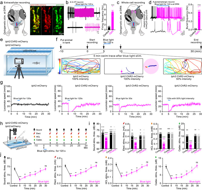
The state can be induced without alarm substance
Sustained activation of raphe serotonin neurons induces Dc bursting, reduced swimming and lower aversive-response thresholds without alarm substance, unlike controls. Persistence varies with activation conditions, suggesting that a neuromodulator can maintain a state beyond an initial cue.
Source: Zhao Y et al. · Nature communications · CC BY
Original figure and English caption ↗
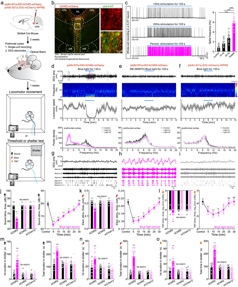
Mice also move less while responding more readily
In male mice, sustained activation shifts prefrontal EEG toward low-frequency delta activity with less movement. Putative excitatory cells classified by spike waveform burst in phase with that rhythm. Aversive-response thresholds fall and shelter stays lengthen; 5-HT7 antagonism prevents corresponding changes.
Pulsed stimulation instead increases movement without changing the EEG power spectrum. Activation pattern matters: recruiting the same neuronal group need not produce a single behavioral outcome.
Source: Zhao Y et al. · Nature communications · CC BY
Original figure and English caption ↗
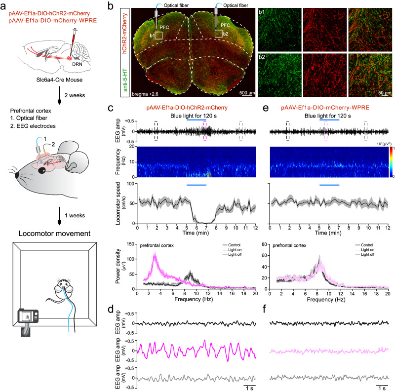
Prefrontal terminals reproduce key state features
Activating serotonergic terminals in prefrontal cortex also produces low-frequency synchronized EEG and reduced movement. This functionally links raphe input to the forebrain; the terminal experiment directly measures EEG and movement rather than repeating every threat-response test.
Source: Zhao Y et al. · Nature communications · CC BY
Original figure and English caption ↗
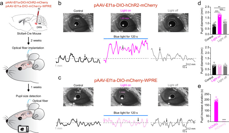
The eyes provide another sign of the state
Pupillometry in head-fixed mice adds another feature: raphe activation enlarges pupils and increases their fluctuations, without a significant change in controls. Fish are four to five weeks old and of mixed sex; mice are two-month-old males.
The study describes vigilance as coordinated changes: less bodily movement, synchronized neural rhythms and greater responsiveness to the outside world. Related serotonergic regulation in two species shows why slowing down need not mean becoming less alert.
Source: Zhao Y et al. · Nature communications · CC BY
Original figure and English caption ↗