Why does fear return after extinction? Locus coeruleus input reshapes an amygdala trace
A mouse study finds that an intervention after extinction reduces returning fear and links locus coeruleus input, dopamine D2 signaling and amygdala engram activity.
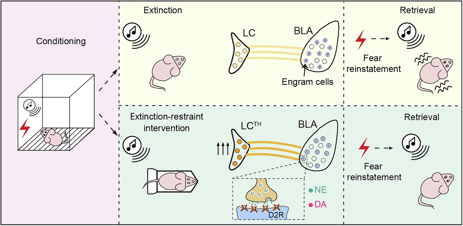
A tone once predicted danger, but repeated tones without shock gradually reduce freezing in mice. New shocks can nevertheless bring fear back. What remains in the brain after extinction? By following basolateral amygdala cells recruited during initial learning, researchers traced a post-extinction intervention to locus coeruleus input and dopamine D2 signaling.
Restraint after extinction reduces reinstatement, amygdala engram reactivation and optically evoked freezing, with contributions from locus coeruleus projections and local D2 signaling.
Contents · 6 sections
After fear subsides, the original ensemble can still evoke freezing
After tone–shock pairing, the tone alone evokes freezing. Repeated tones without shock reduce that response through extinction, but unsignaled shocks can reinstate it. The contrast prompted the team to examine the cells behind the behavior.
The team tagged basolateral amygdala cells active during conditioning as a fear engram. Direct optical activation still increased freezing after extinction, and reinstatement recruited the tagged ensemble again. The response had subsided while the cells retained the ability to drive it.
An intervention after extinction weakens returning fear
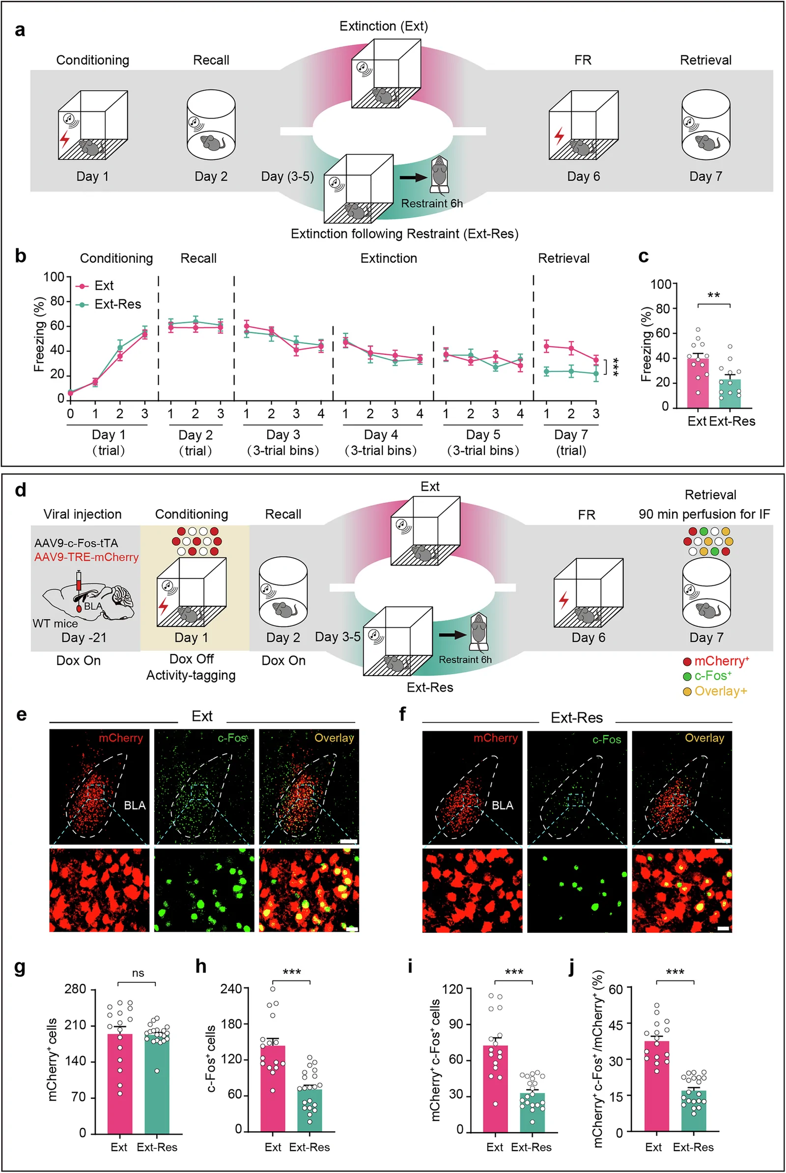
Restraint followed extinction on three successive days, before unsignaled shocks tested reinstatement. The combined group froze less and showed less reactivation of tagged BLA cells, with similar tagged-cell counts. The change concerned renewed recruitment of the ensemble.
The main experiment used six hours of restraint, while ninety minutes also reduced reinstatement. Motor, grip, object-recognition and tested anxiety-related performance remained comparable. The next question was whether the original ensemble itself had become less able to drive fear.
Source: Li T et al. · Experimental & molecular medicine · CC BY
Original figure and English caption ↗
Direct engram activation also produces a weaker response
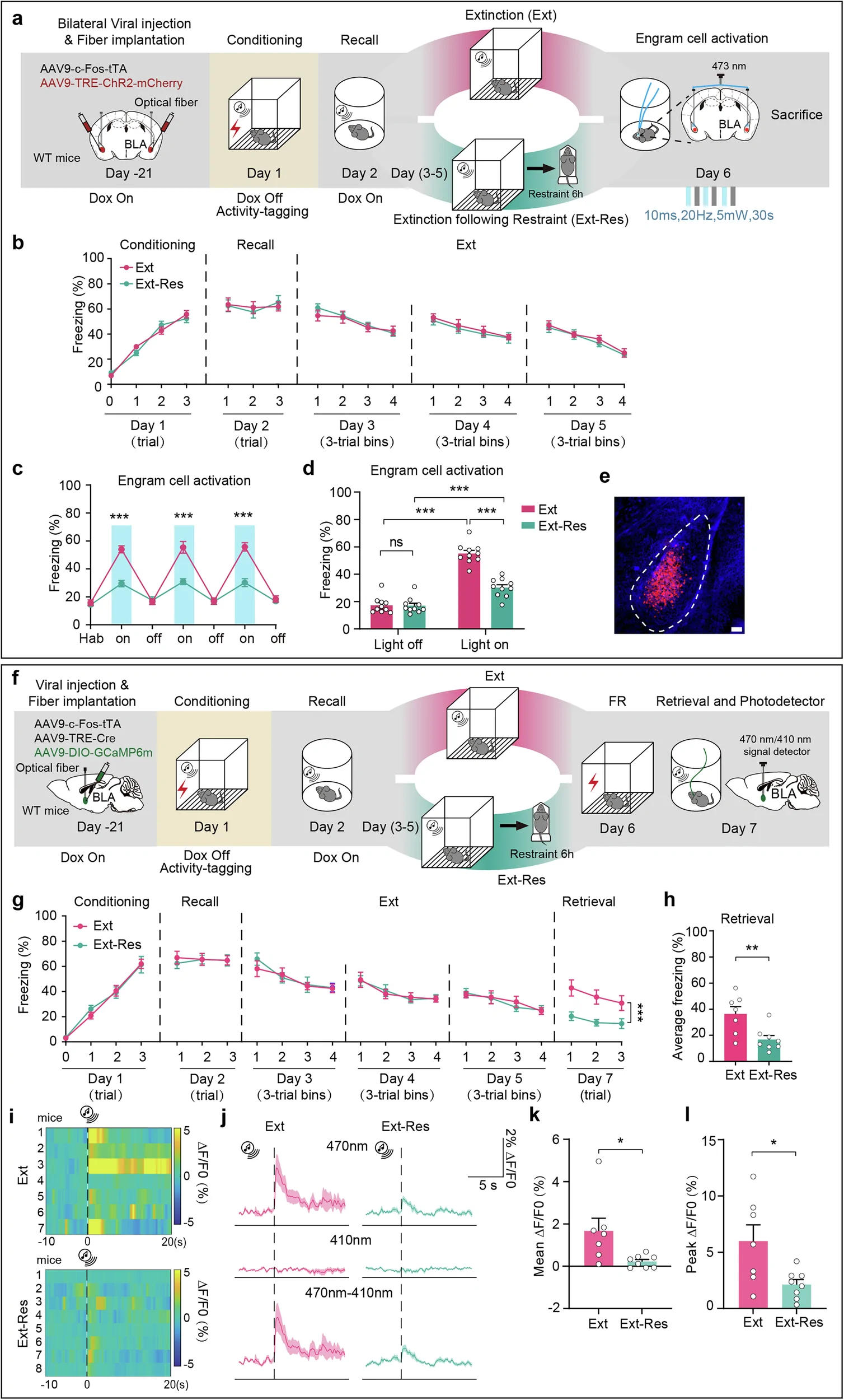
The researchers bypassed tone perception and retrieval by directly stimulating the tagged BLA cells. Extinction-only mice retained pronounced light-evoked freezing, whereas the combined group responded much less. The same ensemble now drove a weaker behavioral output.
Calcium recordings showed the same direction: tone-evoked mean and peak engram responses decreased. Weaker freezing and lower activity linked the behavioral effect to functional changes in a specific ensemble and motivated a search for its upstream regulator.
Source: Li T et al. · Experimental & molecular medicine · CC BY
Original figure and English caption ↗
Locus coeruleus input provides an upstream link
Restraint increased activity labeling in tyrosine-hydroxylase-positive locus coeruleus cells. Tracing connected them to BLA fear engrams, and activating LC cells during recording lowered engram calcium activity. The brainstem node became an upstream candidate.
Activating BLA-projecting LC cells after extinction reproduced lower subsequent freezing, while inhibiting them during combined restraint raised freezing again. The complementary manipulations strengthened the link between LC input and the intervention’s effect.
From locus coeruleus input to dopamine and D2 receptors
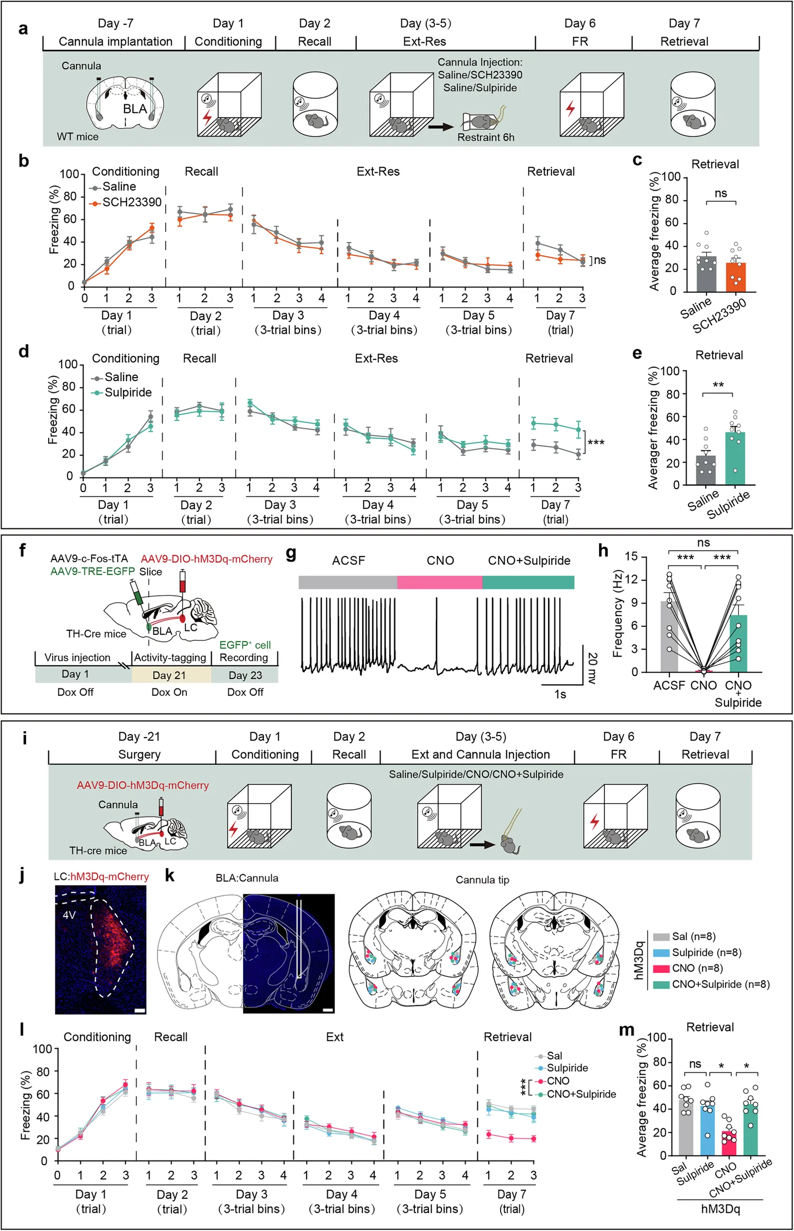
LC is strongly associated with norepinephrine, but another signal emerged here. Restraint increased ensemble-associated dopamine-sensor responses, while the corresponding norepinephrine readout did not significantly change over the compared periods. LC activation raised dopamine signals, and local BLA dopamine after extinction reduced later freezing.
Transmitters differed across timing and recording scope: supplementary bulk BLA norepinephrine responses were faster than the ensemble-associated dopamine change. D2 blockade weakened both the combined intervention and LC activation effects and opposed reduced engram firing; D1 blockade did not significantly change the intervention’s effect.
Source: Li T et al. · Experimental & molecular medicine · CC BY
Original figure and English caption ↗
A window for changing memory after extinction
The study follows a familiar problem—why fear returns—into identifiable cells, brainstem input and local receptor signaling. Timing stands out: altering a tagged ensemble after extinction can change later fear expression. How D2 signaling affects synapses and cellular plasticity is the next question.
The evidence comes from adult mice in conditioned fear and reinstatement tests. Longer follow-up, other relapse paradigms and engram-subtype studies will clarify durability and scope. Human traumatic memories and clinical interventions require independent investigation.