Remembering a new partner after sleep: social memory in spindle rhythms
From mouse thalamic circuits to children’s sleep recordings, a study follows the connection between spindles and socially relevant memory.
After meeting a partner, how does the brain retain the clues needed to recognize them later? Sleep contributes to consolidation, and its rhythms may organize that process more precisely than duration alone. A 2026 Nature Communications study focuses on NREM sleep spindles, following cells and projections in the mouse sensory thalamic reticular nucleus to investigate social-memory retention. The work also examines children’s face memory and overnight EEG, connecting observations of rhythms, circuits and behavior.
Mouse sensory thalamic reticular PV neurons and their VPM projection regulate spindles and social-memory consolidation. In children’s sleep recordings, spindle features are associated with next-morning face memory.
Contents · 6 sections
Beyond sociability lies memory for individuals
A mouse may approach another mouse willingly yet fail to recognize it later. The study assessed social memory through choices between familiar and novel animals, alongside initial sociability, object memory and overall activity. Together, these measures made remembering individual partners a distinct question to investigate.
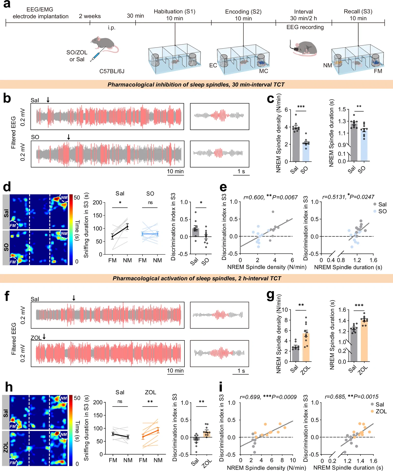
Pharmacological conditions that reduced spindle density and duration impaired social memory after thirty minutes, while spindle-enhancing conditions supported retention at two hours. Sociability, object memory and locomotion remained similar. Because the drugs also affected sleep latency and other sleep features, the researchers turned to rhythm-generating cells and circuits for a more specific connection.
Source: Cui D et al. · Nature communications · CC BY-NC-ND
Original figure and English caption ↗
Sensory TRN PV cells are recruited during spindles
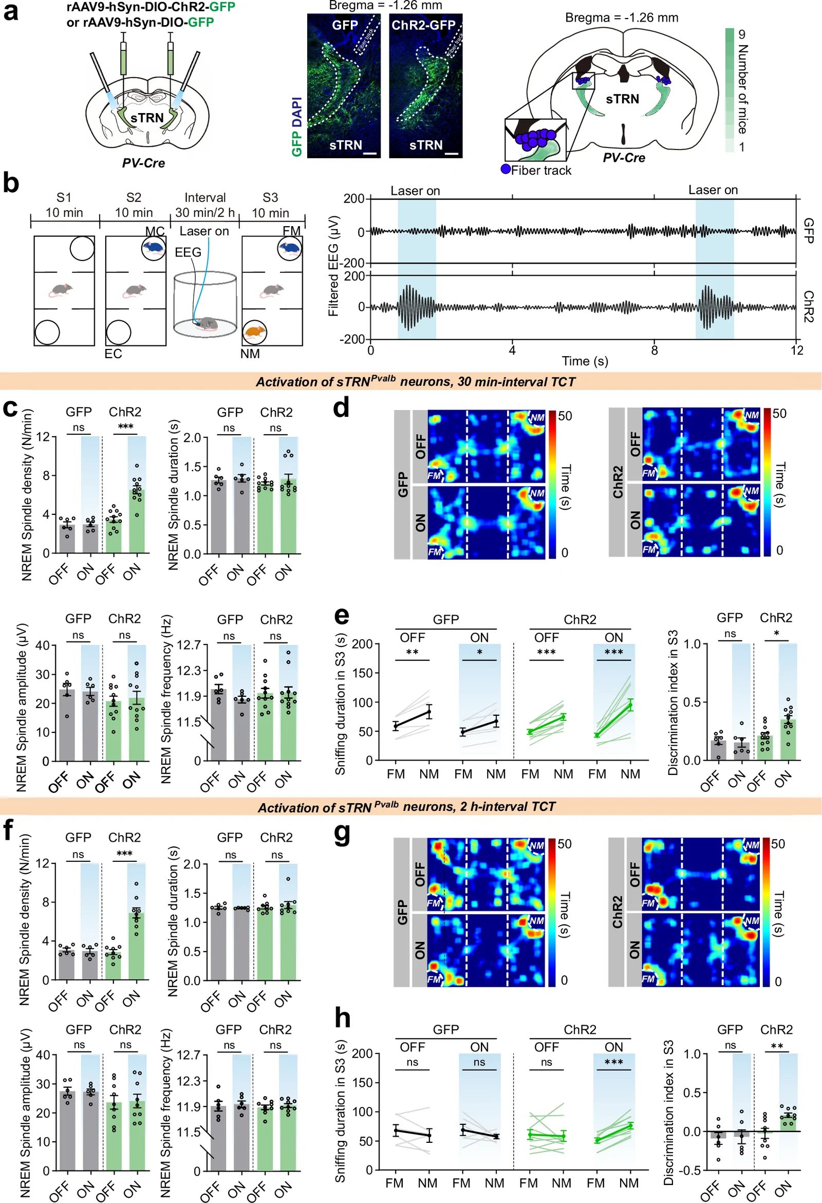
Simultaneous EEG, EMG and calcium recordings showed that PV neurons in the sensory thalamic reticular nucleus, or sTRN, were recruited during spindles and associated with sleep transitions. Suppressing these cells during the interval after an encounter reduced spindle density and duration and impaired later social memory. Suppressing the corresponding limbic TRN population did not produce the same result.
Spindle-like stimulation during the memory interval increased sTRN activity, raised spindle density and improved memory. Duration, amplitude and intrinsic frequency remained similar, with the main rhythmic change being more frequent events; sleep latency also changed. The connection between cellular activity and sleep rhythms thus took on a more specific form.
Source: Cui D et al. · Nature communications · CC BY-NC-ND
Original figure and English caption ↗
VPM and PF contribute at different memory stages
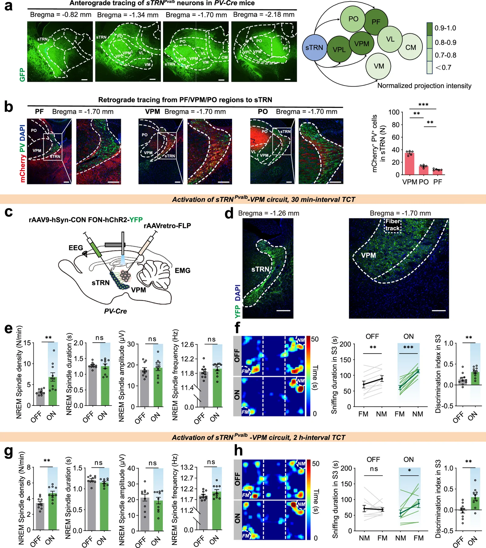
sTRN PV neurons project to several thalamic nuclei. The projection to the ventroposteromedial nucleus, or VPM, emerged as a lead for consolidation. Increasing its activity during the memory interval raised spindle density and later novel-mouse preference, while sociability and object memory remained similar. A defined anatomical connection thus linked the rhythm with social memory.
The projection to the parafascicular nucleus, or PF, showed a different role. The same stimulation during consolidation did not significantly change spindles or social memory, whereas stimulation during encoding or retrieval improved memory. From encountering a partner to retaining information during sleep and recognizing them later, thalamic outputs contributed differently across memory stages.
Source: Cui D et al. · Nature communications · CC BY-NC-ND
Original figure and English caption ↗
Weaker transmission leaves room for compensation
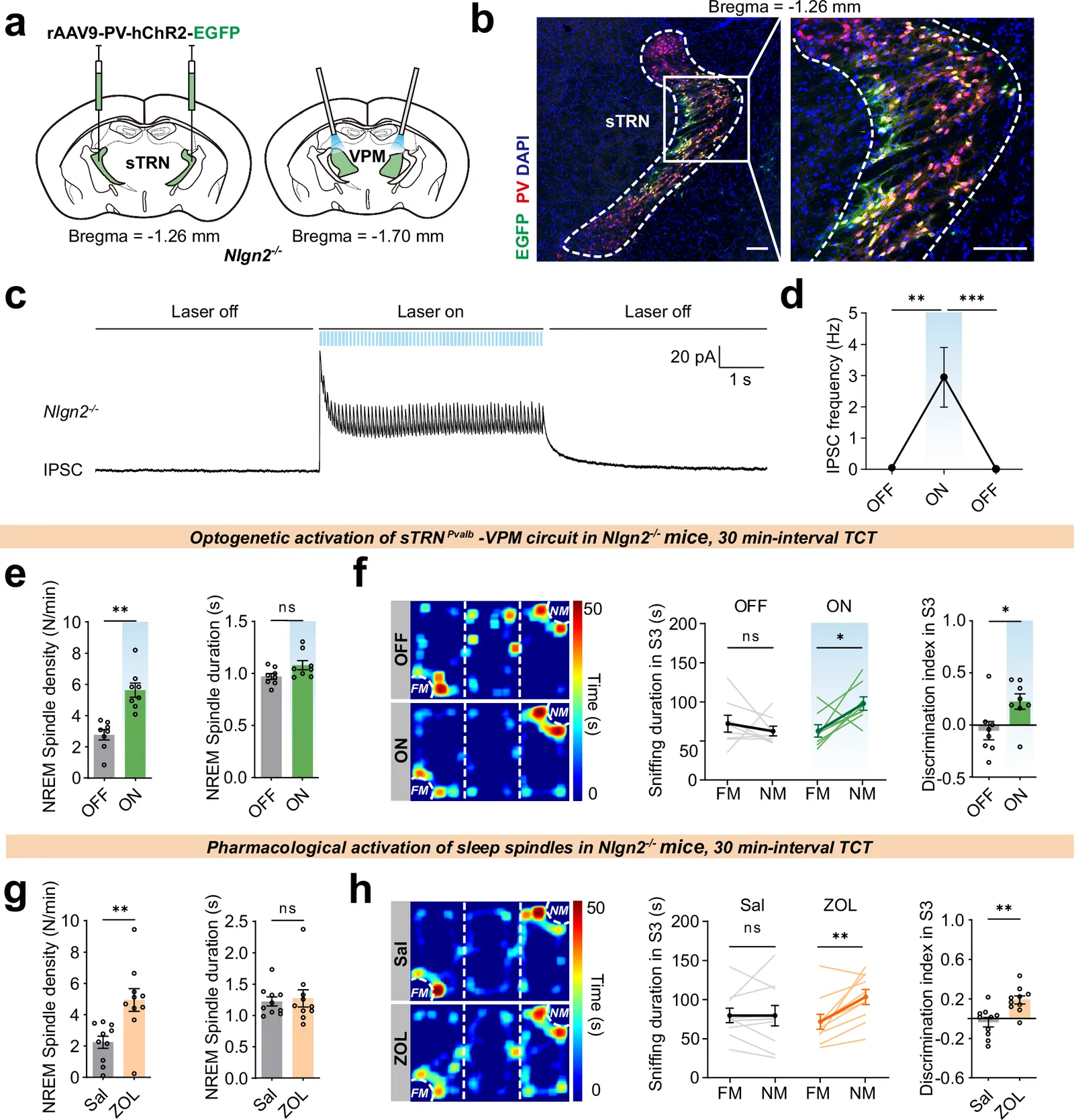
Nlgn2-deficient mice showed weaker inhibitory sTRN–VPM transmission, reduced spindle density and impaired social memory, with preserved sociability and spindle duration. These findings placed the memory phenotype in the identified connection, linking altered sleep rhythms with synaptic transmission.
Spindle-like terminal stimulation increased VPM inhibitory current frequency and spindle density and improved social memory. Pharmacological spindle enhancement also improved the phenotype. This defined memory outcome was therefore modifiable, making connection function and rhythmic organization useful entry points for investigating compensation.
Source: Cui D et al. · Nature communications · CC BY-NC-ND
Original figure and English caption ↗
Spindle features relate to next-morning face memory
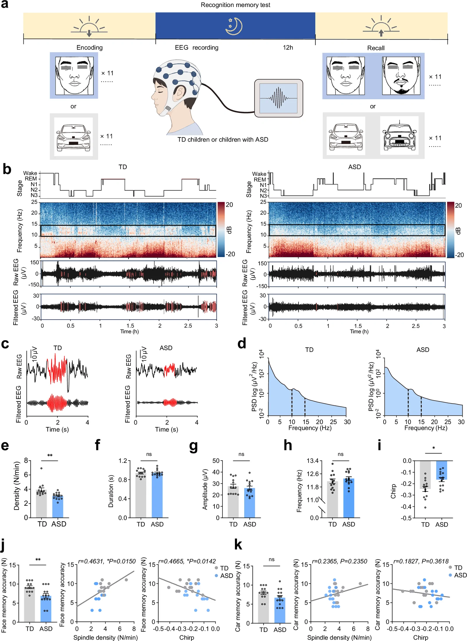
The observational component included fourteen children with ASD and thirteen typically developing children. Bedtime encoding, next-morning face and car recognition, and overnight EEG connected sleep with memory. Overall sleep and major sleep stages were comparable, but the ASD group had lower spindle density and absolute chirp values and poorer face recognition, with comparable car memory.
Spindle features correlated with face memory, linking detailed overnight rhythmic features with next-day socially relevant memory. Mouse partner recognition and children’s face recognition approach memory through different tasks. The observations inform one another and motivate further study of sleep rhythms and recognition of individuals.
Source: Cui D et al. · Nature communications · CC BY-NC-ND
Original figure and English caption ↗
Sleep features open a broader research window
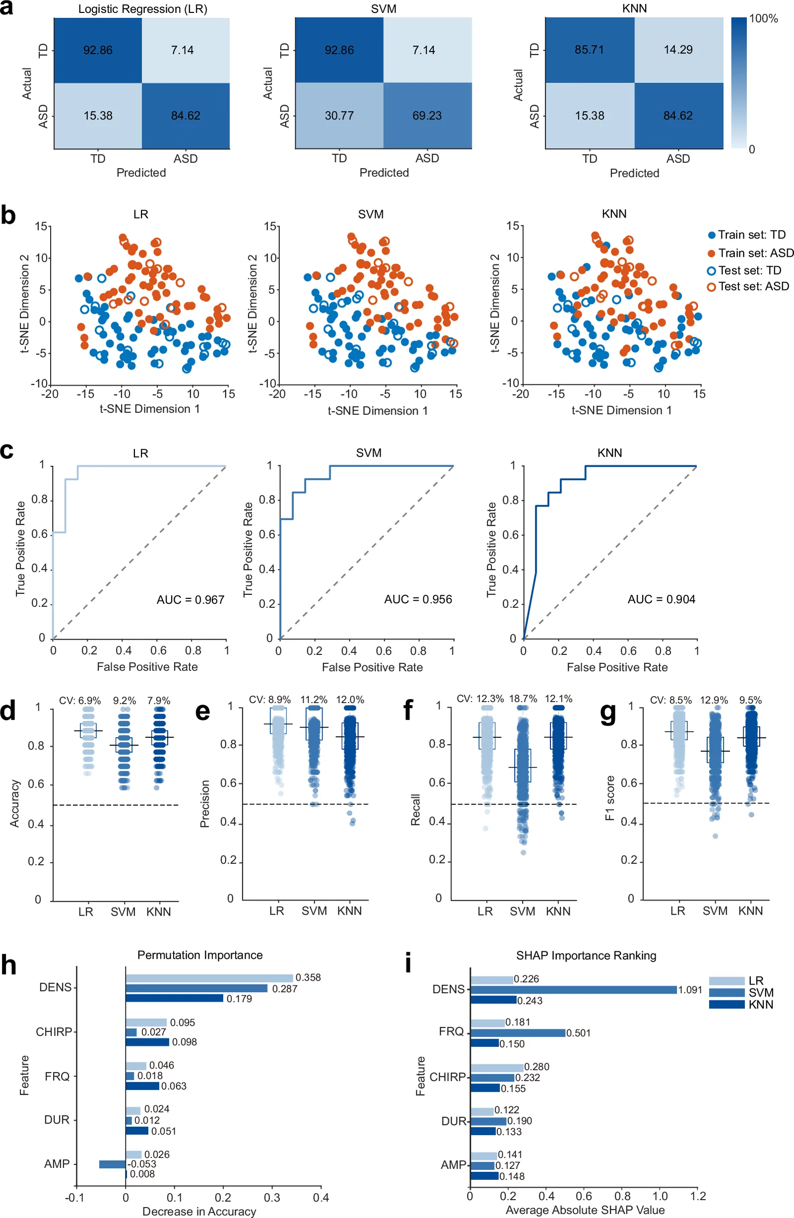
The study also explored classification information in sleep features. A larger EEG dataset included sixty-eight children with ASD and sixty-eight controls, using an eighty/twenty training/holdout split to compare models. Logistic regression achieved 88.9% accuracy in the within-cohort holdout set, with spindle density and chirp important features. These results identify rhythmic candidates for investigation across cohorts.
Main circuit experiments used male mice, with female validation of pharmacological memory findings. The children’s samples had defined age, cognitive and comorbidity ranges. Cross-cohort studies and work on how spindles coordinate with other memory rhythms will extend this picture. The study establishes a framework connecting brief sleep rhythms with recognition of a partner after waking.
Source: Cui D et al. · Nature communications · CC BY-NC-ND
Original figure and English caption ↗