Why can anxiety remain when nasal symptoms improve? From patient follow-up to brain–immune clues
Starting with differing patient symptom trajectories, a study follows amygdala activity, nasal sympathetic and immune changes, and IL-33 signaling in mice.
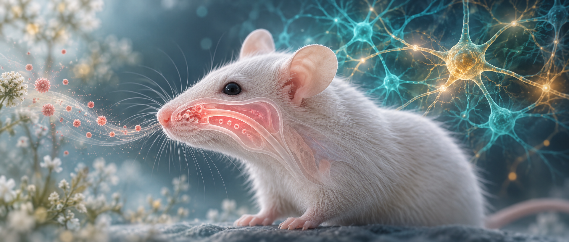
When local symptoms improve but another difficulty does not change alongside them, how can the difference be understood? A 2026 Journal of Neuroinflammation study first follows patients with chronic rhinosinusitis, then examines a mouse nasal-inflammation model. Nasal symptoms and anxiety scores follow different trajectories in patients. Animal experiments connect basolateral amygdala, or BLA, ventral hippocampus, or vHPC, and peripheral immune measures. From clinical observations to modifiable brain–immune components, the study investigates states that are related yet can change separately.
Mouse BLA excitatory cells and vHPC-projecting populations contribute to anxiety-related behavior and nasal immune changes. Nasal IL-33 neutralization improves some peripheral measures, behavior and BLA excitability.
Contents · 6 sections
Nasal and anxiety scores follow different trajectories
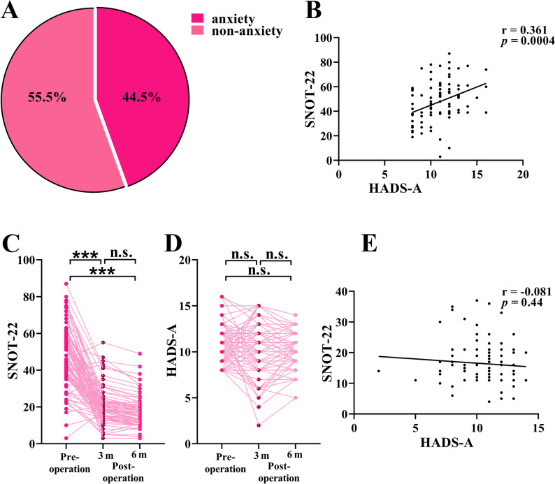
Among 209 patients, 44.5% met the study’s HADS-A anxiety-symptom criterion before surgery, and anxiety scores correlated with SNOT-22 nasal scores. After surgery and local steroid treatment, nasal scores improved at three months and remained improved at six months. Anxiety scores did not significantly change, and postoperative correlations were not significant.
The differing score trajectories prompted investigation of mechanisms beyond local symptom severity. Anxiety and nasal state could be related before treatment yet change differently afterward. This clinical observation became the starting point for animal work on links between central activity and peripheral inflammation.
Source: Zhang X et al. · Journal of Neuroinflammation · CC BY-NC-ND
Original figure and English caption ↗
Higher BLA activity accompanies altered exploration
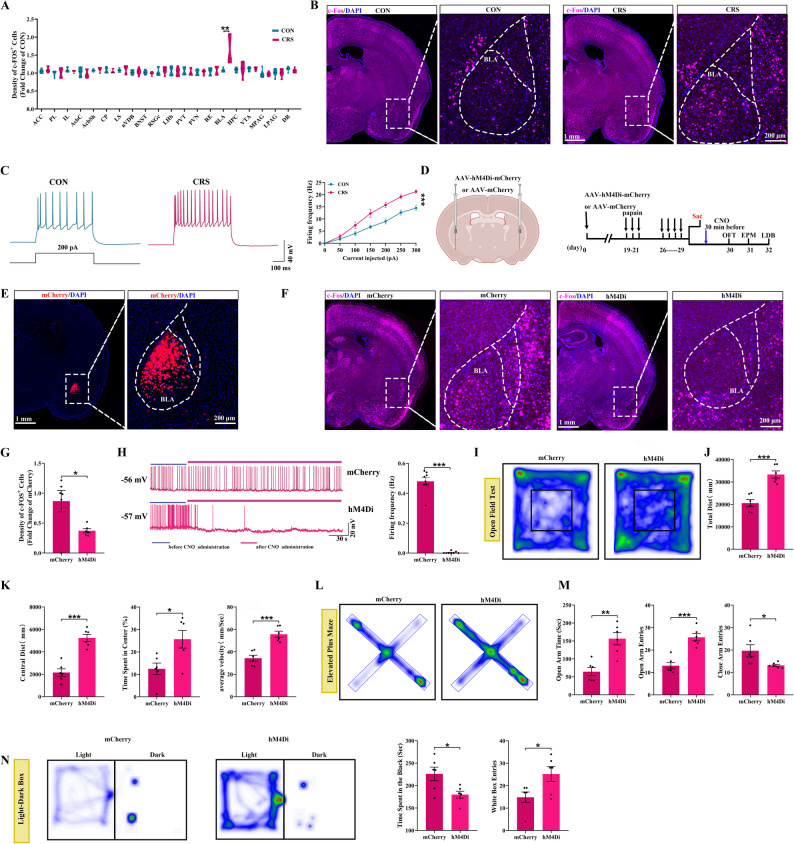
In the papain nasal-inflammation model, eosinophils, type-two cytokines and local sympathetic-related measures increased. Open-field, elevated-maze and light–dark behavior changed, with reduced movement and speed. Depression-related, conditioned-fear and social measures did not show the same significant abnormalities, giving the model a defined behavioral profile.
Among twenty examined regions, BLA activity labeling stood out, with greater cellular excitability. Suppression improved exploration and avoidance, whereas activation worsened the corresponding outcomes. Several behavioral measures changed alongside restored movement, linking the region with the model state and motivating more specific cellular studies.
Source: Zhang X et al. · Journal of Neuroinflammation · CC BY-NC-ND
Original figure and English caption ↗
Excitatory input events increase with similar individual responses
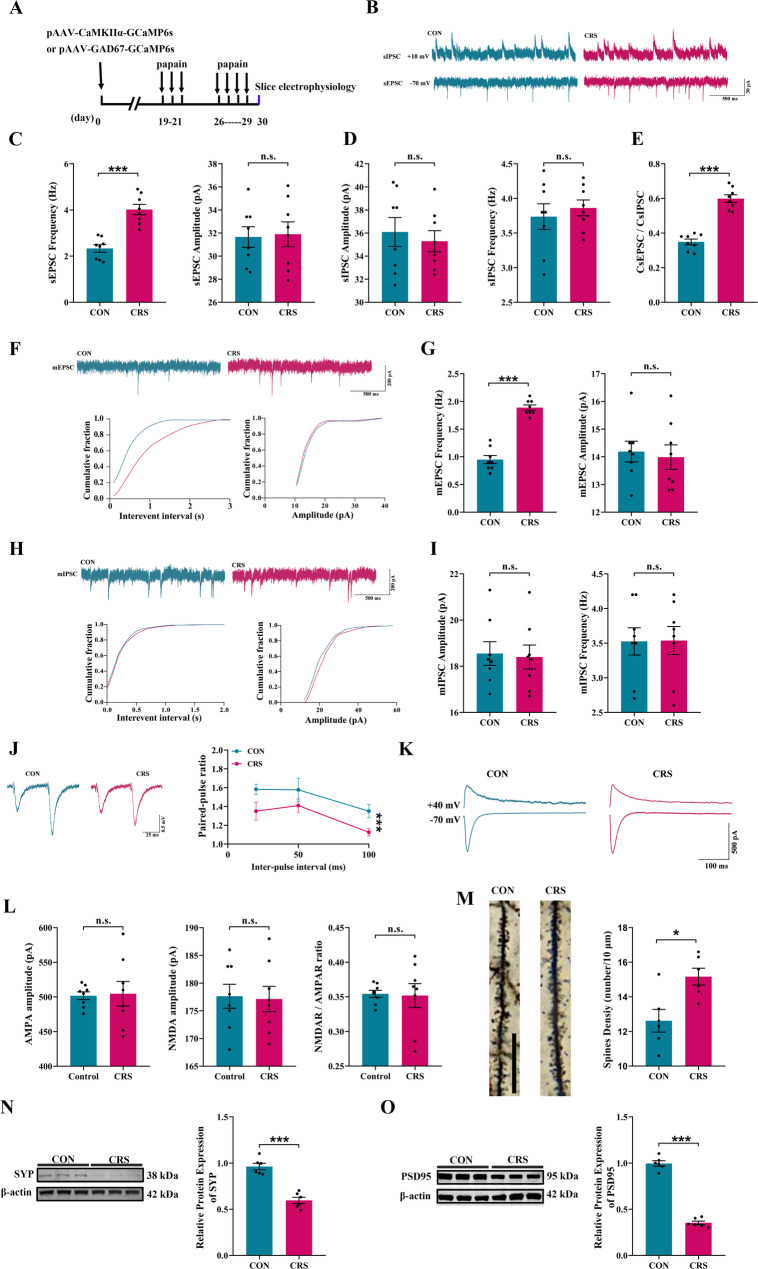
BLA CaMKIIα-positive cells received more excitatory-current events with similar amplitudes; inhibitory currents did not show matching changes. Paired-pulse and dendritic-spine findings also supported input and structural changes, while AMPA and NMDA peak currents remained similar. The measures outlined stronger input drive rather than uniform increases across every cellular readout.
The population showed stronger calcium activity during open-arm transitions, and removing it attenuated related behaviors. Local inputs, activity and functional changes converged on BLA excitatory cells as a node connecting nasal inflammation with behavior. Their output targets became the next question.
Source: Zhang X et al. · Journal of Neuroinflammation · CC BY-NC-ND
Original figure and English caption ↗
Hippocampal-projecting cells also influence nasal measures
Tracing linked BLA, vHPC and premammillary regions, or PMN. Suppressing vHPC-projecting BLA CaMKIIα cells improved anxiety-related behavior and reduced overall PMN excitability. Nasal sympathetic fibers, norepinephrine, IL-33 and eosinophil measures also decreased.
A centrally located population with a defined output identity was thus connected with peripheral immune changes. The findings outlined a functional network from BLA cells through other regions to nasal state. Resolving downstream cells and outputs at each step will help explain coordination of behavior with local inflammation.
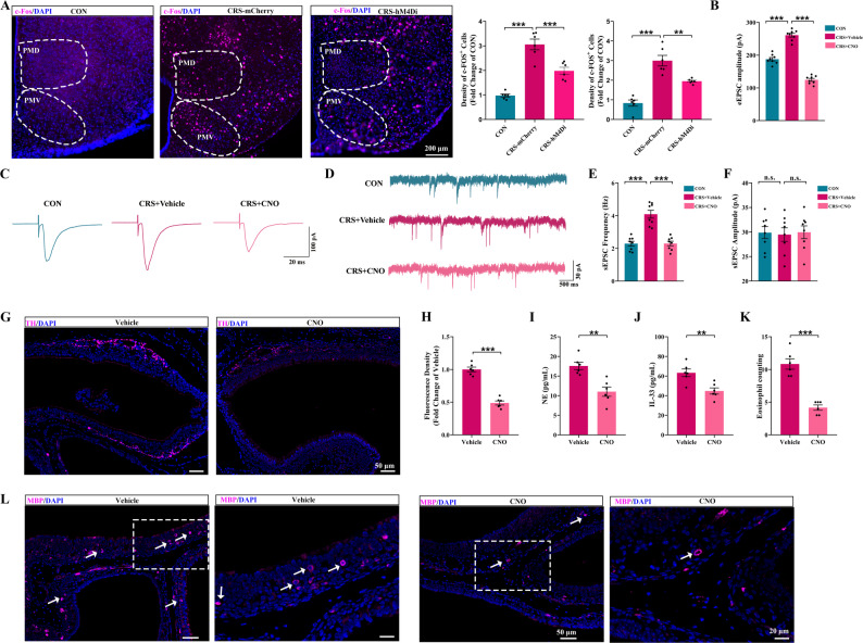
Source: Zhang X et al. · Journal of Neuroinflammation · CC BY-NC-ND
Original figure and English caption ↗
IL-33 modulation accompanies central and nasal changes
The model also showed changes in barrier-related proteins and permeability. Nasal IL-33 neutralization reduced type-two inflammation and eosinophil infiltration, improved behavior and attenuated BLA hyperexcitability. Peripheral immune modulation and central responses changed together, making the influence of nasal signals on brain state a question for further study.
From local immune measures to central excitability, the study gained functional clues spanning both ends of the relationship. Specific signaling routes still need localization and may involve several immune and sensory components. Following transport, cellular reception and indirect effects will refine the mechanism beyond whole-system responses.
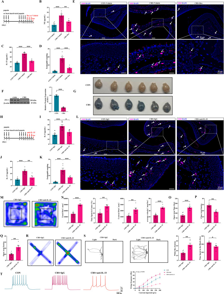
Source: Zhang X et al. · Journal of Neuroinflammation · CC BY-NC-ND
Original figure and English caption ↗
Related states can also change separately
The differing clinical trajectories and animal brain–immune findings together suggest multilayer relationships between local inflammation and behavior. The work observes associations as well as cases where improvement at one end leaves the other relatively unchanged, motivating searches for regulatory sites at different levels.
Mechanistic experiments used male mice, mainly in the papain model, while patient follow-up provided a separate symptom observation. Studies across sexes, clinical subtypes and more precise signaling routes will extend the picture. The work brings central cells and peripheral immunity into one traceable framework for understanding symptoms that coexist yet change differently.