Why can alternating bilateral light weaken alcohol reward memory? An inhibitory cue from superior colliculus to VTA
A mouse study shows that 4 Hz alternating bilateral visual stimulation recruits inhibitory input from the superior colliculus to VTA dopamine neurons, attenuating alcohol place preference.
A central obstacle in treating alcohol use disorder (AUD) is the persistence of cue- and context-induced reward memories that drive craving and relapse. Alternating bilateral sensory stimulation (ABS)—the core component of eye movement desensitization and reprocessing (EMDR) therapy—is widely used for post-traumatic stress disorder and has shown promise in reducing substance craving, yet its effective stimulation parameters and underlying neural circuit mechanisms have remained largely unknown. On June 24, 2024, Prof. Bo Tian and Associate Prof. Pei Zhang's team at Tongji Medical College, Huazhong University of Science and Technology (with co-first authors Jie Lei and Pei Zhang) published a study in Cell Reports titled "Alternating bilateral sensory stimulation alleviates alcohol-induced conditioned place preference via a superior colliculus-VTA circuit." Using an ethanol-induced conditioned place preference (CPP) paradigm in adult male mice alongside whole-brain c-Fos mapping, in vivo fiber photometry, monosynaptic and trans-synaptic viral tracing, in vivo multi-channel electrophysiology, and chemogenetics, the authors discovered that 30 minutes of 4-Hz ABS—but not 1-Hz ABS, 8-Hz ABS, or 4-Hz synchronous flashing light—selectively recruits a predominantly inhibitory superior colliculus (SC)-to-ventral tegmental area (VTA) pathway that suppresses anticipatory VTA dopamine neuron activity and abolishes ethanol reward preference.
This study demonstrates that 30 minutes of 4-Hz alternating bilateral visual stimulation (ABS) selectively recruits a predominantly GABAergic superior colliculus (SC)-to-VTA dopamine neuron pathway to suppress anticipatory VTADA calcium activity and abolish ethanol conditioned place preference, providing a circuit mechanism for non-invasive sensory neuromodulation in addiction therapy.
Contents · 7 sections
Thirty Minutes of 4-Hz Alternating Bilateral Stimulation Selectively Attenuates Ethanol Conditioned Place Preference
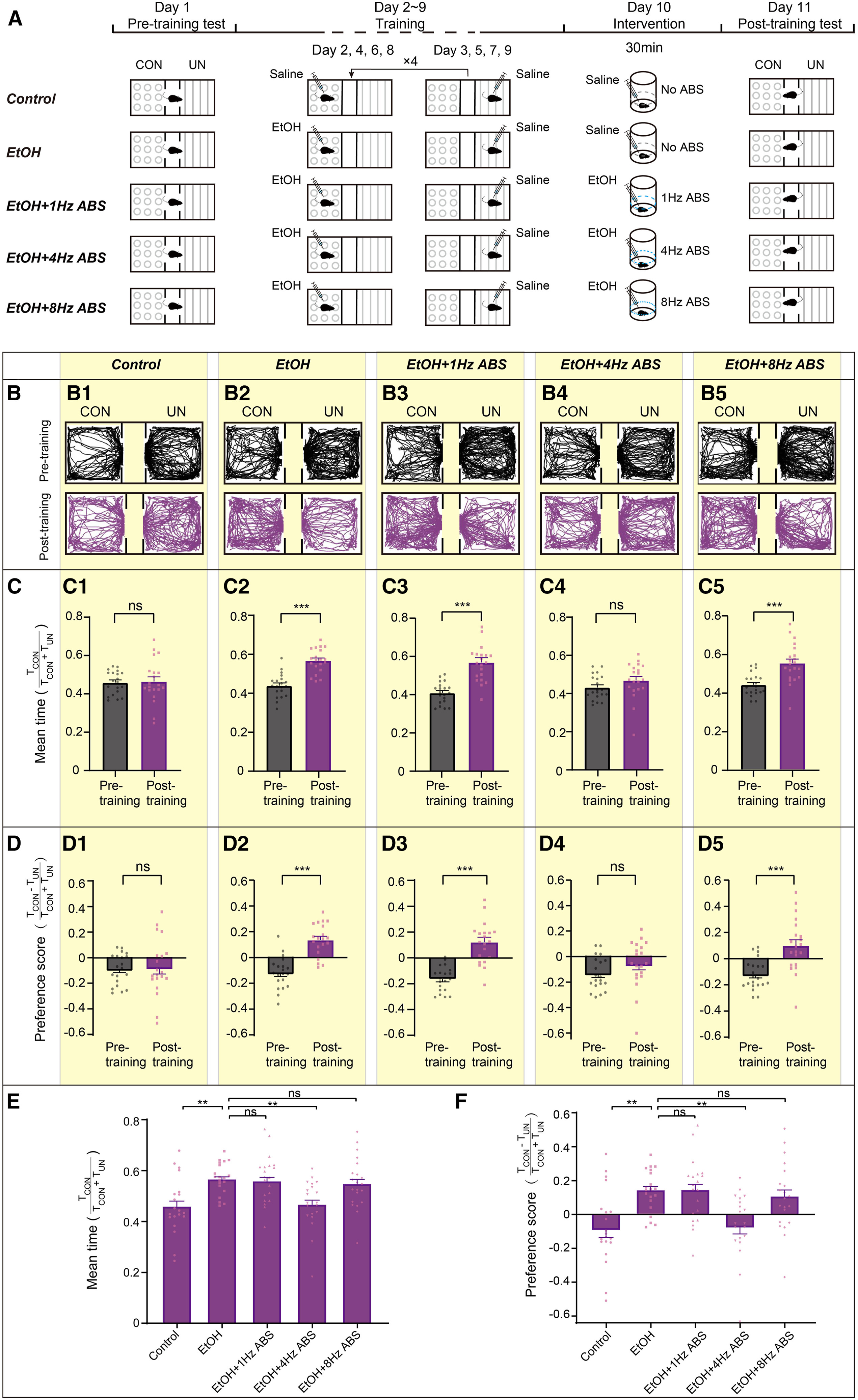
The researchers first established an 8-day biased three-chamber ethanol (2 g/kg, i.p.) conditioned place preference (CPP) paradigm in adult male C57BL/6J mice. Following alternating conditioning sessions, ethanol-paired mice displayed a robust preference for the ethanol-paired chamber on day 9 (Test 1). On day 10, mice were placed in a cylindrical chamber surrounded by a horizontal LED strip that illuminated sequentially from left to right for 30 minutes to deliver alternating bilateral visual stimulation (ABS).
Frequency-comparison experiments (n = 20–21 per group) revealed that only 4-Hz ABS significantly reduced conditioned-chamber occupancy and CPP scores on day 11 (Test 2), whereas neither 1-Hz nor 8-Hz ABS had a significant effect; delivering 30 minutes of 4-Hz ABS prior to daily conditioning sessions similarly prevented ethanol CPP acquisition. Open-field, elevated plus maze, and rotarod assays confirmed that 4-Hz ABS did not alter locomotion, anxiety-like behavior, or motor coordination.
Source: et al. · Cell Reports · Original figure reproduced for academic literature review under fair scholarly commentary; copyright remains with the authors/publisher.
Original figure and English caption ↗
Spatial Alternation Is Required for 4-Hz Visual Stimulation to Attenuate Ethanol Reward Memory
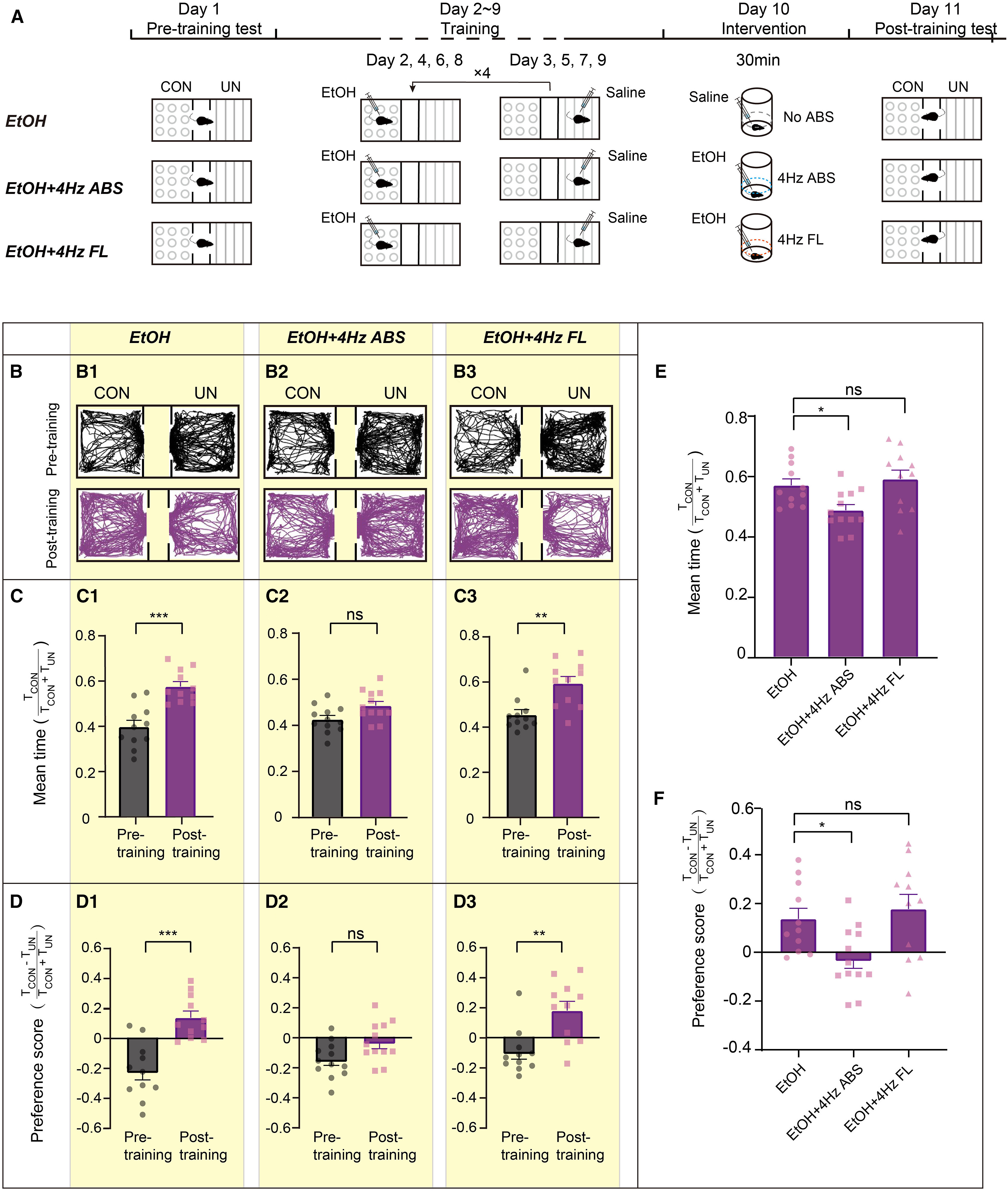
To determine whether the therapeutic effect of 4-Hz ABS stems simply from the 4-Hz temporal frequency or requires spatially alternating bilateral visual motion, the team designed a 4-Hz synchronous flashing light (4-Hz FL) control paradigm in which all LEDs along the horizontal ring flashed simultaneously at 4 Hz for 30 minutes, preserving temporal frequency while eliminating bilateral spatial alternation.
Behavioral testing showed that ethanol-conditioned mice exposed to 30 minutes of 4-Hz synchronous flashing light (n = 11–12 per group) maintained high conditioned-side occupancy and CPP scores on day 11 that were indistinguishable from unstimulated controls (p = 0.7749), demonstrating that bilateral spatial alternation is essential for attenuating ethanol CPP.
Source: et al. · Cell Reports · Original figure reproduced for academic literature review under fair scholarly commentary; copyright remains with the authors/publisher.
Original figure and English caption ↗
4-Hz ABS Suppresses Anticipatory Calcium Surges in VTA Dopamine Neurons Prior to Entering the Ethanol Context
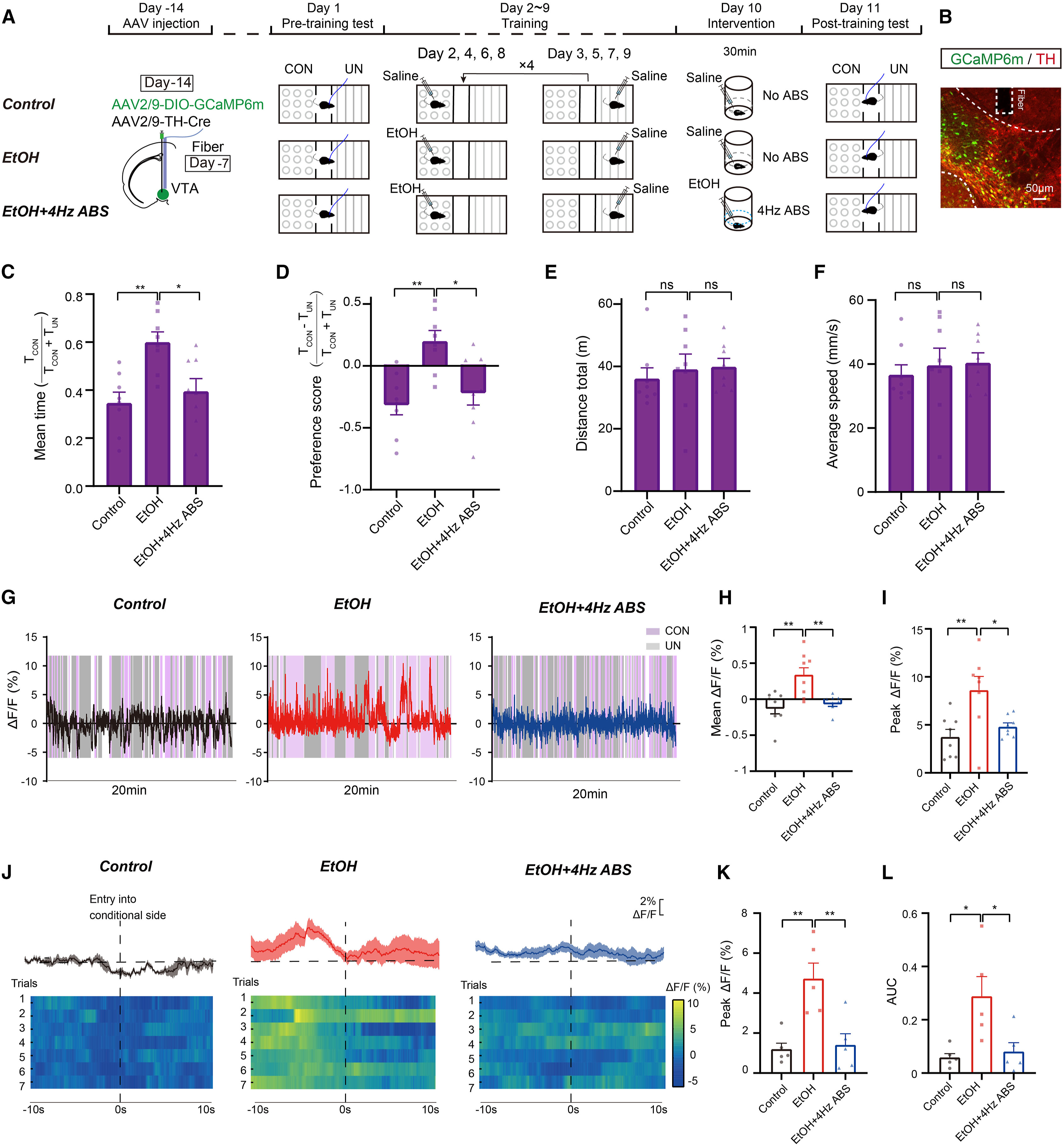
Ventral tegmental area dopamine (VTADA) neurons are central encoders of addiction cue anticipation and reward preference. The researchers co-injected AAV2/9-TH-Cre and AAV2/9-EF1α-DIO-GCaMP6m into the VTA (with ~90.5% of GCaMP6m-expressing cells confirmed as TH-positive dopamine neurons), implanted an Inper ceramic optical fiber above the VTA, and used an Inper fiber photometry system (acquired with Inper Studio and analyzed with Inper Data Process using 488 nm LED excitation and 410 nm control wavelengths) to track VTADA calcium dynamics throughout CPP testing.
Fiber photometry revealed that ethanol-conditioned control mice exhibited significantly elevated session-wide mean and peak VTADA ΔF/F compared with saline controls, alongside a pronounced anticipatory calcium surge (elevated peak ΔF/F and area under the curve, AUC) in the 10-second window immediately preceding entry into the ethanol-paired chamber—a surge absent before entering the saline chamber. Following 30 minutes of 4-Hz ABS, both session-wide VTADA calcium elevations and the pre-entry anticipatory calcium surge before entering the ethanol chamber were abolished without any change in total travel distance or movement speed.
Source: et al. · Cell Reports · Original figure reproduced for academic literature review under fair scholarly commentary; copyright remains with the authors/publisher.
Original figure and English caption ↗
Whole-Brain c-Fos Mapping Identifies the Superior Colliculus as the Key Node Selectively Recruited by 4-Hz ABS
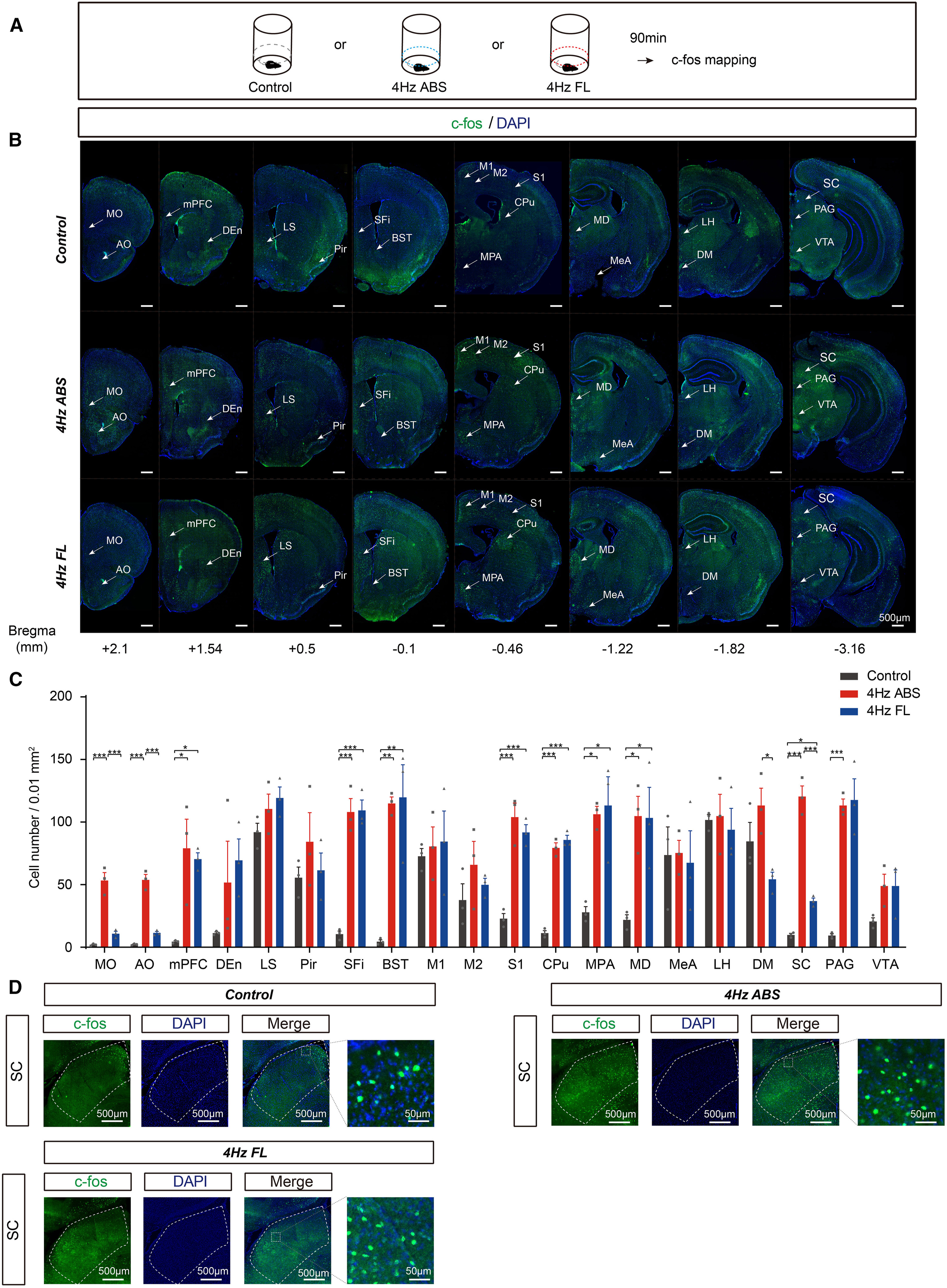
To identify the upstream brain regions mediating the effects of 4-Hz ABS, the team harvested brains 90 minutes after unstimulated Control, 4-Hz ABS, or 4-Hz synchronous flashing light (4-Hz FL) exposure and systematically quantified c-Fos immunostaining across 23 candidate brain areas spanning eight rostrocaudal coronal levels (+2.1 mm to -3.16 mm from bregma).
Whole-brain comparisons showed that canonical visual pathway structures—including the lateral geniculate nucleus (LGN), primary visual cortex (V1), and secondary visual cortex (V2)—were activated to similar degrees by both 4-Hz ABS and 4-Hz FL. By contrast, the superior colliculus (SC) displayed the largest c-Fos increase across the entire brain following 4-Hz ABS and was activated significantly more strongly by 4-Hz ABS than by 4-Hz FL (with VTA c-Fos levels decreasing after 4-Hz ABS). Complementary staining in vGAT-tdTomato mice confirmed that the vast majority of SC neurons recruited by 4-Hz ABS are GABAergic inhibitory neurons.
Source: et al. · Cell Reports · Original figure reproduced for academic literature review under fair scholarly commentary; copyright remains with the authors/publisher.
Original figure and English caption ↗
Viral Tracing Confirms That VTA Dopamine Neurons Receive Predominantly GABAergic Monosynaptic Inputs from the SC
To dissect the direct anatomical connectivity between the SC and VTA, the authors first co-injected AAV2/9-hSyn-Cre and the anterograde presynaptic marker AAV2/9-DIO-Synaptophysin-EGFP into the SC, observing dense EGFP-labeled SC presynaptic terminals closely apposed to TH-positive dopamine somata in the VTA (with anterograde trans-synaptic AAV2/1-hSyn-Cre tracing further showing that 73.3% of innervated VTA neurons are TH-positive).
Next, monosynaptic retrograde rabies tracing was performed by injecting AAV2/9-EF1α-DIO-TVA-EGFP and AAV2/9-EF1α-DIO-RVG into the VTA of DAT-Cre mice followed by RV-EnvA-ΔG-DsRed. Quantitative immunofluorescence co-labeling demonstrated that among SC neurons providing direct monosynaptic input to VTADA starter cells, 58.7% (315/536) were GABA-positive inhibitory neurons whereas only 26.7% (147/552) were CaMKIIα-positive excitatory neurons, establishing that the SC-to-VTADA pathway is predominantly GABAergic.
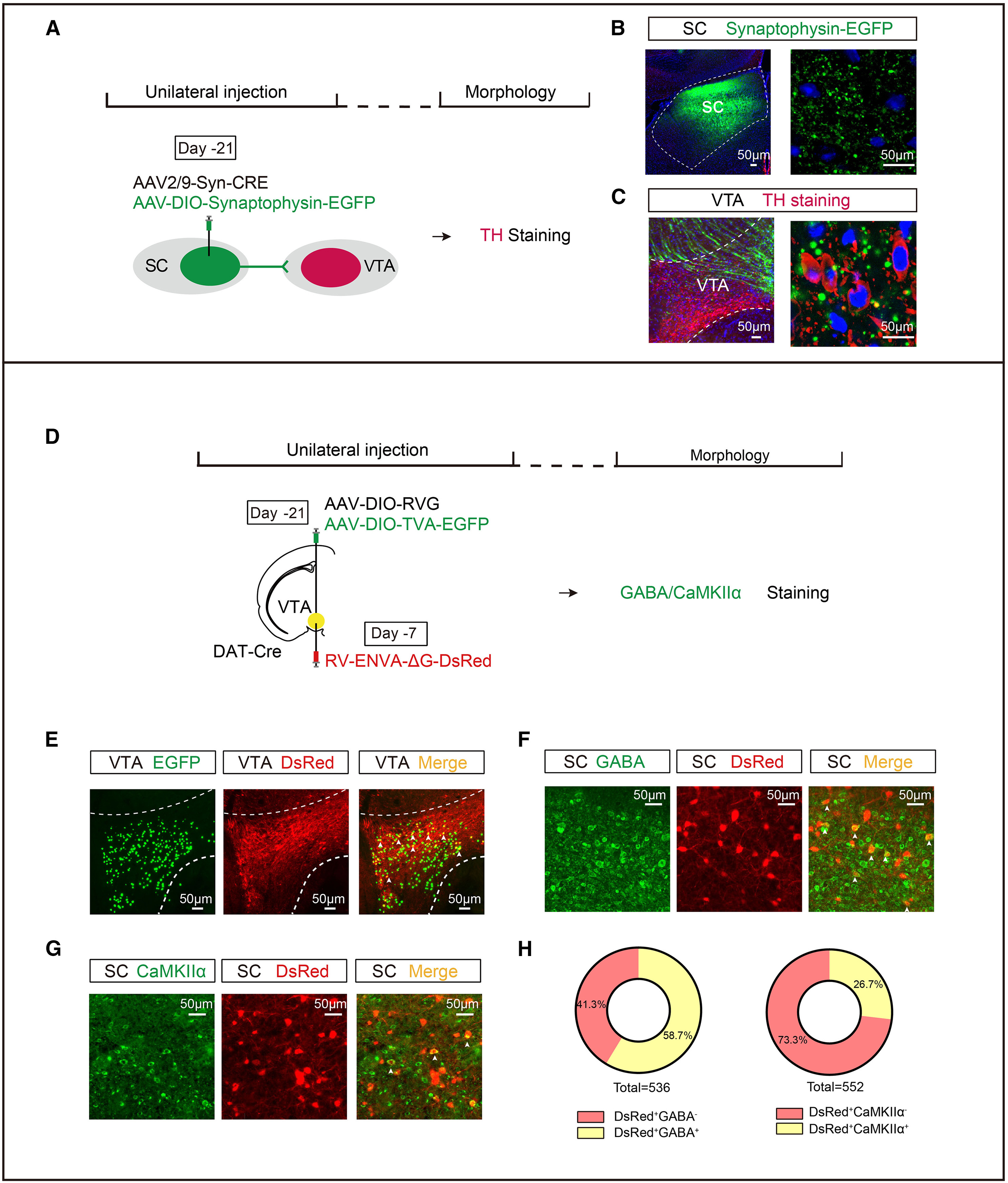
Source: et al. · Cell Reports · Original figure reproduced for academic literature review under fair scholarly commentary; copyright remains with the authors/publisher.
Original figure and English caption ↗
Chemogenetic Inhibition of SC-Recipient VTA Dopamine Neurons Mimics 4-Hz ABS to Abolish Ethanol CPP
Given that 4-Hz ABS recruits predominantly GABAergic SC neurons to suppress downstream VTADA neurons, the authors asked whether directly inhibiting SC-recipient VTADA neurons could mimic the behavioral effect of 4-Hz ABS without light stimulation. Using an anterograde trans-synaptic intersectional strategy, they injected AAV2/1-TH-Cre bilaterally into the SC and AAV2/9-EF1α-DIO-hM4Di-mCherry (or control DIO-mCherry) bilaterally into the VTA, selectively expressing hM4Di in SC-recipient VTADA neurons (VTADA←SC).
Sixteen-channel in vivo microelectrode array recordings confirmed that intraperitoneal CNO (1 mg/kg) significantly suppressed the firing rate of putative VTA dopamine neurons characterized by broad spike waveforms and low baseline firing (<10 Hz). Administering CNO alone on day 10 without any visual stimulation significantly reduced conditioned-chamber occupancy and CPP scores on day 11 in the hM4Di+CNO group compared with mCherry+Saline, mCherry+CNO, and hM4Di+Saline controls, demonstrating that inhibiting SC-recipient VTADA neurons is sufficient to mimic the therapeutic effect of 4-Hz ABS.
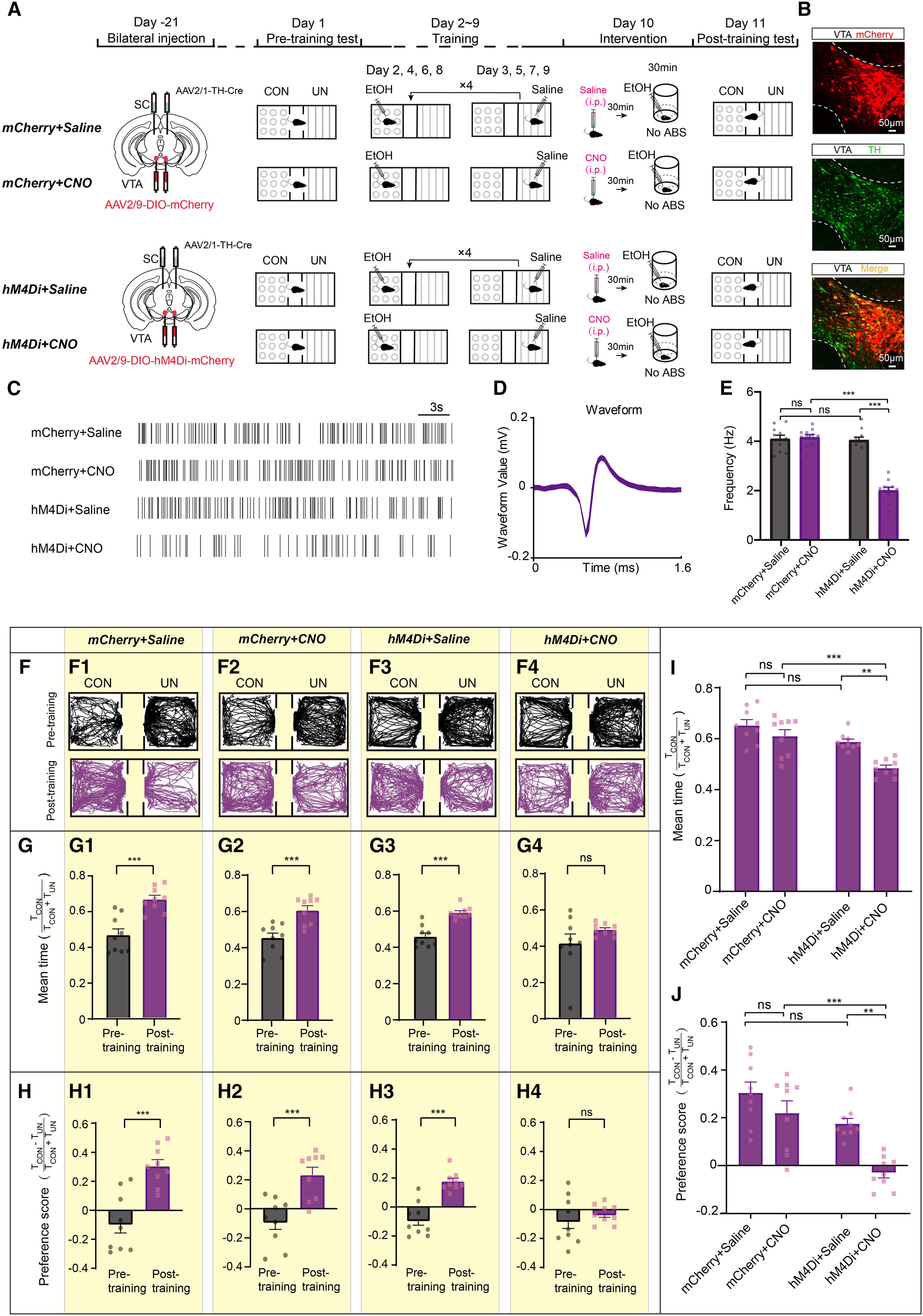
Source: et al. · Cell Reports · Original figure reproduced for academic literature review under fair scholarly commentary; copyright remains with the authors/publisher.
Original figure and English caption ↗
Activating SC-Recipient VTA Dopamine Neurons Reverses the Therapeutic Effect of 4-Hz ABS on Ethanol CPP
Finally, to test whether suppression of the SC-to-VTADA pathway is necessary for the therapeutic effect of 4-Hz ABS, the team injected AAV2/1-TH-Cre bilaterally into the SC and the excitatory chemogenetic virus AAV2/9-EF1α-DIO-hM3Dq-mCherry (or control DIO-mCherry) bilaterally into the VTA. In vivo multi-channel electrophysiological recordings confirmed that CNO administration significantly elevated the firing rate of SC-recipient VTADA neurons.
When CNO or saline was administered 30 minutes prior to the 30-minute 4-Hz ABS session on day 10, retesting on day 11 showed that ethanol place preference was successfully abolished by 4-Hz ABS in the mCherry+Saline, mCherry+CNO, and hM3Dq+Saline groups. In contrast, mice in the hM3Dq+CNO group—whose SC-recipient VTADA neurons were chemogenetically activated during 4-Hz ABS—retained a strong preference for the ethanol-paired chamber, completely reversing the therapeutic effect of 4-Hz ABS and establishing the causal necessity of the SC-to-VTADA circuit.
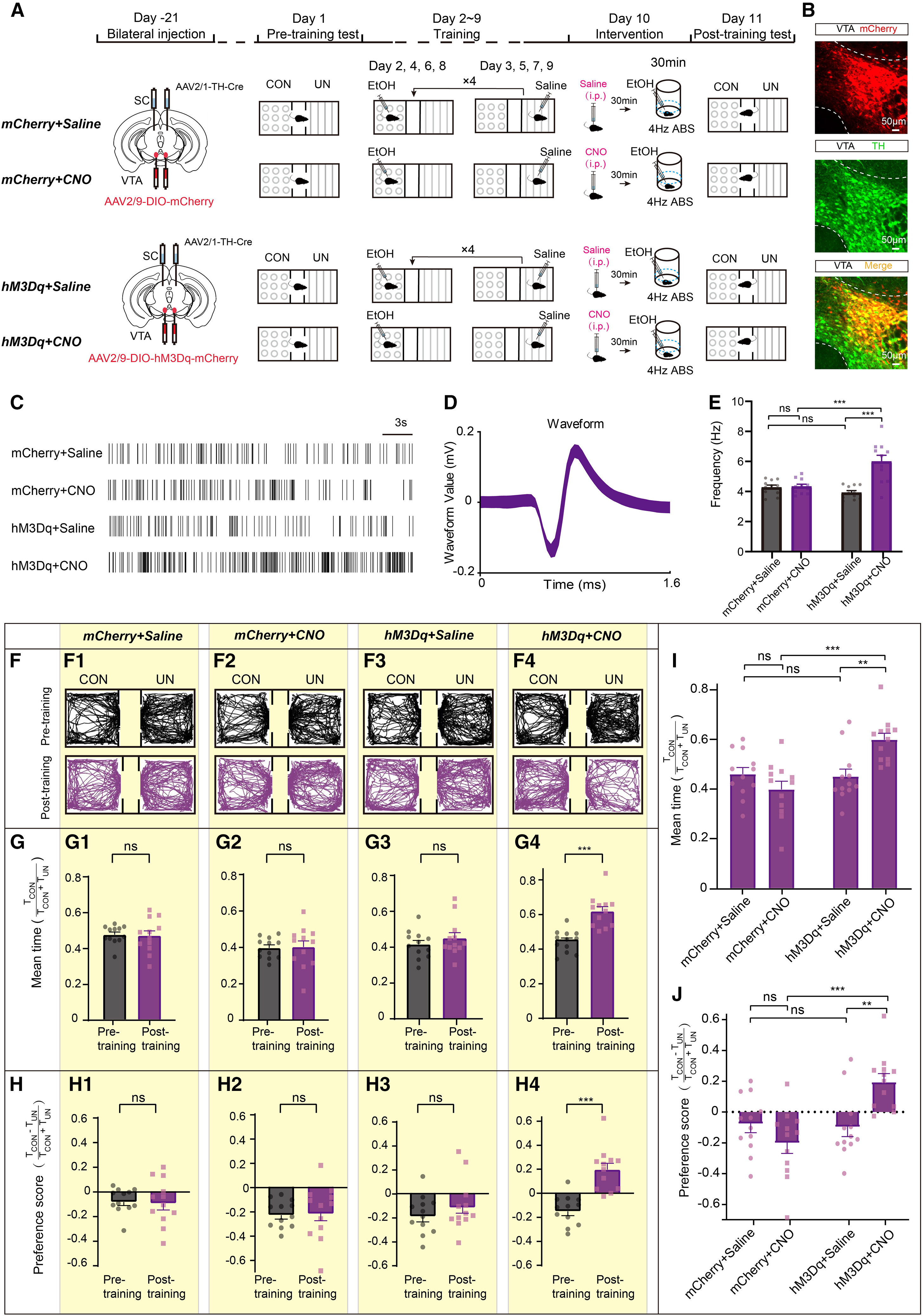
Source: et al. · Cell Reports · Original figure reproduced for academic literature review under fair scholarly commentary; copyright remains with the authors/publisher.
Original figure and English caption ↗