Why activating and inhibiting the same cells both reduce flight
Rostral zona-incerta SST-labeled cells participate in visual-threat responses in male mice. Inhibition reduces flight, while activation increases immobility and lowers heart rate, with a thalamic Re output contributing to defensive responses.
An expanding overhead dark disk sends a mouse back to shelter, modeling a looming visual threat. A study published online in Neuroscience Bulletin on October 19, 2022 finds a contrast: inhibiting a rostral zona-incerta (ZIr) population reduces flight, but activating it also reduces flight. The underlying responses differ. Activation additionally increases immobility and lowers heart rate, suggesting a different defensive pattern. The team identifies the cells by somatostatin (SST) labeling and follows output toward thalamic nucleus reuniens (Re), alongside superior-colliculus-related inputs.
SST-labeled ZIr cells and their Re output participate in tested defensive responses. Activation accompanies freezing-like signs, while inhibition alters threat-related flight and hiding.
Contents · 7 sections
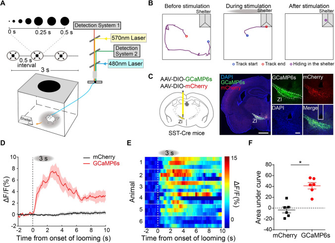
A visual threat increases SST-population calcium
Mice typically flee toward a corner shelter during overhead looming. Calcium increases in the SST-labeled ZIr population without a corresponding control-fluorescence change. A sweeping visual stimulus that induces freezing also increases this population’s calcium signal.
Normal locomotion does not produce a corresponding increase, and neighboring PV-labeled cells do not show the same looming response. Population activity relates to the tested visual-defense contexts without yet identifying individual cells responsible for flight versus freezing.
Source: Lin S et al. · Neuroscience bulletin · CC BY
Original figure and English caption ↗
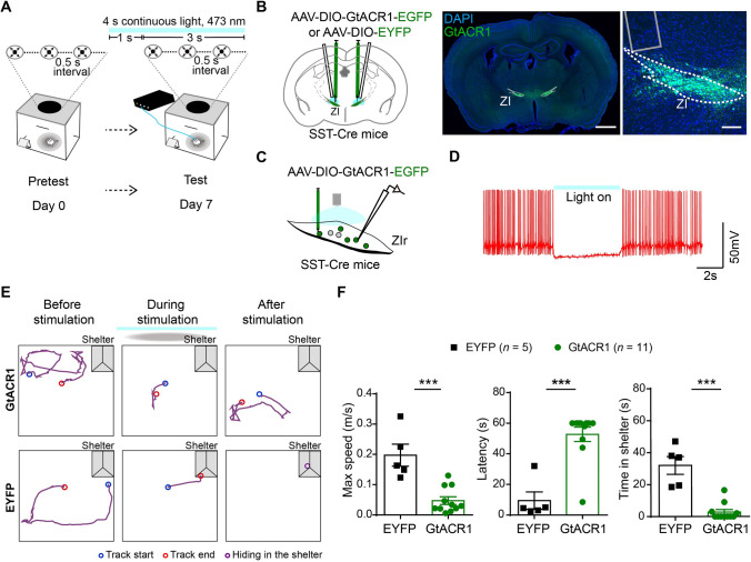
Inhibiting the population reduces flight and hiding
Optogenetic inhibition lowers maximum flight speed, increases shelter-return latency and reduces subsequent hiding time. Defensive actions to the visual threat change, supporting participation of this population in the tested response.
With chemogenetic inhibition, mice still flee; maximum speed and return latency do not change significantly, while hiding time decreases. Effects differ across inhibition approaches rather than every defensive action disappearing together.
Source: Lin S et al. · Neuroscience bulletin · CC BY
Original figure and English caption ↗
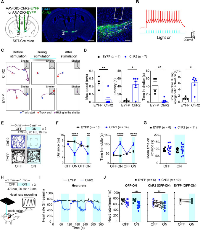
Activation reduces flight but increases immobility
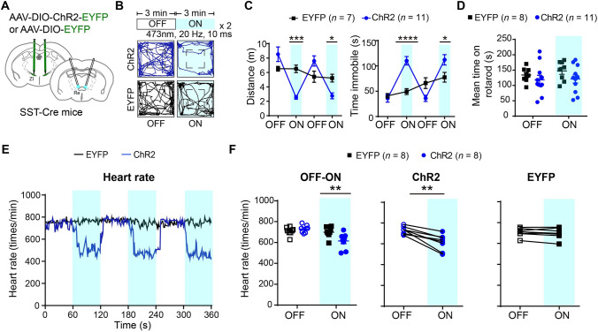
Activation likewise slows shelter return and reduces hiding, but markedly increases immobility during stimulation. In an open field without the external visual threat, activation also reduces movement and increases immobility. Reduced flight here accompanies a different behavioral pattern.
Heart rate falls while rotarod coordination does not change significantly. Together, immobility and bradycardia lead the authors to interpret a freezing-like passive defensive response, with rotarod performance providing an additional comparison for reduced movement.
Source: Lin S et al. · Neuroscience bulletin · CC BY
Original figure and English caption ↗
A direct inhibitory connection reaches thalamic Re
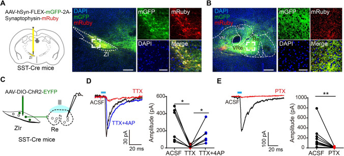
Tracing finds dense SST-labeled output in midline thalamus, particularly Re. Predominantly glutamatergic Re participates in memory, stress and visual-related processes, motivating the next tests.
Activating ZIr terminals in Re evokes inhibitory currents in slices. Pharmacological isolation and GABA-receptor blockade support a direct monosynaptic GABAergic connection. Activity in ZIr can influence Re through this inhibitory output, identifying a specific target for subsequent behavioral tests.
Source: Lin S et al. · Neuroscience bulletin · CC BY
Original figure and English caption ↗
Re-terminal activation also produces freezing-like signs
Activating these terminals locally in Re reduces open-field distance, increases immobility and lowers heart rate, again without significant rotarod changes. Corresponding population and local-output results support a contribution to passive-defense-like responses.
The population also projects elsewhere, and the authors propose that branches or subgroups could contribute to different defensive actions. Re-output results more specifically explain why increased population activity can make mice stop rather than flee faster.
Source: Lin S et al. · Neuroscience bulletin · CC BY
Original figure and English caption ↗
Terminal inhibition differs with and without threat
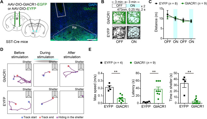
Inhibiting Re terminals does not significantly change ordinary open-field distance. During looming, local inhibition lowers maximum flight speed, slows shelter return and reduces hiding. Stable baseline locomotion can coexist with altered threat responses.
The comparison makes context important: the output influences defensive behavior beyond a single movement switch. Although both activation and inhibition reduce flight, accompanying immobility, heart-rate and baseline-locomotion findings distinguish their effects.
Source: Lin S et al. · Neuroscience bulletin · CC BY
Original figure and English caption ↗
Superior-colliculus input adds a visual link
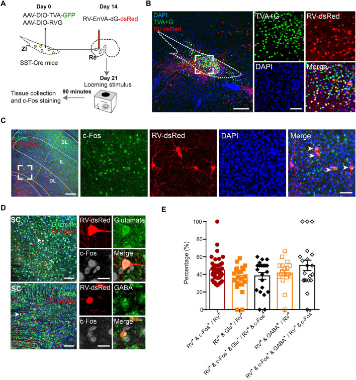
Tracing upstream of Re-projecting SST cells identifies related input cells in intermediate and deep superior-colliculus layers. Some carry c-Fos after looming, and the population includes glutamatergic- and GABA-related cells. This adds a candidate visual input to the ZIr–Re connection.
The superior-colliculus evidence chiefly comes from tracing and activity markers without cell-class-specific input manipulation. The study analyzes adult male mice with defensive responses in pretesting, and its looming task mainly elicits flight. It reveals diverse behavioral and bodily responses while leaving branch-specific organization of flight and freezing unresolved.
Source: Lin S et al. · Neuroscience bulletin · CC BY
Original figure and English caption ↗