When social encounters become frightening: a basal forebrain–habenular connection
After aversive social learning, mice avoid unfamiliar partners. A study traces an excitatory basal forebrain–habenular pathway and distinguishes altered population responses from synaptic changes.
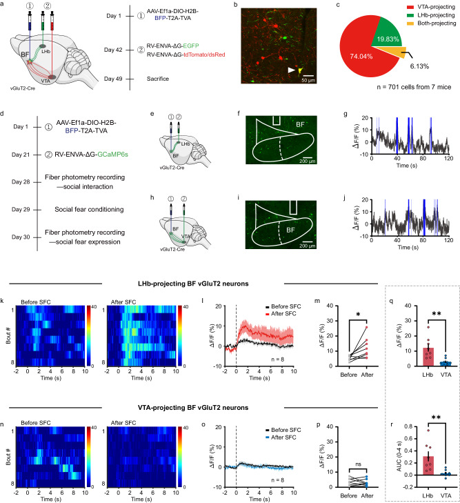
An unfamiliar partner can invite exploration, but after aversive learning the same encounter may provoke hesitation and retreat. A study published in Nature Communications on May 13, 2024 examines social fear expression in adult male mice. It identifies a glutamatergic basal forebrain–lateral habenula connection: more relevant cells increase activity during social encounters after conditioning, and attenuating this input reduces avoidance and vigilant postures. Synaptic measures change without a corresponding increase in the intrinsic excitability measures tested.
A glutamatergic basal forebrain–lateral habenula pathway contributes to social fear expression. Conditioning enhances population responses and changes synaptic measures; pathway inhibition reduces avoidance without establishing erasure of fear memory.
Contents · 7 sections
Avoidance extends to a new partner
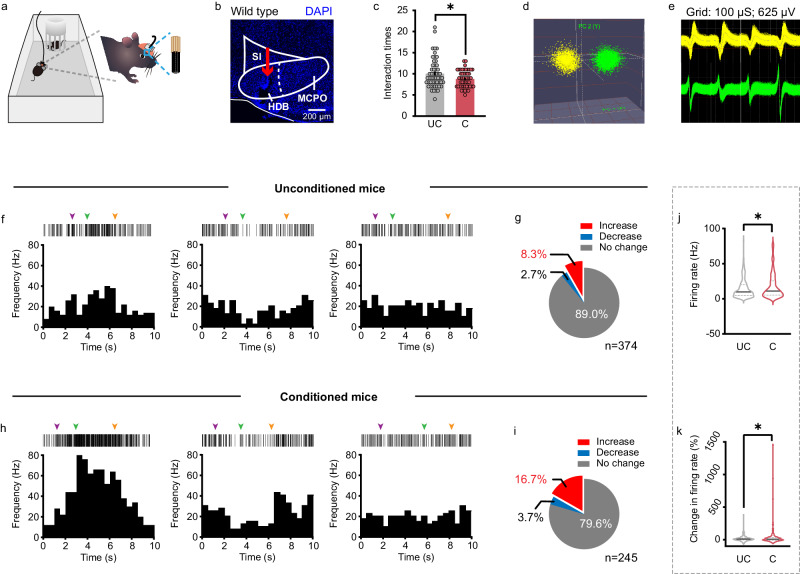
After social fear conditioning, mice reduced contact with unfamiliar partners and more often adopted stretched postures. Avoidance thus generalized beyond the original social stimulus. Open-field locomotor and anxiety-related measures did not show corresponding significant changes.
Basal forebrain recordings revealed increases, decreases and unchanged firing during social approaches. Conditioned mice had a higher proportion of increasing cells and higher average firing than unconditioned controls, linking the region to altered population responses.
Source: Wang J et al. · Nature communications · CC BY
Original figure and English caption ↗
A stronger response in glutamatergic populations
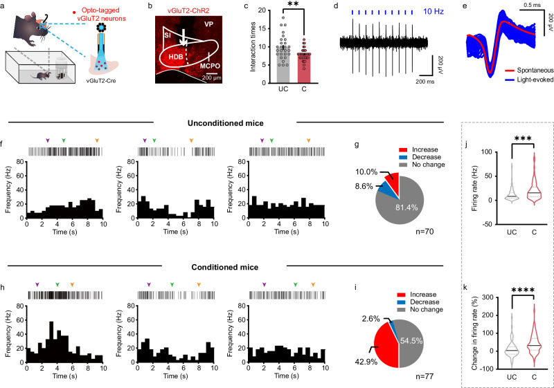
Among basal forebrain cell classes, glutamatergic populations showed prominent calcium responses during social fear expression compared with investigation of an empty cage. Cell identification combined with single-unit recordings refined this population-level clue.
Among recorded glutamatergic cells, the fraction with increased firing during social interaction rose from around 10% in controls to approximately 43% after conditioning. Average firing and baseline-relative changes also increased, while substantial response diversity remained.
Source: Wang J et al. · Nature communications · CC BY
Original figure and English caption ↗
Attenuating activity increases social approach
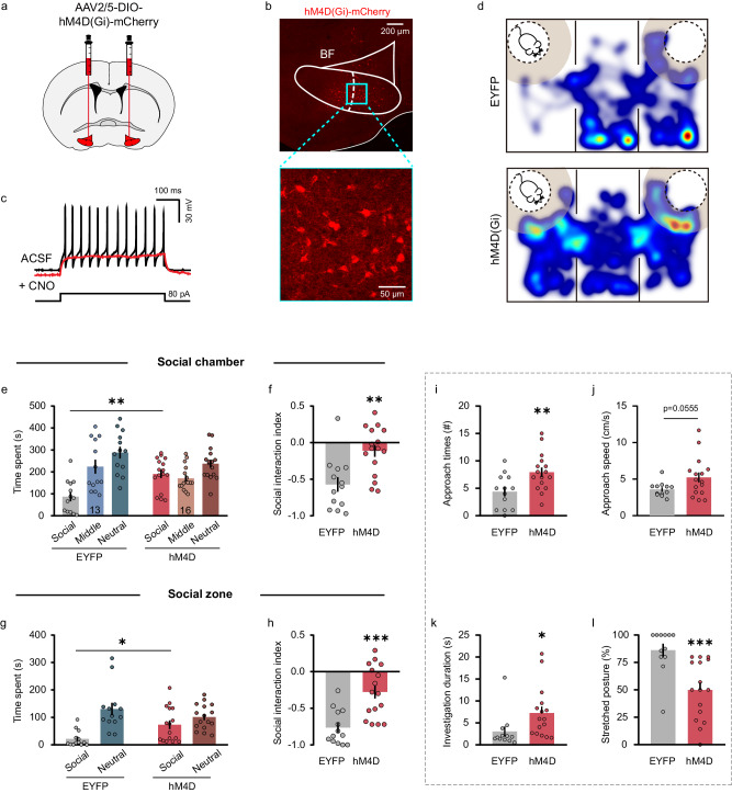
Inhibiting basal forebrain glutamatergic cells increased social-zone time, approach frequency and individual investigation duration while reducing stretched postures. Approach speed did not differ significantly in this comparison, and open-field measures remained unchanged.
A complementary inhibition strategy produced related social effects. In naive mice, inhibition did not similarly increase social measures. The contribution therefore depended on prior experience, and other cell classes did not show the same improvement pattern.
Source: Wang J et al. · Nature communications · CC BY
Original figure and English caption ↗
Outputs from one region reach different targets
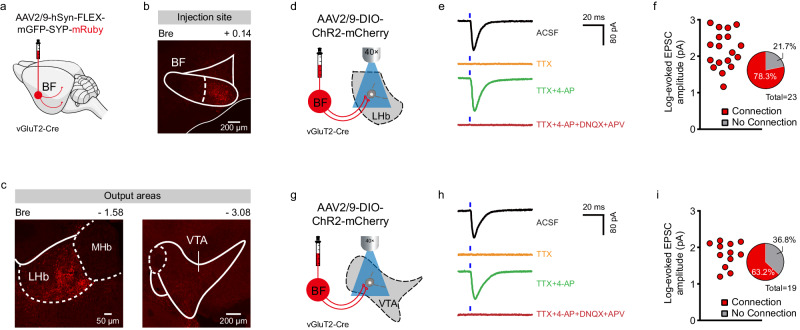
Glutamatergic basal forebrain outputs reached lateral habenula and VTA, and slice recordings supported excitatory synaptic inputs in both regions. Connectivity established a substrate; comparison across behavioral states was needed to identify the more relevant pathway.
Both targets participate in affective or motivational processes, but that did not establish identical roles for their basal forebrain inputs. The study therefore separated upstream populations by their projection targets and compared their social responses.
Source: Wang J et al. · Nature communications · CC BY
Original figure and English caption ↗
The habenula-projecting population responds more strongly
Tracing found largely separate projection populations with a smaller overlapping fraction. Comparing recordings before and after conditioning, social calcium peaks increased in habenula-projecting cells but not significantly in VTA-projecting cells.
Terminal recordings supported the distinction. Different state-dependent responses separated basal forebrain output networks, with social fear-associated enhancement more evident along the habenular direction.
Source: Wang J et al. · Nature communications · CC BY
Original figure and English caption ↗
Attenuating habenular input reduces avoidance
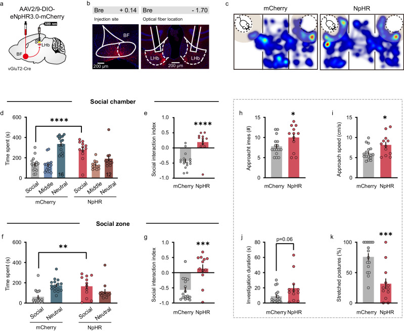
Inhibiting glutamatergic basal forebrain terminals in lateral habenula increased social occupancy, approach frequency and speed while reducing stretched postures in conditioned mice. Individual investigation duration did not differ significantly, and naive animals did not show the same social changes.
Inhibiting the corresponding VTA projection did not produce the same significant improvement. Additional activation of the habenular projection in already conditioned mice also failed to significantly worsen fear measures, showing that activation and inhibition were not simple symmetric effects.
Source: Wang J et al. · Nature communications · CC BY
Original figure and English caption ↗
Changes emerge in connectivity rather than intrinsic measures
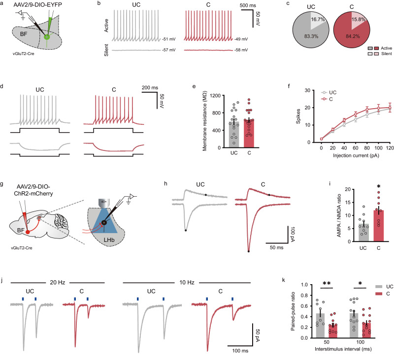
Stronger behavioral responses did not imply universally higher excitability. Resting potential, membrane resistance and evoked spike counts did not differ significantly in slices. By contrast, increased AMPA/NMDA current ratios and reduced paired-pulse ratios suggested contributions from postsynaptic and presynaptic changes.
The study connects social responses, projection-specific behavioral effects and synaptic changes into a circuit-level account of fear expression in male mice. Human therapeutic effects remain untested. The upstream information shaping this pathway and downstream habenular routes influencing behavior also remain unresolved.
Source: Wang J et al. · Nature communications · CC BY
Original figure and English caption ↗