Returning to a sheltered passage: how an amygdala cell group modulates avoidance
An adult-mouse study follows transition-related signals in MeA Tac1 cells, linking ventral-tegmental input and hypothalamic output to anxiety-like behavior in exposed spaces.

In an elevated plus maze, open arms lack side walls while closed arms have them. How does brain activity change as a mouse moves between these spaces? A study published on July 28, 2025 in EMBO Reports finds that Tac1-expressing medial-amygdala (MeA) cells become active when mice return from open to closed arms. Enhancing these cells increases exploration of exposed or bright spaces, while suppressing them increases avoidance. Following ventral-tegmental-area (VTA) input and ventrolateral ventromedial-hypothalamic (VMHvl) output expands the transition signal into a behavioral regulatory pathway.
MeA Tac1 activity bidirectionally influences exploration choices across anxiety-related tasks. Cells linking VTA input with VMHvl output, together with downstream GABA transmission, contribute to these changes.
Contents · 6 sections
A signal accompanies return to the closed arm
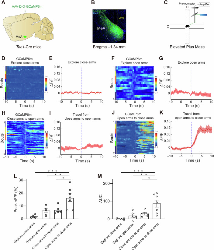
The study first records MeA Tac1 activity aligned with entry into different regions. Calcium signals rise on return from an open to a closed arm, but equivalent changes are absent during the opposite transition or continued exploration within either arm.
The transition itself becomes informative. The signal does not merely mark occupancy of an open arm; it accompanies return to a more sheltered space. This links the population with anxiety-related transitions and motivates active manipulation to ask whether it influences choices.
Source: Wang Y et al. · EMBO reports · CC BY
Original figure and English caption ↗
Enhancement increases exploration of exposed spaces
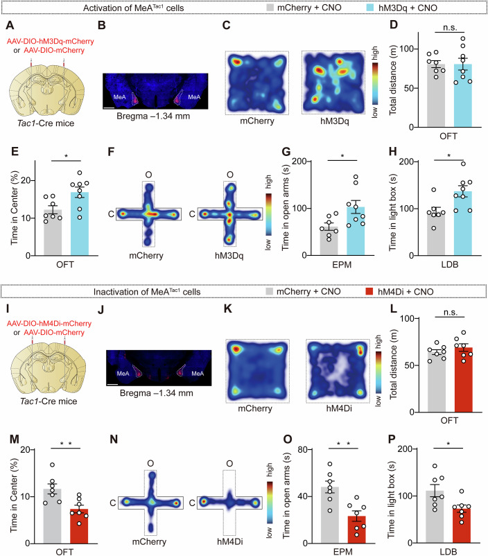
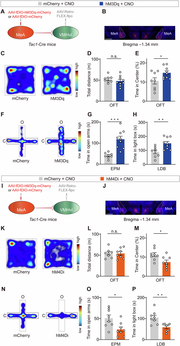
Enhancing MeA Tac1 activity increases time in the open-field center, elevated-maze open arms and light–dark-box bright chamber. Suppression produces the opposite direction across the three tasks. Comparable total open-field distance focuses the effects on spatial choices rather than simply more or less movement.
Tail-suspension immobility and sucrose preference show no clear changes alongside anxiety-related exploration. In females, MeA Tac1 suppression also reduces center, open-arm and bright-chamber exploration. The task pattern concentrates the population’s role on particular avoidance-related behaviors.
Source: Wang Y et al. · EMBO reports · CC BY
Original figure and English caption ↗
VTA and MeA show different transition-related signals
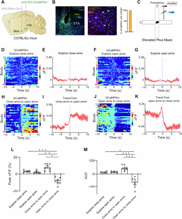
Tracing identifies inputs to MeA Tac1 from several regions, with labeled VTA cells showing GABAergic markers. VTA contains inhibitory cells as well as its frequently discussed dopaminergic population. The study next records activity in this upstream region.
VTA GABA calcium signals rise on entering an open arm and fall on returning to a closed arm, while continued exploration within either remains broadly unchanged. Together with the MeA return signal, this reveals distinct activity directions across the transition rather than uniform enhancement along the pathway.
Source: Wang Y et al. · EMBO reports · CC BY
Original figure and English caption ↗
The side of the connection matters
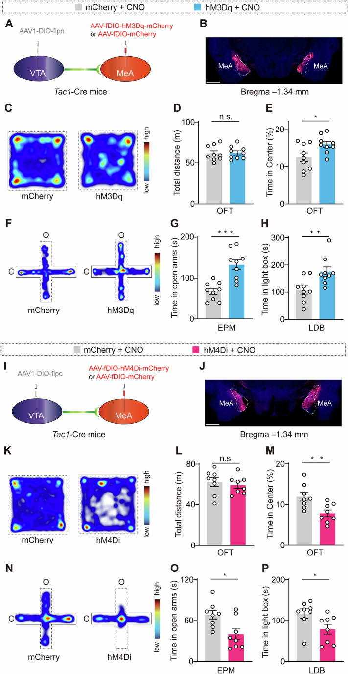
The study distinguishes two manipulations. Enhancing VTA-input-recipient MeA Tac1 cells increases center, open-arm and bright-chamber exploration, while suppressing these recipients reduces it. This matches modulation of the overall MeA Tac1 population, linking the VTA-connected recipients to behavioral regulation.
By contrast, enhancing MeA-projecting VTA cells increases avoidance, while suppressing them reduces it. Opposite behavioral directions for upstream input and downstream recipients fit a model of inhibitory input regulating the MeA population. In this model, stronger upstream inhibition suppresses the MeA population supporting exposed-space exploration, whereas weaker upstream input allows recipients to contribute more. Serial inhibition can produce different activity and behavioral directions at neighboring nodes, explaining why one region’s activity level cannot summarize the whole pathway.
Source: Wang Y et al. · EMBO reports · CC BY
Original figure and English caption ↗
MeA output contributes through an inhibitory connection
Selecting VMHvl-projecting MeA Tac1 cells shows that enhancement increases exposed-space exploration and suppression reduces it, with comparable total open-field distance. This output-defined population extends the MeA effect toward hypothalamus, matching modulation of the overall MeA Tac1 population.
Slice recording shows that activating MeA terminals evokes inhibitory postsynaptic currents in VMHvl cells, supporting a direct GABAergic connection. Behavioral and synaptic observations converge: MeA cells involved in exposed-space choices can deliver inhibitory signals to hypothalamus.
Source: Wang Y et al. · EMBO reports · CC BY
Original figure and English caption ↗
Beyond the cell label, GABA transmission provides a key lead
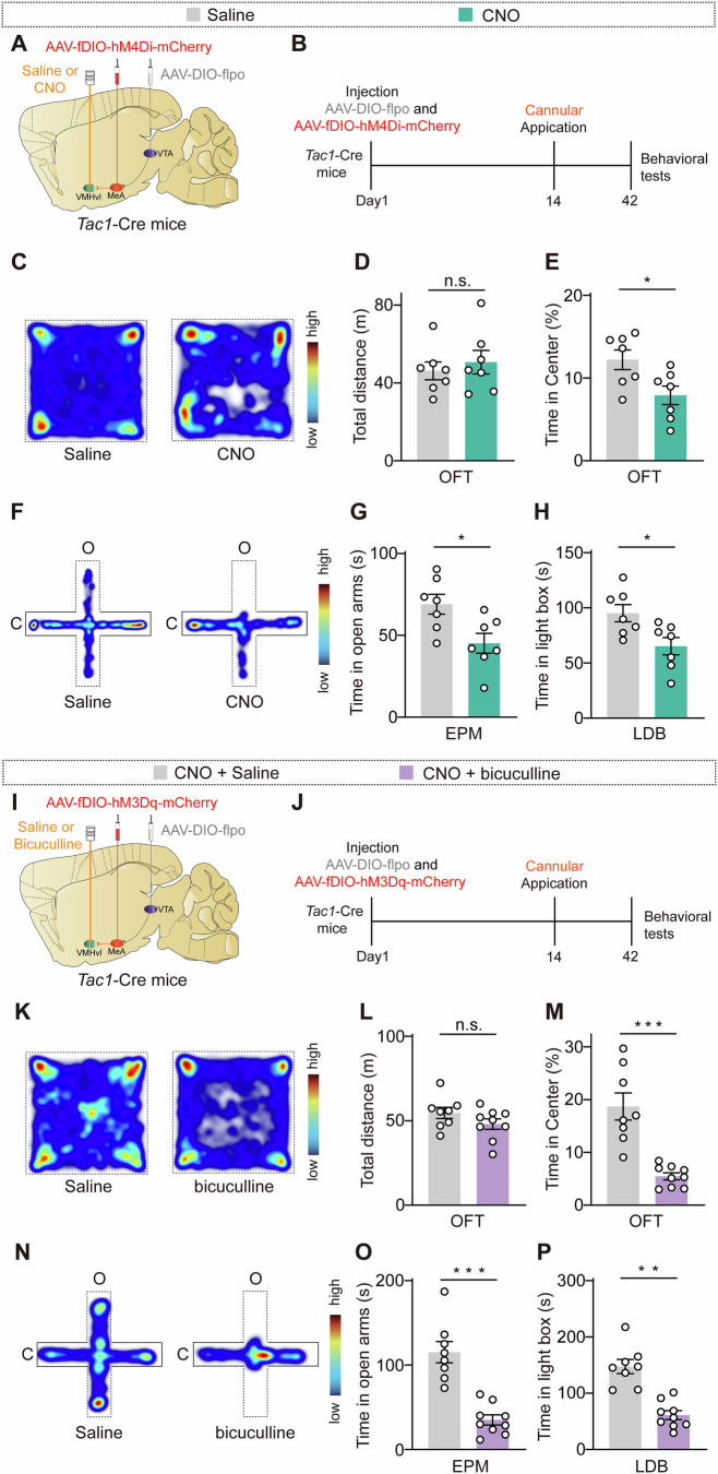
Tac1 contributes to production of neuropeptides including substance P, but reducing its expression in MeA leaves the tested exploration measures broadly unchanged. In contrast, VMHvl GABA_A receptor blockade reduces center, open-arm and bright-chamber exploration. Blocking these downstream receptors during enhancement of VTA-input-recipient MeA cells likewise accompanies greater avoidance.
Suppressing these input recipients’ output in VMHvl also reduces exposed-space exploration. The experiments link upstream input, MeA cells and hypothalamic inhibitory transmission. These observations come from adult mice, with main pathway experiments in males, describing network participation in choices between exploration and avoidance of exposed spaces.
Source: Wang Y et al. · EMBO reports · CC BY
Original figure and English caption ↗