Pain sensitivity persists as affect-related behavior improves: a cortical–amygdala connection
A male-mouse nerve-injury model links somatosensory-cortical and amygdala cells, and their CCK, to later behavioral changes, revealing a separation between sensory hypersensitivity and affect-related responses.
Chronic pain and low mood often coexist, but how does the brain connect persistent sensory changes to affect-related behavior? A study published on April 21, 2025 in Scientific Reports follows four weeks after nerve injury in male mice. Mechanical hypersensitivity appears first; reduced sucrose preference and increased swimming immobility emerge later. Examining the connection between hindlimb somatosensory cortex and basolateral amygdala reveals an intriguing separation: suppressing related cells improves the two affect-related measures while mechanical pain thresholds remain broadly unchanged.
Somatosensory-cortical cells projecting to the amygdala and cells receiving their input contribute to affect-related behavior after chronic pain, with effects distinguishable from maintenance of mechanical hypersensitivity.
Contents · 6 sections
Hypersensitivity precedes affect-related changes
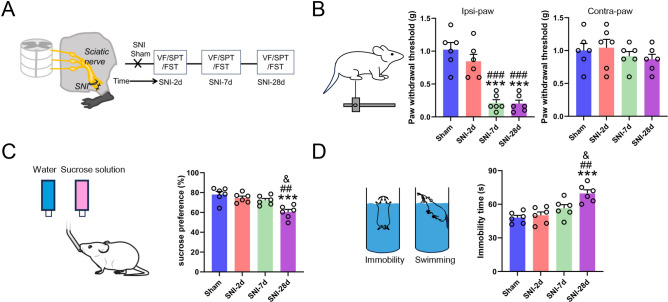
On day seven after injury, mechanical thresholds fall in the affected hindpaw and remain low on day twenty-eight; thresholds in the other paw stay broadly stable. Two affect-related measures clearly change on day twenty-eight: sucrose preference falls and swimming immobility increases.
The timing difference lets the study follow potential brain links as the condition progresses. Sensory information continues to be processed, but which cells change alongside later behavior? Sucrose preference provides a view of reward choice, while swimming immobility provides another behavioral measure in a stressful situation; together they describe the later changes.
Source: Chen G et al. · Scientific reports · CC BY-NC-ND
Original figure and English caption ↗
Somatosensory cortex activates earlier, amygdala later
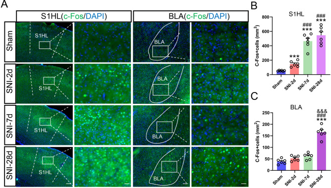
The hindlimb region of primary somatosensory cortex, S1HL, processes hindlimb sensation. Contralateral S1HL shows more activity-marker-positive cells on days seven and twenty-eight. Activity markers in the basolateral amygdala, BLA, clearly rise only on day twenty-eight, accompanying the later behavioral changes.
The amygdala integrates sensory and affect-related information, making the connection between these regions worth following. Rather than an instantaneous switch, persistent sensory change accompanies later alterations in another processing network. The study next identifies the cellular connection between the regions.
Source: Chen G et al. · Scientific reports · CC BY-NC-ND
Original figure and English caption ↗
Cortical inputs reach predominantly CCK-positive cells
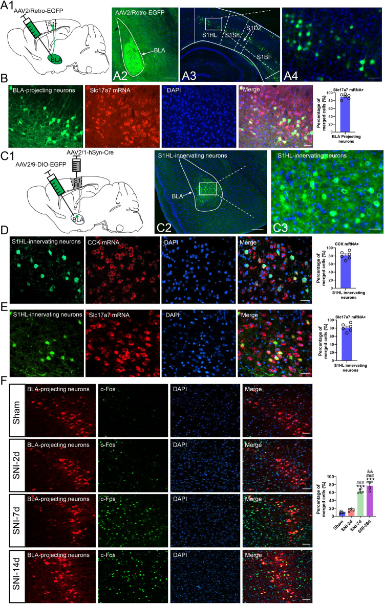
Tracing shows that somatosensory-cortical cells projecting to BLA lie mainly in layer five and mostly express a glutamatergic marker. From the receiving side, most BLA cells receiving S1HL input express the neuropeptide CCK and many also carry a glutamatergic marker. A regional arrow becomes a connection between identified cellular populations.
S1HL cells projecting to the amygdala show stronger activity markers at the later stage. CCK identifies many recipient cells and introduces a molecular question: does expression of this neuropeptide contribute to behavioral change after persistent pain?
Source: Chen G et al. · Scientific reports · CC BY-NC-ND
Original figure and English caption ↗
Stronger responses to sensory input and swimming stress
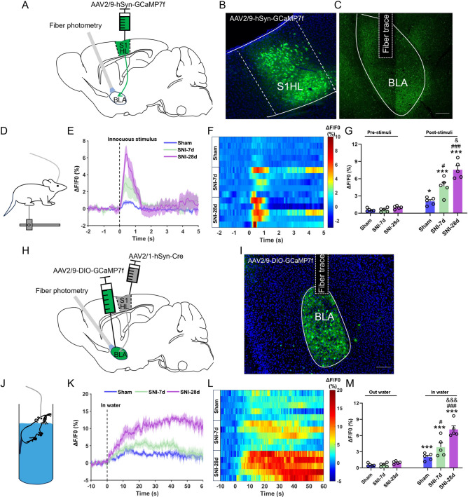
In vivo calcium recordings show that light mechanical stimulation evokes responses in S1HL terminals projecting to BLA. Responses are stronger on day seven and largest on day twenty-eight. A relatively mild sensory input leaves a larger activity-associated signal in this projection.
A separate recording examines BLA recipient cells. Entering the swimming condition evokes responses in all groups, with stronger responses in mice twenty-eight days after injury. Sensory and stress-related events reveal changes at the two ends separately, adding event-linked dynamics to the static activity-marker findings.
Source: Chen G et al. · Scientific reports · CC BY-NC-ND
Original figure and English caption ↗
Related-cell inhibition leaves mechanical thresholds low
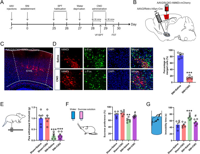
At the fourth week, when both kinds of change are present, suppressing BLA-projecting S1HL cells raises sucrose preference and reduces swimming immobility. The affected-paw mechanical threshold does not clearly improve. The same inhibition does not clearly change these three measures in uninjured controls, linking the improvement to the post-injury state. These cells thus contribute to later behavioral changes, with improvement occurring without a parallel reduction in mechanical hypersensitivity.
Suppressing BLA cells receiving S1HL input produces the same pattern. Tests at both ends support a specific link: cortical–amygdala-related networks contribute to later affect-related behavior accompanying persistent sensory change, while mechanical thresholds remain at their post-injury level.
Source: Chen G et al. · Scientific reports · CC BY-NC-ND
Original figure and English caption ↗
Reducing CCK attenuates later behavioral changes
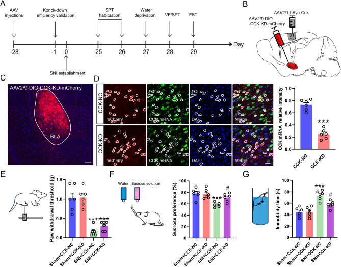
The study next reduces CCK expression in BLA recipient cells before nerve injury. After injury, these mice show improved sucrose preference and swimming immobility relative to controls, while mechanical hypersensitivity persists. A neuropeptide associated with cellular identity is thereby linked to later behavior.
This male-mouse study connects sensory processing, amygdala cells and CCK, with the divergence of behavioral measures providing a particularly informative finding. Why the connection strengthens after persistent injury remains to be explained; the results identify cellular and molecular contributors to affect-related changes accompanying chronic pain.
Source: Chen G et al. · Scientific reports · CC BY-NC-ND
Original figure and English caption ↗