One brain region, two tasks: how ACC outputs divide social behavior and attention
A mouse study finds that anterior cingulate outputs to zona incerta and limbic thalamic reticular nucleus separately modulate partner interaction and attention-task performance.

Interacting with a partner recruits attention, while an attention task involves waiting, choosing and controlling responses. How does the brain divide work across these related behaviors? A 2026 Science Advances study examines the anterior cingulate cortex, or ACC, a region often implicated in both social behavior and attention. Projection mapping, cellular characterization and pathway modulation reveal two minimally overlapping populations targeting zona incerta, or ZI, and limbic thalamic reticular nucleus, or lTRN. Multiple functions within one region thus acquire a more detailed cellular organization.
In mouse tasks, ACC→ZI mainly modulates partner investigation, while ACC→lTRN modulates attention-task performance and premature responding. Enhancing each output improves the corresponding behavior in Shank3b-deficient mice.
Contents · 6 sections
Social interaction and correct choices both recruit ACC
CaMKII-positive ACC population calcium increased during partner contact and decreased during retreat, without matching changes during digging, rearing or grooming. In the five-choice serial reaction task, mice waited for a visual cue and selected the correct aperture. ACC signals rose during waiting and cue phases, mainly on correctly completed trials. The region was recruited in both kinds of task.
Suppressing the broad ACC excitatory population reduced partner investigation and correct performance and increased premature responding, while measures including locomotion and object memory remained similar. Regional effects were observed in both sexes, with sex differences in some attention and premature-response measures. Shared recruitment then led to a deeper question: which outputs perform the work in each task?
Two populations connect with different targets
Activity-dependent labeling outlined two task-associated output patterns: socially recruited ACC cells prominently targeted ZI, whereas attention-recruited cells prominently targeted lTRN. Dual retrograde labeling showed about 1.61% overlap between the populations. Output targets thus identified largely separate populations with a small shared component.
The map made ACC function more concrete. Neurons within the same cortical region may participate in different behaviors through their distinct connections. Comparing cellular properties and then observing behavior during modulation of each output connected the anatomical organization with functional specialization.
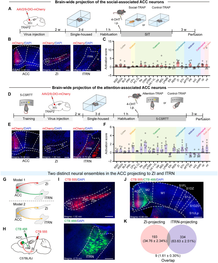
Source: Wang F et al. · Science Advances · CC BY-NC
Original figure and English caption ↗
Different outputs have different cellular profiles
The populations had similar resting potentials, but lTRN-projecting cells had lower rheobase, higher membrane resistance and greater firing under stimulation. ZI-projecting cells had longer, more complex dendrites. Electrical and structural features thus showed cellular differences accompanying the projection organization.
Optically evoked excitatory currents in target regions were sensitive to synaptic blockade, supporting functional glutamatergic connections. Transcript analysis also identified differentially expressed genes, adding molecular features to the two populations. Their dendrites, firing and expression profiles offer leads for studying how each participates in network computation.
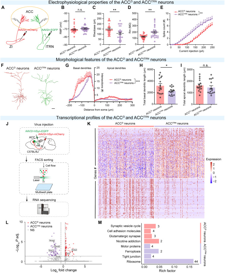
Source: Wang F et al. · Science Advances · CC BY-NC
Original figure and English caption ↗
The ZI output influences partner investigation
Suppressing ACC terminals in ZI reduced sniffing of a mouse-containing cup, sociability indices and contact during free interaction. Suppressing ACC→lTRN did not significantly change the corresponding social readouts. The two outputs affected the same behavioral questions differently, placing social specialization within a defined projection.
Light controls and baseline behavior measures helped relate the changes to projection activity rather than a general reduction in movement. Convergent effects across partner-investigation tasks clarified the ZI output’s contribution. The investigation then turned to attention tasks to ask whether the other output performed different work.
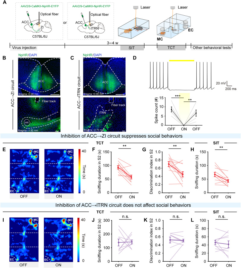
Source: Wang F et al. · Science Advances · CC BY-NC
Original figure and English caption ↗
The lTRN output influences task performance
In the five-choice task, ACC→lTRN inhibition reduced correct performance and increased premature responding. Related changes appeared with modulation across the whole trial or restricted to waiting or cue phases. ACC→ZI inhibition did not significantly change these measures. Together with the social findings, the outputs showed a double dissociation across the tasks.
The task combines waiting, visual selection and response control. The lTRN output therefore relates to how animals use cues and regulate response timing. Behavioral specialization aligned with anatomical organization, providing direct clues to how the ACC organizes behavior through different connections.
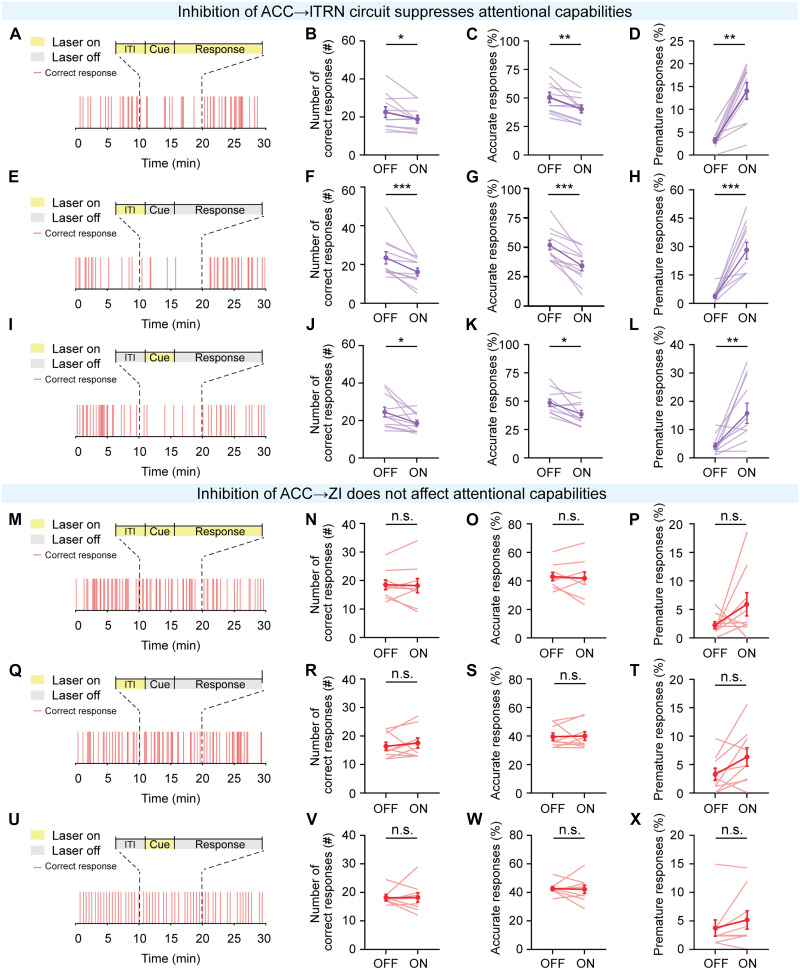
Source: Wang F et al. · Science Advances · CC BY-NC
Original figure and English caption ↗
Enhancing each output improves its corresponding behavior
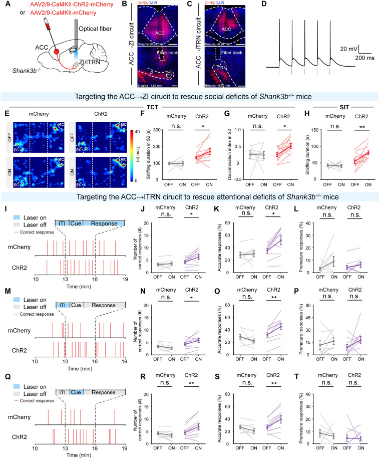
Shank3b-deficient mice showed both social and attention-task deficits, with attention problems more evident later in the task. Both ACC populations had altered excitatory inputs and dendritic spines, with differing details across ZI and lTRN connections. Enhancing ACC→ZI improved social behavior, while enhancing ACC→lTRN improved correct performance, extending the parallel-output findings to a genetic model.
Most projection-behavior and model-improvement experiments used males, while regional interventions and some cellular analyses included both sexes. Full specialization across sexes and coordination of social behavior and attention during natural interaction will extend the work. The identified cellular and connection organization offers a more detailed, testable account of multiple functions within one region.
Source: Wang F et al. · Science Advances · CC BY-NC
Original figure and English caption ↗