Once histamine reaches the hippocampus, how do astrocytes shape anxiety-like behavior?
A mouse study traces histaminergic input to ventral hippocampal CA1, showing that astrocytic H3 receptors regulate anxiety-like behavior through calcium signaling and GABA release.
Anxiety disorders rank among the most prevalent psychiatric conditions worldwide, yet conventional anxiolytics remain limited by sedation and dependence liabilities. Central histaminergic neurons originate exclusively in the hypothalamic tuberomammillary nucleus (TMN) and project broadly across the brain, but how histamine acts within specific downstream targets—and whether astrocytic histamine H3 receptors (H3Rs) participate in emotional regulation—has remained unclear. On March 5, 2026, Yulan Li, Yi Wang, Zhong Chen, Shumin Duan, and colleagues published a study titled "Astrocytic H3 receptors regulate anxiety through GABA signaling" in Neuron. Combining genetically encoded histamine sensors, circuit-specific optogenetics, cell-type-specific Hrh3 deletion, and in vivo extracellular GABA imaging, the authors demonstrate that histamine released from TMN-to-vCA1 projections selectively activates astrocytic—rather than neuronal—H3Rs in ventral CA1, triggering intracellular Ca2+ elevations and astrocytic GABA release to suppress anxiety via GABAA receptors.
Histaminergic projections from the tuberomammillary nucleus to ventral hippocampal CA1 (TMN->vCA1) selectively release histamine during anxiogenic exploration, activating astrocytic H3 receptors to drive intracellular Ca2+ elevations and GABA release that suppress anxiety via GABAA receptors.
Contents · 5 sections
Region-Specific Activation of vCA1 Histaminergic Input During Anxiogenic Exploration
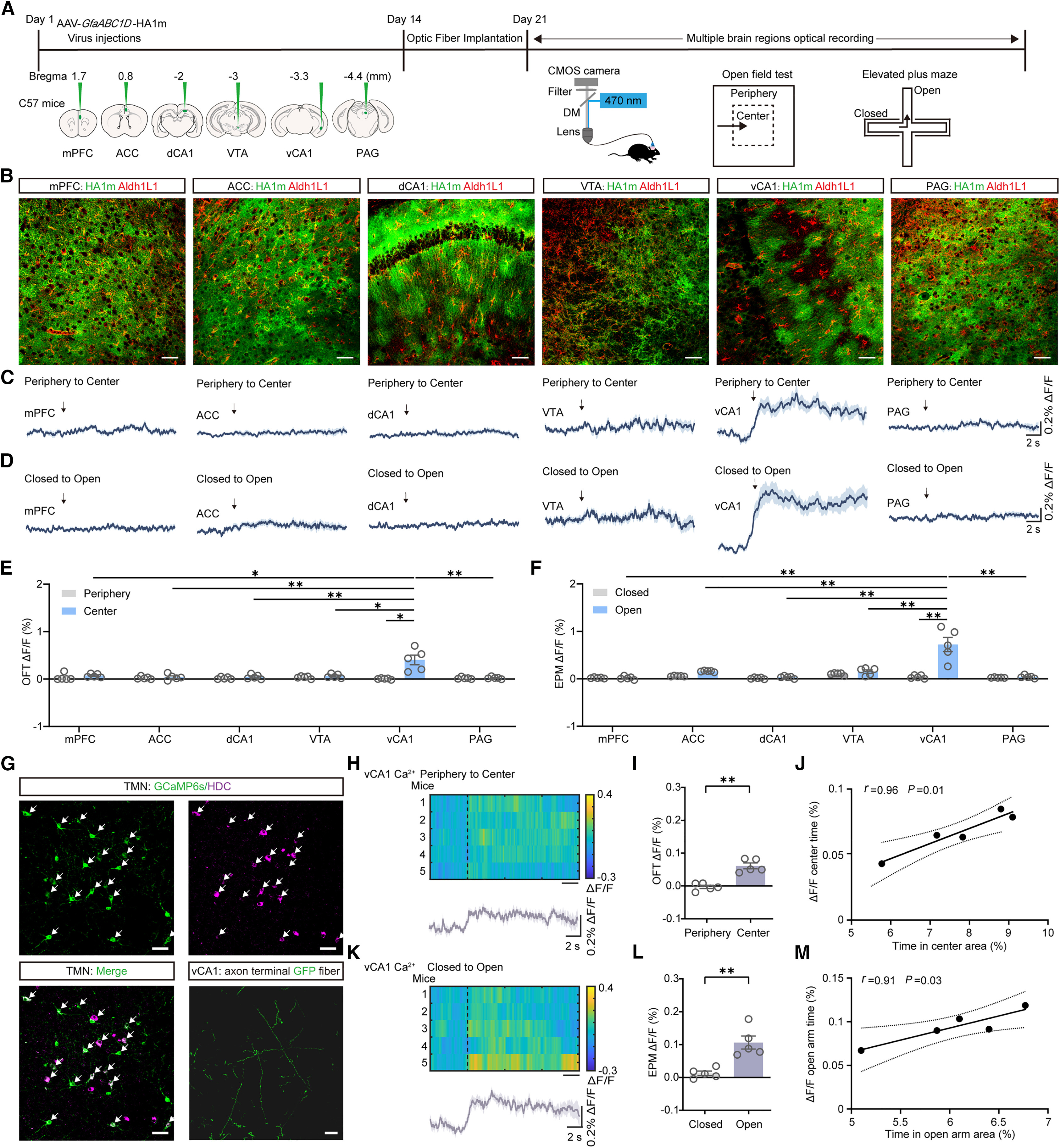
To track real-time brain histamine dynamics during anxiety-related behaviors, the authors expressed the genetically encoded fluorescent histamine sensor HA1m across the mPFC, ACC, dCA1, VTA, vCA1, and PAG and performed in vivo fiber photometry during the open field test (OFT) and elevated plus maze (EPM). Extracellular histamine levels surged selectively in vCA1 when mice transitioned into the OFT center or EPM open arms, whereas no significant changes occurred in the other five brain regions.
Control recordings with the binding-deficient mutant sensor HA1mut showed no signal fluctuations during arena transitions, ruling out motion artifacts, while acute social defeat attacks similarly evoked rapid histamine transients in vCA1. Recording axonal Ca2+ signals from inducible HDC-CreERT2 mice expressing AAV-DIO-axon-GCaMP6s in the TMN confirmed that TMN->vCA1 histaminergic terminals are strongly activated upon entry into the OFT center and EPM open arms, with terminal Ca2+ amplitude positively correlating with open-arm exploration time.
Source: Yulan Li et al. · Neuron · © Elsevier Inc.
Original figure and English caption ↗
TMN-to-vCA1 Histaminergic Circuitry Bidirectionally Regulates Anxiety via H3 Receptors
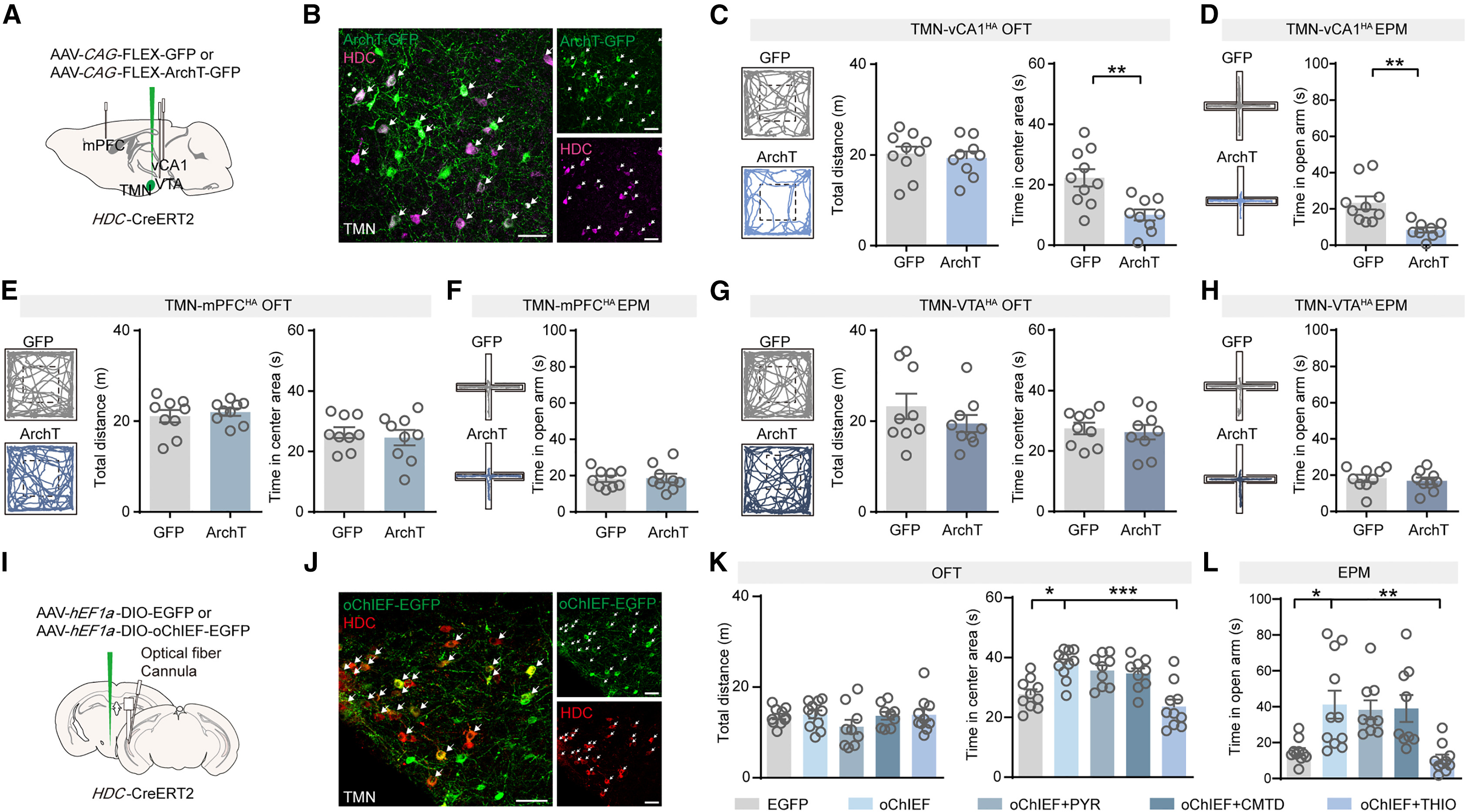
To establish causality, the authors expressed the inhibitory opsin ArchT in TMN histaminergic neurons of HDC-CreERT2 mice and delivered yellow light to terminals in vCA1, mPFC, or VTA. Optogenetic inhibition of TMN->vCA1 terminals significantly reduced OFT center time and EPM open-arm exploration without altering general locomotion, whereas inhibiting TMN->mPFC or TMN->VTA projections had no effect on anxiety-like behaviors.
Conversely, 20 Hz blue-light stimulation of oChIEF-expressing TMN->vCA1 terminals evoked local histamine release and markedly increased OFT center time and EPM open-arm exploration. Intra-vCA1 microinjection of the selective H3R antagonist thioperamide (THIO) completely abolished this optogenetic anxiolytic effect, whereas neither the H1R antagonist pyrilamine (PYR) nor the H2R antagonist cimetidine (CMTD) blocked it, pinpointing local H3Rs as the essential downstream mediator.
Source: Yulan Li et al. · Neuron · © Elsevier Inc.
Original figure and English caption ↗
Astrocytic—Rather Than Neuronal—H3 Receptors in vCA1 Govern Anxiolytic Regulation
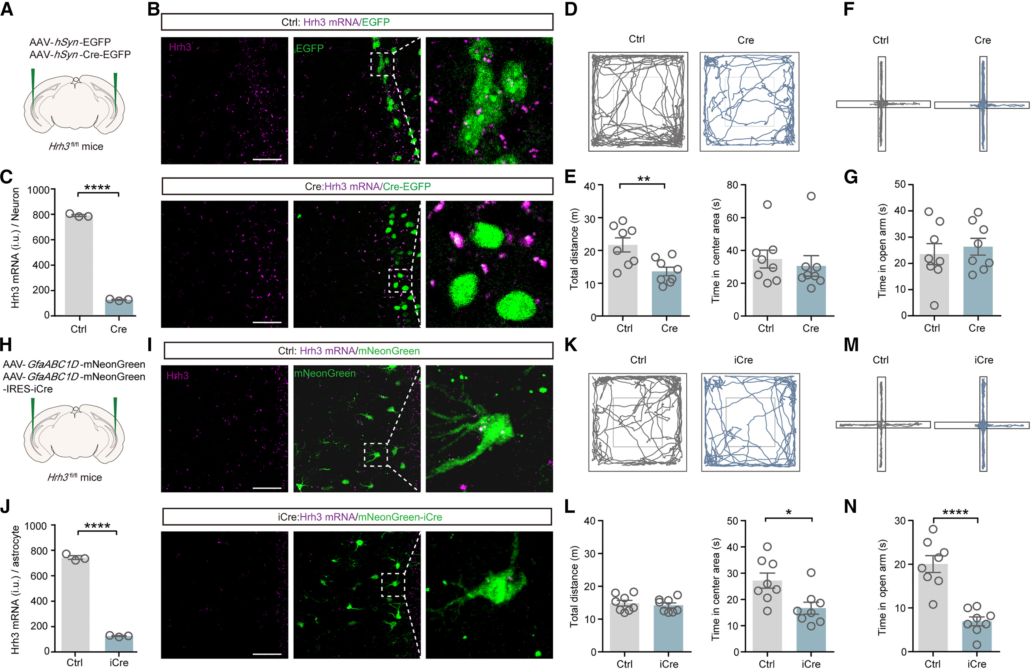
RNAscope in situ hybridization combined with immunostaining revealed that Hrh3 mRNA is expressed in 88.79% of GFAP-positive vCA1 astrocytes alongside neurons. To dissect cell-type-specific contributions in Hrh3fl/fl mice, the authors selectively ablated Hrh3 in vCA1 neurons using AAV-hSyn-Cre or in vCA1 astrocytes using AAV-GfaABC1D-mNeonGreen-IRES-iCre. Deleting neuronal Hrh3 left OFT and EPM anxiety behaviors unchanged, whereas astrocytic Hrh3 knockdown significantly reduced OFT center time and EPM open-arm exploration without affecting novel object recognition or contextual fear memory.
Furthermore, conditional knockdown of astrocytic H1R or H2R in vCA1 had no impact on anxiety-like behaviors. In gain-of-function experiments using astrocyte-specific OptoH3R expression or chronic Hrh3 overexpression in wild-type and Aldh1l1-CreERT2 mice, acute light-evoked astrocytic OptoH3R signaling robustly increased both OFT center and EPM open-arm exploration, while chronic astrocytic H3R overexpression significantly increased EPM open-arm exploration time without notably altering OFT center exploration, confirming bidirectional control of anxiety by astrocytic H3Rs.
Source: Yulan Li et al. · Neuron · © Elsevier Inc.
Original figure and English caption ↗
H3R-Dependent Astrocytic Calcium Signaling Mediates Histamine's Anxiolytic Action
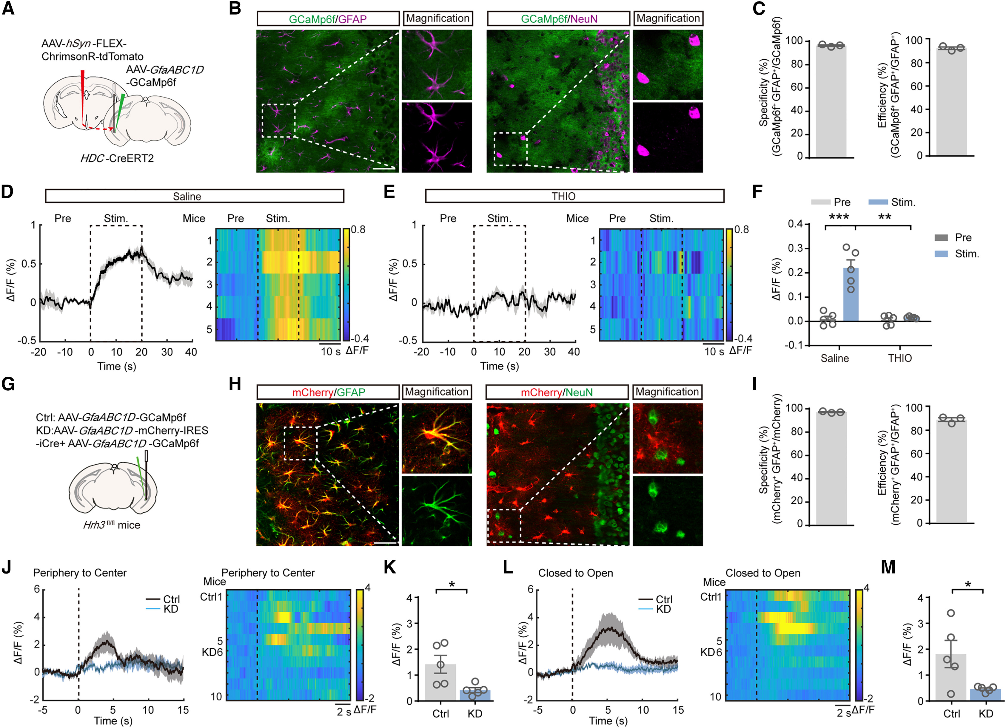
Although H3Rs are classically Gi/o-coupled, astrocytic Gi/o signaling can mobilize intracellular Ca2+ via the PLC-IP3R2 cascade. Dual-color fiber photometry in HDC-CreERT2 mice expressing ChrimsonR in the TMN and GCaMP6f in vCA1 astrocytes revealed that red-light activation of TMN->vCA1 terminals reliably evoked astrocytic Ca2+ elevations that were abolished by the H3R antagonist THIO. Astrocytic Ca2+ transients also surged when mice entered the OFT center or EPM open arms; these behavior-locked Ca2+ responses were blunted by astrocytic Hrh3 knockdown but remained intact when surrounding vCA1 neurons were chemogenetically silenced with hM4Di.
Two-photon Ca2+ imaging in acute brain slices confirmed that histamine and the H3R agonist immepip directly triggered vCA1 astrocytic Ca2+ elevations in the presence of tetrodotoxin (TTX), an effect abolished by astrocytic Hrh3 knockdown, the PLC inhibitor U73122, or IP3R2 knockout. Behaviorally, optogenetic elevation of vCA1 astrocytic Ca2+ was anxiolytic, whereas attenuating astrocytic Ca2+ with the plasma membrane Ca2+ pump hPMCA2w/b induced anxiety and completely blocked the anxiolytic effect of TMN->vCA1 stimulation.
Source: Yulan Li et al. · Neuron · © Elsevier Inc.
Original figure and English caption ↗
Astrocyte-Derived GABA Release and GABAA Signaling Reverse Chronic Stress-Induced Anxiety
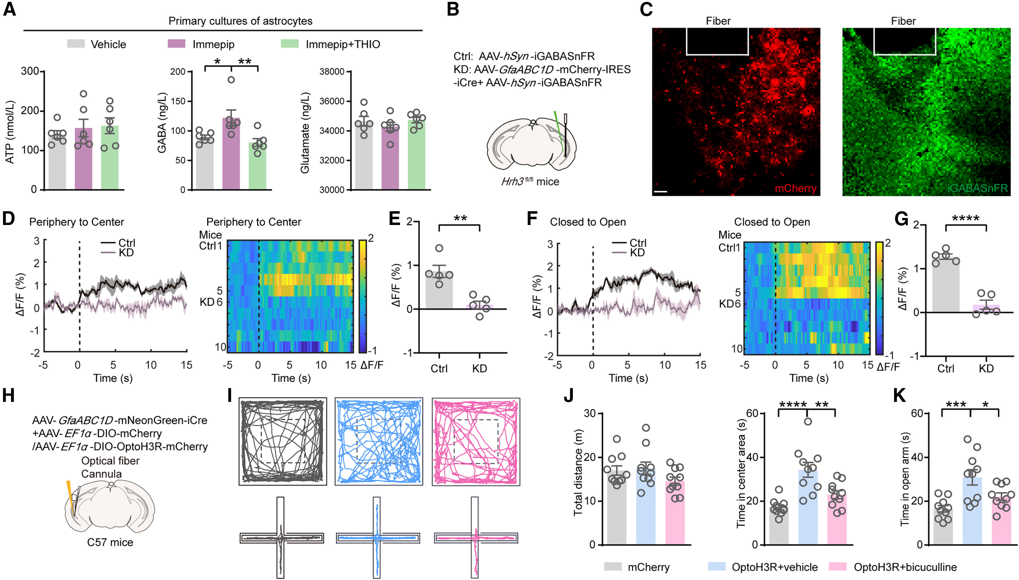
To identify the downstream gliotransmitter, the authors assayed cultured astrocytes by ELISA and found that the H3R agonist immepip selectively stimulated GABA release—without altering ATP or glutamate levels—in a THIO-sensitive manner. In vivo fiber photometry with the extracellular GABA sensor AAV-hSyn-iGABASnFR showed robust vCA1 GABA surges upon entry into the OFT center and EPM open arms in control mice, which were significantly blunted in mice with conditional astrocytic Hrh3 knockdown; moreover, intra-vCA1 microinjection of the GABAA receptor antagonist bicuculline abolished the anxiolytic effects of astrocytic OptoH3R activation or Hrh3 overexpression.
Finally, in a 10-day chronic social defeat stress (CSDS) model, stressed mice exhibited pronounced anxiety-like behaviors alongside a compensatory upregulation of vCA1 astrocytic Hrh3. Both acute optogenetic stimulation of astrocytic OptoH3R and chronic viral overexpression of Hrh3 in vCA1 astrocytes effectively rescued OFT and EPM exploration in CSDS mice. Together, these findings delineate a previously unrecognized TMN histamine -> vCA1 astrocytic H3R -> Ca2+ -> GABA -> GABAA receptor pathway that gates anxiety and offers a glial target for anxiolytic intervention.
Source: Yulan Li et al. · Neuron · © Elsevier Inc.
Original figure and English caption ↗