Nanoparticle-assisted magnetic stimulation improves sociability in model mice
Magnetic nanoparticles combined with an external field increase mouse PVN activity and local oxytocin signals. Sociability and other behavioral readouts improve in a valproate-associated model.
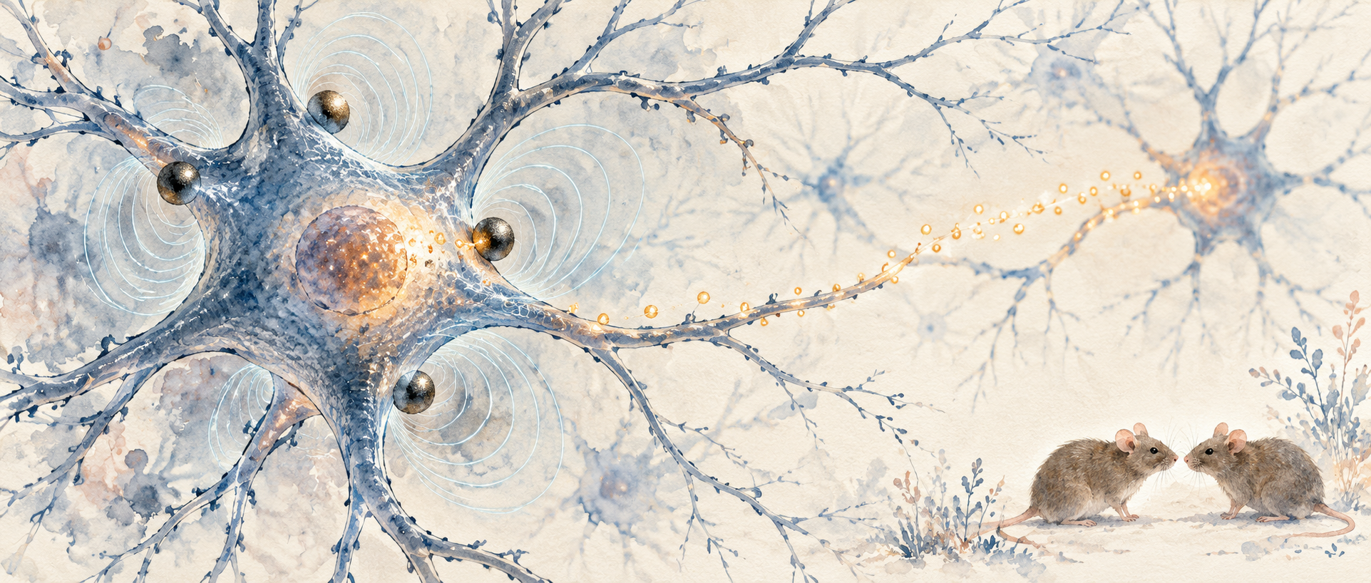
How can modulation engage a region deep within the brain? A study in Neuroscience Bulletin brings magnetic nanoparticles into the process. With particles already located in the hypothalamic paraventricular nucleus, magnetic stimulation increases associated neural activity and oxytocin signals and improves sociability in model mice. The findings connect a material-assisted approach with neural and behavioral responses.
Nanoparticle-assisted stimulation increases PVN activity and oxytocin signals and improves social preference in model mice. Oxytocin receptor inhibition removes the clear social improvement, supporting this signal’s contribution to the behavioral change.
Contents · 7 sections
Materials in the target contribute to field responses
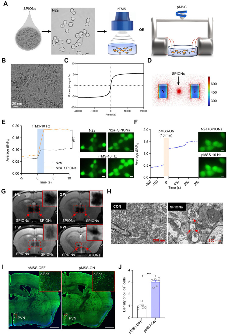
Conventional magnetic stimulation faces challenges in localization and resolution for deep targets. This strategy uses superparamagnetic iron oxide nanoparticles so that local material and an external field act together. With particles in PVN, stimulation increases activity markers there while the observed superficial cortical response is small.
The particles remain locally detectable during the brain observation period and can be taken up by cells. Targeting therefore depends on material distribution. The subsequent neural and behavioral findings arise from the combination of a field with particles already present at the target.
Source: Liu S et al. · Neuroscience bulletin · CC BY
Original figure and English caption ↗
Neural responses vary with stimulation conditions
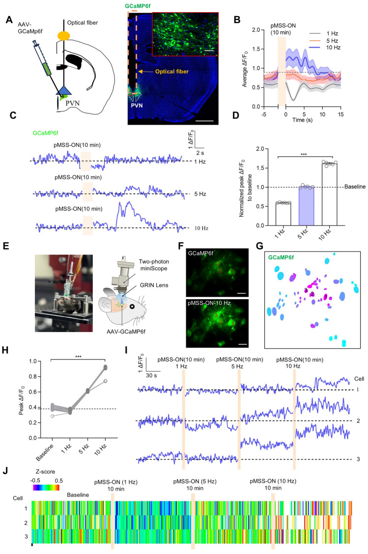
PVN population calcium recordings show different response directions across frequencies: lower-frequency conditions reduce responses, higher-frequency conditions increase them, and the intermediate condition produces a smaller overall change. Single-cell observations also reveal condition-dependent differences. The field does not operate as a uniformly excitatory switch.
The team follows the condition with clearer activity enhancement into oxytocin and behavioral measurements. This moves the question from whether the material alters cellular responses to which neuronal state changes accompany particular biological outcomes.
Source: Liu S et al. · Neuroscience bulletin · CC BY
Original figure and English caption ↗
Local oxytocin signals increase
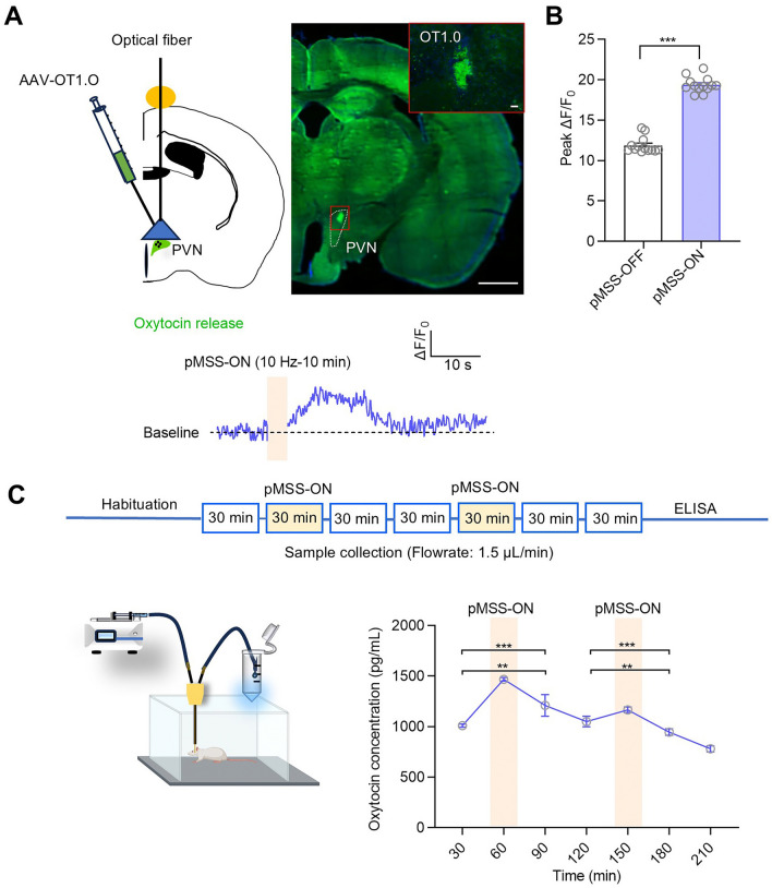
PVN is a neuroendocrine region associated with oxytocin. Following activity enhancement, the local oxytocin sensor signal rises, and microdialysis measurements also show increased oxytocin. Two readouts connect the neural response with a molecule involved in social behavioral regulation.
Gene and protein measurements show higher oxytocin expression and increases in several associated signaling proteins. These molecular changes accompany the release readouts: stimulation is associated with both stronger local signals and changes in cellular expression states.
Source: Liu S et al. · Neuroscience bulletin · CC BY
Original figure and English caption ↗
Neurites change alongside activity
Cultured PVN neurons show longer dendrites and axons during sustained observations. Dendritic spine density in brain tissue also varies with stimulation conditions, decreasing at the lower frequency and increasing at higher frequencies. Cellular structure therefore responds alongside neuronal activity.
The changes are not uniform across spine types. Densities of several types increase, whereas branched spines do not change clearly. Dendritic spines are diverse structures, and an increase in average density can encompass different responses across their forms.
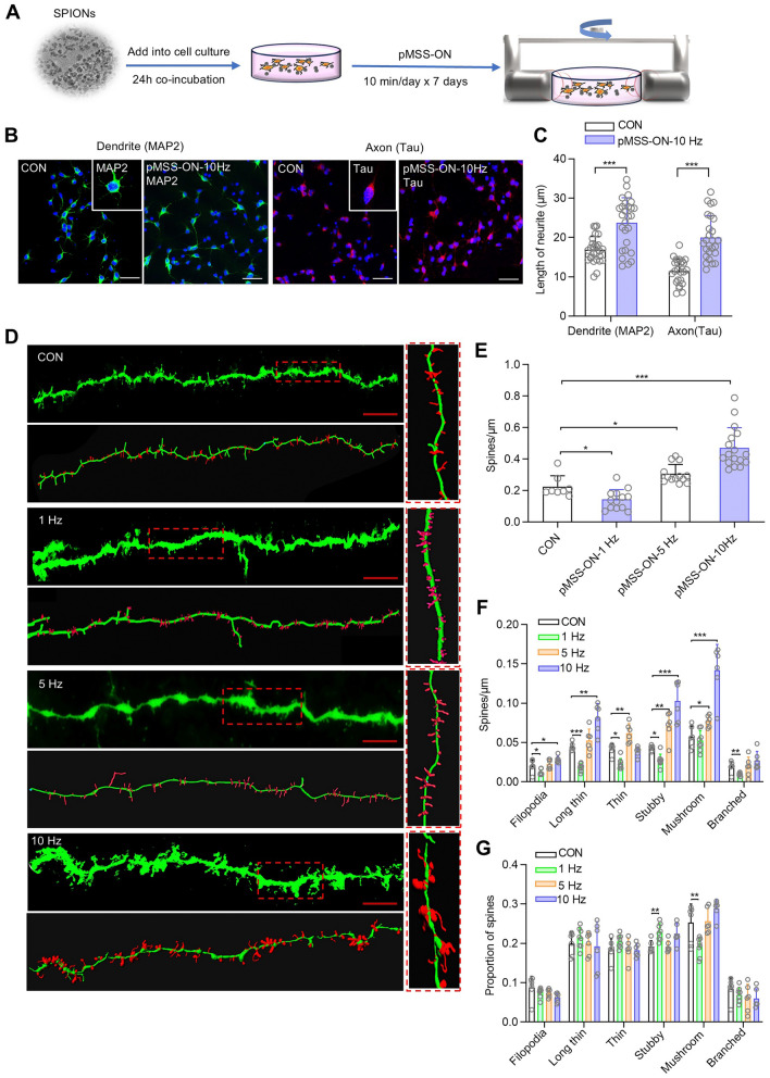
Source: Liu S et al. · Neuroscience bulletin · CC BY
Original figure and English caption ↗
Model mice show greater social preference
In the developmental valproate-exposure model, mice have weaker social preference, less open-field center time and longer self-grooming. After a week of intervention observations, the nanoparticle-assisted stimulation group shows greater social preference, more center time and less repetitive grooming. Particles alone do not produce the same improvement.
Within this model, initially lower PVN oxytocin expression increases after stimulation, and local release measurements also rise. Molecular and behavioral changes in the same study allow the team to ask whether oxytocin signaling contributes to the change in sociability.
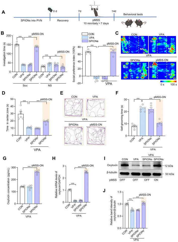
Source: Liu S et al. · Neuroscience bulletin · CC BY
Original figure and English caption ↗
Receptor inhibition removes the clear social improvement
Reducing oxytocin receptor signaling in prefrontal cortex, or inhibiting it more broadly, removes the clear improvement in social preference after stimulation. Other behavioral readouts also differ from stimulation without receptor inhibition. Oxytocin receptor signaling thus contributes to the behavioral effect rather than merely accompanying it.
The finding links activity in a deep region with signal reception elsewhere. Changes associated with PVN can influence a broader network through molecular signaling, with behavior arising from the participation of these connected elements.
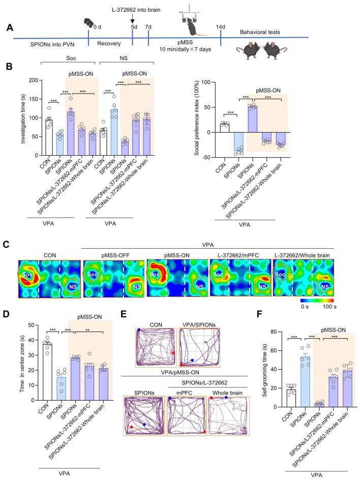
Source: Liu S et al. · Neuroscience bulletin · CC BY
Original figure and English caption ↗
Engaging endogenous signals differs from supplying them
The study also compares external oxytocin with stimulation-associated endogenous signal changes. Under the tested conditions, social preference is higher in the magnetic-stimulation group, while group relationships for open-field and grooming measures differ. How a molecule engages the network may matter for the resulting behavioral readout.
The findings come from male mice and a particular developmental model, with longer-term effects of brain-resident material still unresolved. They connect material-assisted magnetic modulation with endogenous oxytocin signaling and social behavioral changes, providing evidence for how deep neural activity can influence social responses.