Microglial lipid metabolism and emotional changes after stroke
A mouse study links preservation of a lipid signal with reduced inflammation, synaptic engulfment and anxiety-like behavior, alongside changes in hippocampus beyond the lesion.
How can a focal brain injury accompany broader behavioral changes? A 2025 Cell Communication and Signaling study follows a lipid signal from a stroke lesion toward contralateral hippocampus. Palmitoylethanolamide (PEA) metabolism changes alongside microglial inflammation and engulfment, synapses and exploration. Connecting these levels moves the explanation beyond lesion size alone.
Global Naaa deletion or PEA supplementation reduces selected injury and anxiety-like measures in model mice, accompanying preserved contralateral hippocampal synaptic markers and reduced microglial engulfment; IL-18 signaling contributes.
Contents · 7 sections
Similar injury, different exploration
Some mice in a photothrombotic stroke model bury more marbles and spend less time in the open-field center and open arms. The study identifies an anxiety-like subgroup and compares brain changes. These measures describe exploration and defensive responses rather than subjective human feelings.
The stroke subgroups have similar infarct volumes, rotarod performance and total open-field distance. Exploration differences do not simply track lesion size, prompting examination of metabolic and immune states in injured tissue.
PEA rises briefly, then declines
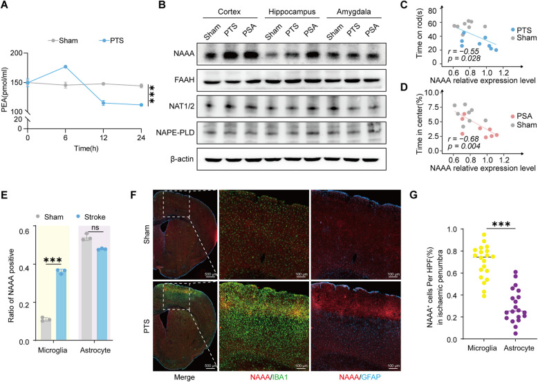
PEA is an endogenous lipid signal involved in cellular stress responses. In ischemic penumbra it rises during the first six hours after stroke and declines by twelve to twenty-four hours, with a larger later reduction in the anxiety-like subgroup. NAAA, an enzyme degrading PEA, increases in injured cortex.
Localization places the increase mainly in microglia; astrocytes in the single-cell data lack the same significant change. Microglia perform immune surveillance and tissue clearance, linking accelerated lipid-signal breakdown with the post-injury immune response.
Source: Yin T et al. · Cell communication and signaling : CCS · CC BY-NC-ND
Original figure and English caption ↗
Global Naaa deletion alters recovery
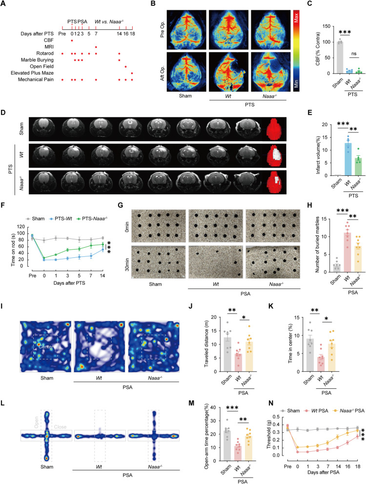
Naaa-deficient mice have similar early blood flow and initial ischemic extent to controls, followed by smaller infarcts and faster rotarod recovery. Marble burying decreases, exposed-space time increases and hindpaw mechanical thresholds rise. Injury, recovery and exploration change together.
Total open-field distance also increases, so behavioral improvement includes movement as well as exploration. Relevant baseline measures do not clearly differ between genotypes, and supplemental experiments also find improvements in anxiety-like measures in female mice.
Source: Yin T et al. · Cell communication and signaling : CCS · CC BY-NC-ND
Original figure and English caption ↗
PEA supplementation changes behavioral outcomes
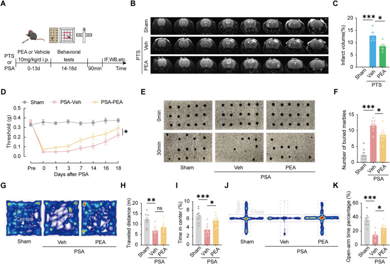
PEA supplementation produces smaller infarcts, higher mechanical thresholds, less marble burying and more exposed-space time. Total open-field distance does not significantly change, making altered exploration choices more apparent.
Supplemental experiments in aged mice also show improvement in selected injury and behavioral measures. Genetic reduction of degradation and external supplementation adjust the same lipid signal through complementary approaches.
Source: Yin T et al. · Cell communication and signaling : CCS · CC BY-NC-ND
Original figure and English caption ↗
Connections decline in contralateral hippocampus
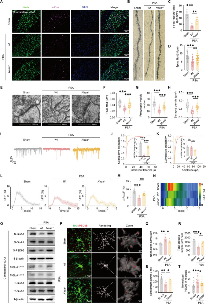
In contralateral ventral hippocampal CA1 of anxiety-like model mice, dendritic spines and synaptic structures decline, alongside activity markers and stimulus-related calcium responses. Naaa deletion improves these measures; miniature excitatory-current frequency rises without a significant amplitude change.
Microglia in the same region enlarge their cell bodies, lose processes and branches, and contain more PSD95 synaptic-marker puncta. Naaa deletion or PEA supplementation reduces engulfment-related features, linking lipid metabolism with cellular interactions around synapses beyond the lesion.
Source: Yin T et al. · Cell communication and signaling : CCS · CC BY-NC-ND
Original figure and English caption ↗
IL-18 links immune state with synaptic changes
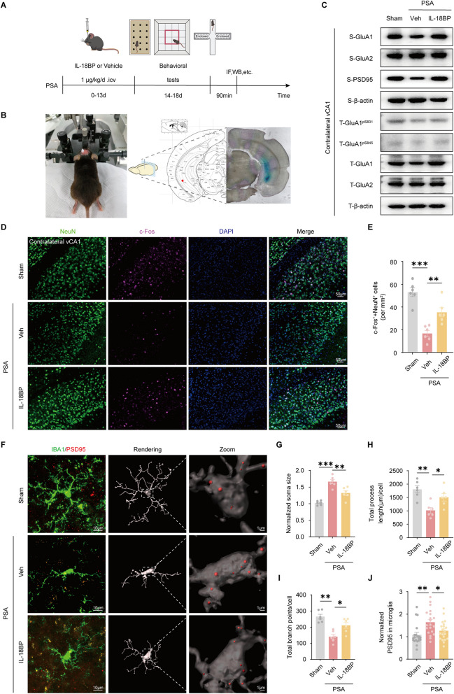
Mitochondrial and inflammasome-related signals change in injured tissue, and IL-18 increases in the lesion and contralateral hippocampus. Reducing PEA degradation or supplying PEA attenuates inflammatory changes. Inhibiting IL-18 also improves exploration and mechanical responses and reduces synaptic-marker puncta within contralateral hippocampal microglia.
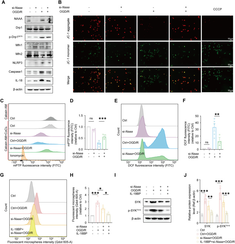
In culture, medium from oxygen–glucose-deprived microglia enhances particle uptake by another microglial population; reducing Naaa or blocking IL-18 weakens this effect. Released signals can influence other cells, while their precise interregional route in the brain awaits direct tracking.
Source: Yin T et al. · Cell communication and signaling : CCS · CC BY-NC-ND
Original figure and English caption ↗
Understanding injury through cellular interactions
Primary-microglial experiments further show reduced mitochondrial-stress and inflammatory readouts after Naaa knockdown, linking tissue-level observations with intracellular lipid signals, energetic state and engulfment functions.
The animal interventions use global deletion or PEA supplementation, with behavioral changes alongside injury recovery. The study shows how stroke-related changes extend from local metabolism to immune-cell–synapse relationships; preserving a lipid signal accompanies changes in these relationships and behavior.
Source: Yin T et al. · Cell communication and signaling : CCS · CC BY-NC-ND
Original figure and English caption ↗