After stress, hypothalamic receptor expression rises with a delay
Days after repeated stress, mice explore exposed areas less and associated hypothalamic responses weaken. Increased GHS-R1a expression links this delayed change with neuronal excitability.
The influence of stress can emerge after the event has ended. A study in Nature Communications finds clearer changes in exploratory behavior days after repeated stress, alongside altered responses in a hypothalamic population. The team identifies delayed increases in the ghrelin-associated receptor GHS-R1a, linking receptor expression with neuronal excitability and anxiety-related behavioral readouts.
Reducing GHS-R1a expression in the targeted VMH population increases open-arm exploration. Increasing expression reduces exploration and suppresses neuronal firing; activating the cells under elevated receptor expression improves this readout.
Contents · 7 sections
Behavior changes become clearer days later
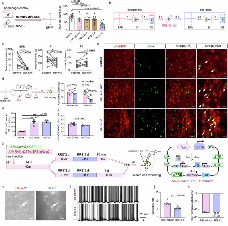
The team observes males at different times after repeated restraint stress. Open-arm exploration does not fall clearly at early measurements but is lower by day six. Social interaction and some passive-coping-related measures also change, revealing a delay between stress exposure and behavioral differences.
The study focuses on dorsomedial and central ventromedial hypothalamus, or VMH. This region contributes to bodily state and defensive responses. Its αCaMKII-expressing population shows increased activity markers after behavioral testing at both early and later measurements. Cells previously recruited by stress nevertheless have lower firing frequency at the later time.
Source: Zhang M et al. · Nature communications · CC BY-NC-ND
Original figure and English caption ↗
The cells still respond on entering exposed areas
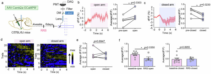
Under baseline conditions, calcium responses rise on entering open maze arms and fall on entering closed arms. Later after stress, entry into an open arm still produces a transient increase, but mean responses during open and closed exploration no longer differ significantly.
Compared with unstressed mice, mean responses during open-arm exploration are lower, while closed-arm responses are comparable. The change is context-dependent: the cells retain responses, but population activity differs in the more exposed setting.
Source: Zhang M et al. · Nature communications · CC BY-NC-ND
Original figure and English caption ↗
More exploration can coexist with acute aversion
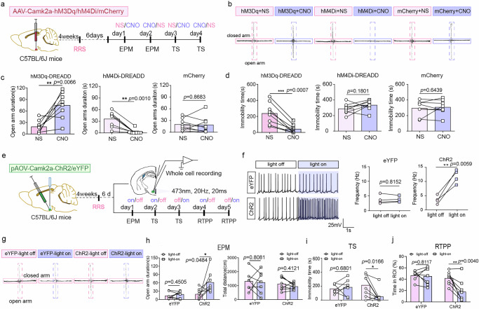
Increasing activity raises open-arm exploration in both unstressed and stressed mice, while reducing activity lowers it. Activation also shortens tail-suspension immobility, whereas inhibition does not produce a symmetric effect on that measure. Total movement distance does not change clearly.
A place-preference task reveals another aspect: activated mice avoid the stimulation-associated area. This coexists with increased open-arm exploration, showing different relationships among avoidance, exploration and passive coping in the tested settings. The population changes particular behaviors without moving all affect-related readouts in one direction.
Source: Zhang M et al. · Nature communications · CC BY-NC-ND
Original figure and English caption ↗
GHS-R1a rises at the later stage
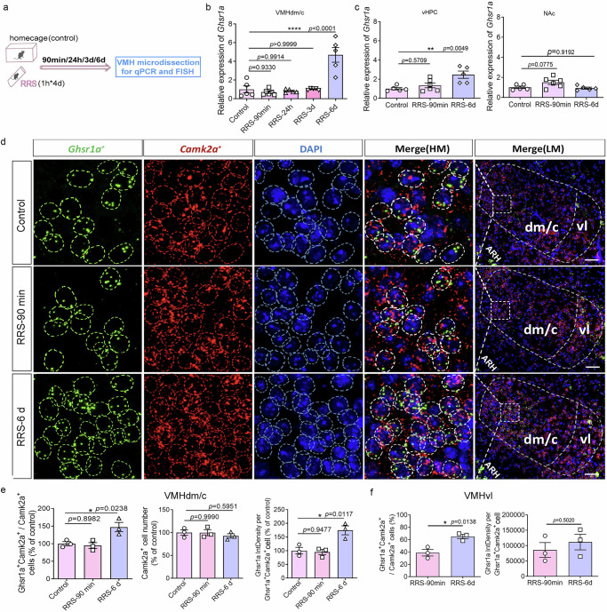
GHS-R1a is associated with the metabolic hormone ghrelin and is expressed across brain regions. After repeated stress, expression in the targeted hypothalamic cells rises by day six, without the same early change. Ventral hippocampus also shows a later increase, whereas corresponding nucleus accumbens comparisons are not significant.
The molecular change follows a similar time course to the behavior and appears within identified cells. The team moves from concurrent changes to manipulating receptor expression and testing whether exploration changes.
Source: Zhang M et al. · Nature communications · CC BY-NC-ND
Original figure and English caption ↗
Changing expression changes exploration
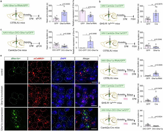
Reducing receptor expression in the targeted hypothalamic population increases open-arm exploration in unstressed and stressed mice; increasing expression lowers it. Two different ways of reducing expression produce consistent directions, supporting a link through intervention.
Increasing receptor expression in hypothalamic inhibitory cells or associated ventral hippocampal cells does not produce the same behavioral change. Its effect depends on region and cell identity, placing the molecular change within a specific network context.
Source: Zhang M et al. · Nature communications · CC BY-NC-ND
Original figure and English caption ↗
More receptor expression accompanies less firing
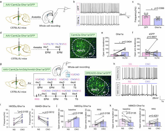
Increased receptor expression lowers spontaneous firing. Local receptor antagonism improves open-arm exploration in these mice without a significant effect in controls. Increasing activity in cells with elevated receptor expression improves open-arm performance under both baseline and stressed conditions.
The reverse intervention is not always symmetric: further inhibiting cells with elevated receptor expression reduces exploration in unstressed mice but has no significant effect in stressed mice. Receptor state, stress history and neuronal activity jointly shape the readout under particular conditions.
Source: Zhang M et al. · Nature communications · CC BY-NC-ND
Original figure and English caption ↗
Cellular state contributes to delayed behavioral changes
The study describes several levels across time after stress: neuronal responses to external conditions, later changes in receptor expression and associated differences in firing and exploration. Changing receptor expression or activity alters the measured behavior.
The findings come from adult males and focus on central receptors and cellular state. Delayed molecular changes connect a stress history with subsequent network responses, and the same population can contribute differently to an animal’s environmental responses across states.