Inhibiting an inhibitor: how CA2 helps recognize a companion
NLG1-deficient male mice approach companions but struggle to distinguish familiar from new ones. CCK-cell regulation of PV cells helps release CA2 pyramidal output.
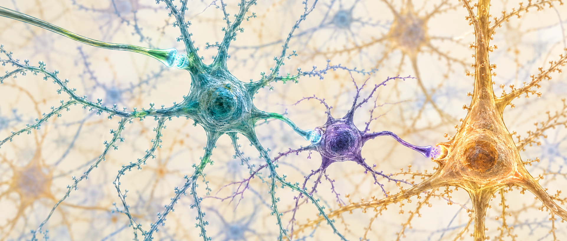
Mice usually explore a new companion differently from one just encountered. NLG1-deficient mice remain sociable but lose the normal novelty preference. In CA2, inhibitory CCK cells can weaken PV-cell restraint and support pyramidal responses, making the recipient of inhibition central to the mechanism.
CA2 CCK cells support social recognition through GABAB-related regulation and CCK signaling. NLG1 loss weakens their output and strengthens PV restraint; PV inhibition improves novelty preference in the model.
Contents · 6 sections
Still approaching, but struggling to recognize familiarity
NLG1, neuroligin-1, is a synapse-associated molecule whose cell-specific effects can shape local connectivity and behavior. CA2 provided a setting to ask which cells change their input and output after its loss.
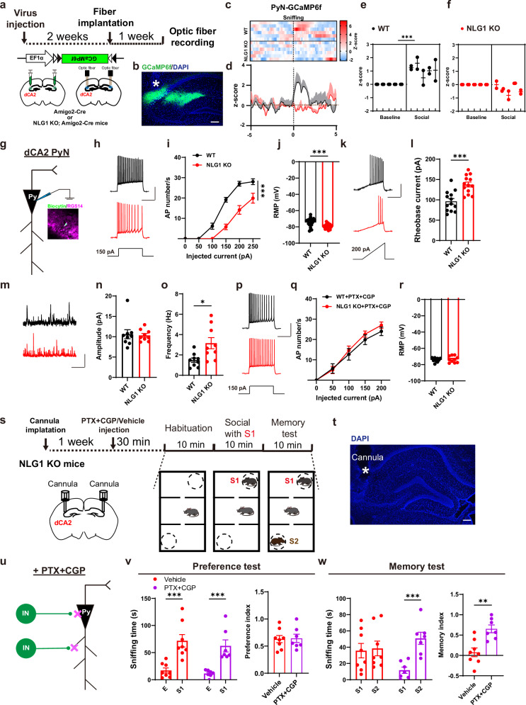
The tasks examined approach to an unfamiliar mouse, then preference between familiar and new companions. NLG1-deficient mice retained approach but lacked normal novelty preference. CA2 pyramidal cells showed weaker social calcium responses, lower excitability and increased inhibitory input.
Local GABA-receptor blockade removed some electrophysiological differences and restored novelty preference. NLG1 reduction in pyramidal or PV cells did not reproduce the deficit, whereas reduction in inhibitory populations did, directing attention to a specific inhibitory member.
Source: Wu M et al. · Nature communications · CC BY-NC-ND
Original figure and English caption ↗
CCK cells emerge as a key population for NLG1 action
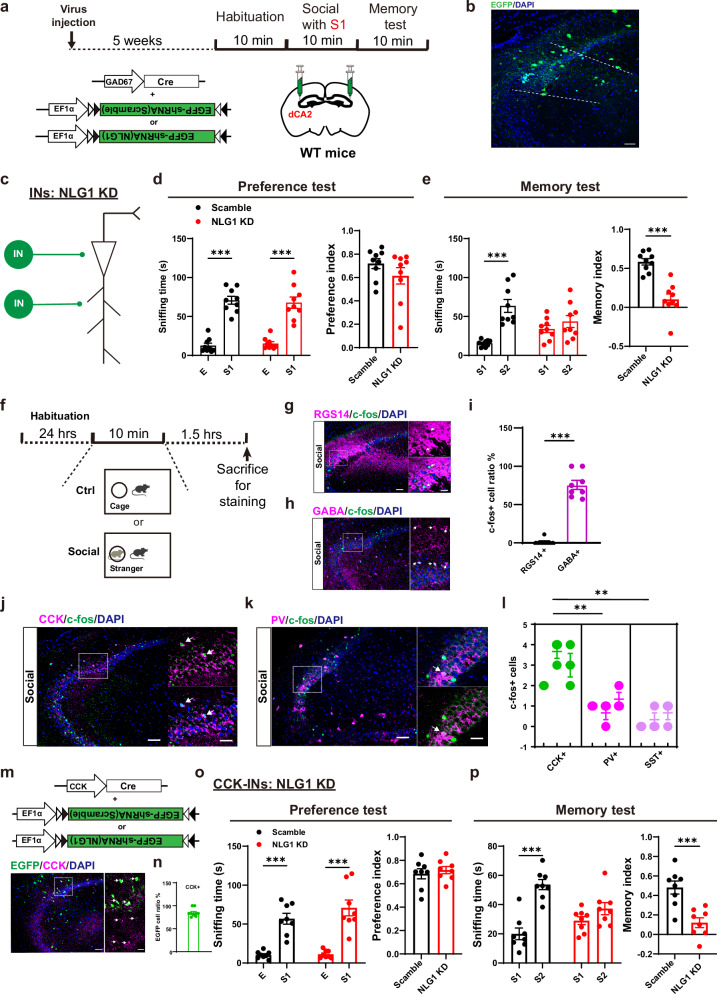
Activity labeling after interaction mainly appeared in inhibitory cells, many CCK-positive. Reducing NLG1 in CCK cells impaired recognition while preserving ordinary approach, localizing a role of this broadly expressed synaptic molecule.
Inhibiting normal CA2 CCK cells weakened recognition and pyramidal social responses; activation did not cause the same impairment. NLG1-deficient CCK cells were still recruited socially but responded less and blocked firing more readily under strong drive.
Source: Wu M et al. · Nature communications · CC BY-NC-ND
Original figure and English caption ↗
Why can inhibiting PV cells help pyramidal output?
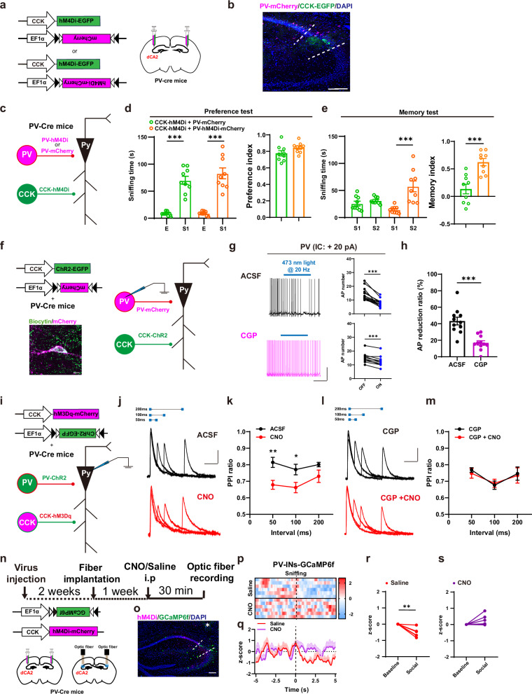
PV cells strongly restrain CA2 pyramidal cells. Sustained high-frequency CCK activation reduced PV firing and inhibitory efficacy, opposed by GABAB antagonism. Single low-frequency stimuli lacked clear fast currents, favoring slower regulation while leaving exact contact sites unresolved.
Behavioral rescue strengthened the chain: simultaneous PV suppression restored recognition impaired by CCK inhibition, and PV suppression also improved the NLG1-deficient model. CCK2 signaling contributed to depolarization, yet CCK2 blockade alone did not impair the task; combined GABAB and CCK2 blockade did.
Source: Wu M et al. · Nature communications · CC BY-NC-ND
Original figure and English caption ↗
Potassium currents and CB1 offer clues to weakened output
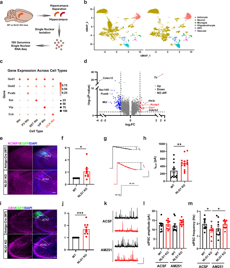
Single-nucleus sequencing identified higher Kcnip1 and Cnr1 expression. Protein and current tests supported stronger A-type potassium current and CB1-associated output modulation, without equivalent sodium or delayed-rectifier current changes. More pronounced abnormalities under high drive suggest input-dependent consequences.
CB1 antagonism removed some PV inhibitory-event differences, but both agonism and antagonism could impair recognition; PV inhibition rescued the antagonist effect. Receptor state and circuit context interacted, with KCNIP1-specific rescue remaining an open question.
Source: Wu M et al. · Nature communications · CC BY-NC-ND
Original figure and English caption ↗
Medial septum provides input to the CCK circuit
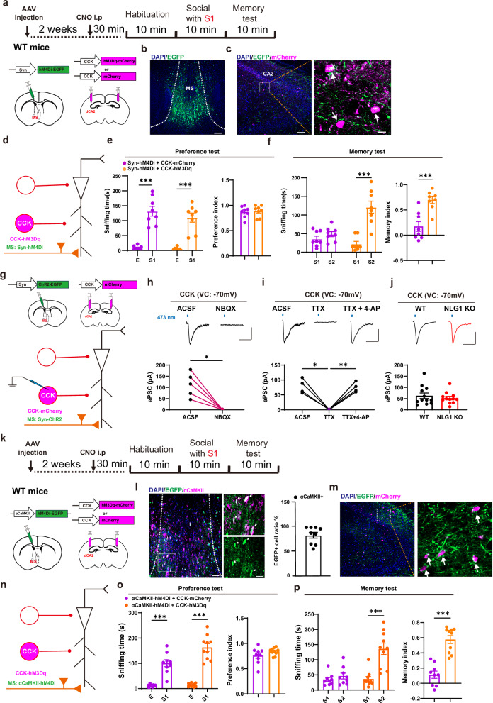
Medial septum supplied direct glutamatergic input that remained comparable after NLG1 loss. Septal or excitatory-population inhibition impaired recognition, rescued by CA2 CCK activation, clarifying the functional link from input to recipient cells.
Together, preserved evoked input accompanies altered CCK processing/output and stronger PV restraint. Recordings and rescues across levels connect recognition changes to specific cellular relationships, identifying one septal route among other hippocampal inputs.
Source: Wu M et al. · Nature communications · CC BY-NC-ND
Original figure and English caption ↗
Recognizing companions requires balanced cellular restraint
The disinhibitory relationship illustrates how an inhibitory cell can support output by restraining another inhibitor. NLG1, CCK, PV and pyramidal-cell relationships distinguish approach from recognition and place a synaptic molecule within a circuit.
The evidence comes from adult males. NLG1 regulation of candidate molecules, exact contacts and peptide/GABA contributions remain open, with some marker overlap. Finer cell and signal resolution may explain how this circuit sustains effective social output.