How stress engages the masseter: a relay between inhibitory cells
Chronic stress increases mouse masseter activity. Inhibiting Vme-projecting amygdala cells reduces muscle activity without simultaneously relieving anxiety-like behavior.
Bodily responses to stress can involve a relatively small muscle. The masseter, which closes the jaw, offers a window into connections between emotional processes and oral movement. A 2024 Frontiers in Cellular Neuroscience study follows the central amygdala (CeA)–mesencephalic trigeminal nucleus (Vme) connection in mice exposed to chronic restraint stress. Counterintuitively, greater upstream inhibitory-cell activity accompanies more excitable downstream excitatory cells; reducing masseter activity also need not improve anxiety-like behavior at the same time.
Vme-projecting CeA GABAergic cells contribute to stress-associated masseter overactivity. Inhibiting this projection-defined population reduces EMG while tested anxiety-like measures remain largely unchanged.
Contents · 6 sections
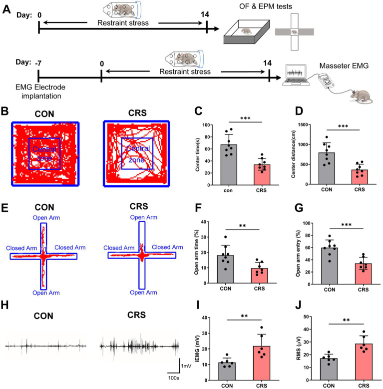
Less exposed-space exploration and more masseter activity
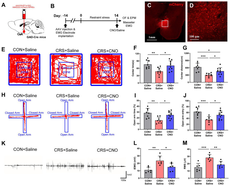
After repeated chronic restraint stress, mice spend less time and travel less distance in the open-field center. Elevated-plus-maze open-arm time and entry proportions also decrease. These behaviors indicate greater exposed-space avoidance, matching the anxiety-like measures used in the study.
Meanwhile, masseter EMG recorded during wakefulness increases in both integrated and root-mean-square measures. Stress is followed by greater electrical activity in the jaw-closing muscle. Rather than measuring sleep bruxism or spontaneous pain, the study asks where this increased activity comes from.
Source: Zhao YJ et al. · Frontiers in cellular neuroscience · CC BY
Original figure and English caption ↗
Inhibitory input reaches another inhibitory population
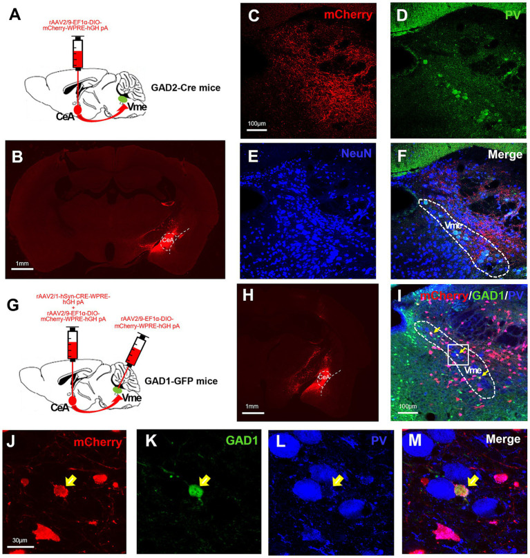
CeA is closely associated with stress-related behavior and bodily responses, while Vme participates in oral movement and proprioception. Tracing shows CeA GABAergic axons reaching Vme. Further transsynaptic labeling identifies input-recipient cells overlapping GABAergic markers rather than the larger pseudounipolar PV-positive population.
Oral movement requires information about the current state of the jaw and masticatory muscles—proprioception. Vme contributes to this processing and connects with the trigeminal motor nucleus. Following the route links stress-associated upstream signals to a brainstem network involved in regulating jaw movement.
Two Vme populations become important: smaller multipolar GABAergic interneurons provide local inhibition, while larger excitatory pseudounipolar cells relate to jaw-movement control. The upstream input reaches a local inhibitory component rather than directly targeting every movement-related cell.
Source: Zhao YJ et al. · Frontiers in cellular neuroscience · CC BY
Original figure and English caption ↗
One population becomes less excitable, the other more excitable
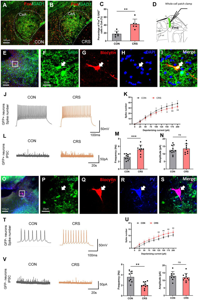
GABAergic activity markers increase in stressed-mouse CeA. In Vme slice recordings, local GABAergic interneurons produce fewer action potentials in response to matched current inputs and receive more frequent inhibitory synaptic-current events. They receive more inhibition and become less excitable.
Excitatory pseudounipolar cells show the opposite combination: more firing under matched input and fewer inhibitory-current events. Current amplitudes remain similar in both populations, with differences concentrated in frequency. This paired pattern suggests disinhibition: suppressing a local brake may allow another population to become more active.
Source: Zhao YJ et al. · Frontiers in cellular neuroscience · CC BY
Original figure and English caption ↗
Muscle activity and exploration improve together
Reducing CeA GABAergic activity increases center time and center travel in stressed mice and improves elevated-plus-maze open-arm measures. Integrated and root-mean-square masseter EMG decline concurrently, showing that this amygdala population affects both muscle and exploration responses after stress.
CeA has multiple output routes. Improvements after broader inhibition need not travel through one downstream connection. To examine the oral-movement-related component, the study next selects CeA cells by their projection to Vme.
Source: Zhao YJ et al. · Frontiers in cellular neuroscience · CC BY
Original figure and English caption ↗
Masseter activity falls while avoidance remains
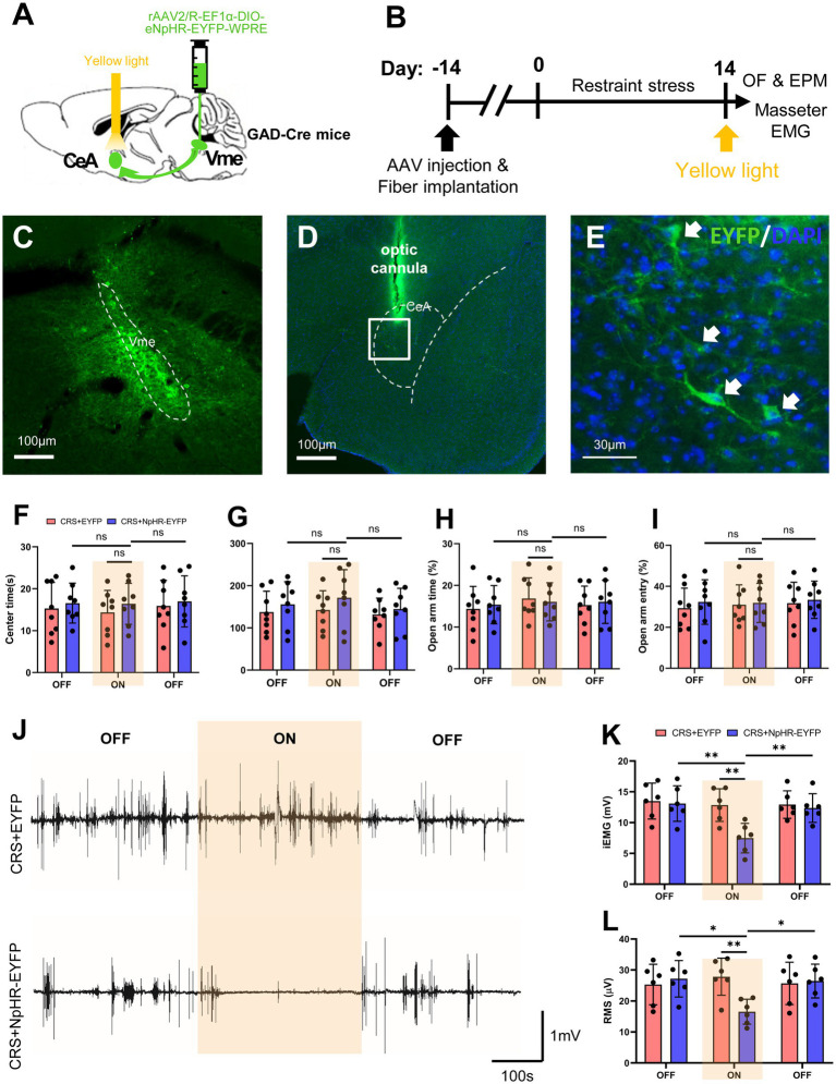
Reducing Vme-projecting CeA GABAergic activity lowers masseter EMG during the intervention, followed by an increase after it ends. This time-linked change strengthens the functional connection between the projection-defined cells and muscle overactivity.
Under the same conditions, center and open-arm measures do not clearly improve; stress-associated exposed-space avoidance remains. Reducing muscle activity and relieving anxiety-like behavior thus diverge. Vme-projecting cells contribute to masseter activity, while behavioral improvements from broader CeA inhibition involve a different pattern of participation.
Source: Zhao YJ et al. · Frontiers in cellular neuroscience · CC BY
Original figure and English caption ↗
Inhibiting inhibition may release downstream activity
GABA generally acts locally to inhibit, but the overall effect of a multistep connection depends on the target. Suppressing another inhibitory population can leave subsequent cells under less restraint. This circuit logic allows increased inhibitory input and greater muscle activity to fit within one possible explanation.
This adult male mouse study links stress-associated masseter overactivity to a defined projection population. Anatomy and electrophysiology support a possible disinhibitory account: upstream GABA input accompanies reduced local-interneuron excitability and more excitable excitatory cells. Bodily and emotion-related responses can share an upstream region yet diverge across its outputs.