How does green light encourage stressed mice to explore open spaces?
Green light during stress increases exploration of exposed spaces in mice. A visual-to-prefrontal pathway shows how excitatory input can recruit local inhibition and temper activity in another cell population.
Light entering the eye carries more than brightness and shape: it can also influence how the brain responds to stress. A mouse study published in Cell Reports in 2026 traces reduced avoidance under green light to connections between the medial secondary visual cortex (V2M) and prelimbic cortex (PrL). Its intriguing turn is that an excitatory visual input can ultimately quiet another excitatory cell population in prefrontal cortex, linking illumination, local networks and stress responses.
Reduced stress-related avoidance under green light accompanies attenuated hyperactivity of PrL glutamatergic neurons. An excitatory V2M-to-PrL projection contributes to the behavioral effect, with local inhibition and CRFR1 signaling also involved; timing matters.
Contents · 7 sections
The effect begins with timing
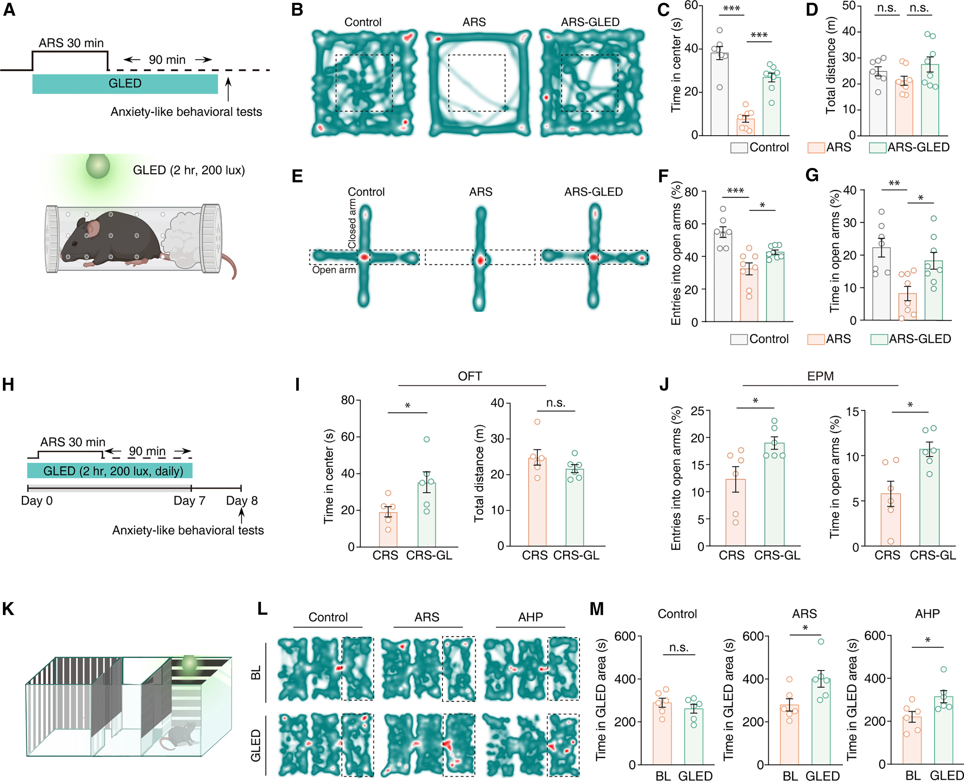
After acute restraint, mice avoid the center of an open field and the open arms of an elevated maze. Green light accompanying stress increases time spent in these exposed areas without significantly changing total distance. Increased exploration also appears during repeated restraint, arguing against a simple increase in general movement.
Timing changes the outcome. Illumination confined to recovery after restraint does not significantly improve avoidance, whereas exposure during stress does. Unstressed mice also fail to show the same preference for a green-light-paired compartment. The effect depends on the animal’s state and experience.
Source: Mingjun Zhang et al. · Cell Reports · CC BY-NC-ND 4.0; © 2026 The Author(s). Published by Elsevier Inc.
Original figure and English caption ↗
Tempering an excessive prefrontal response
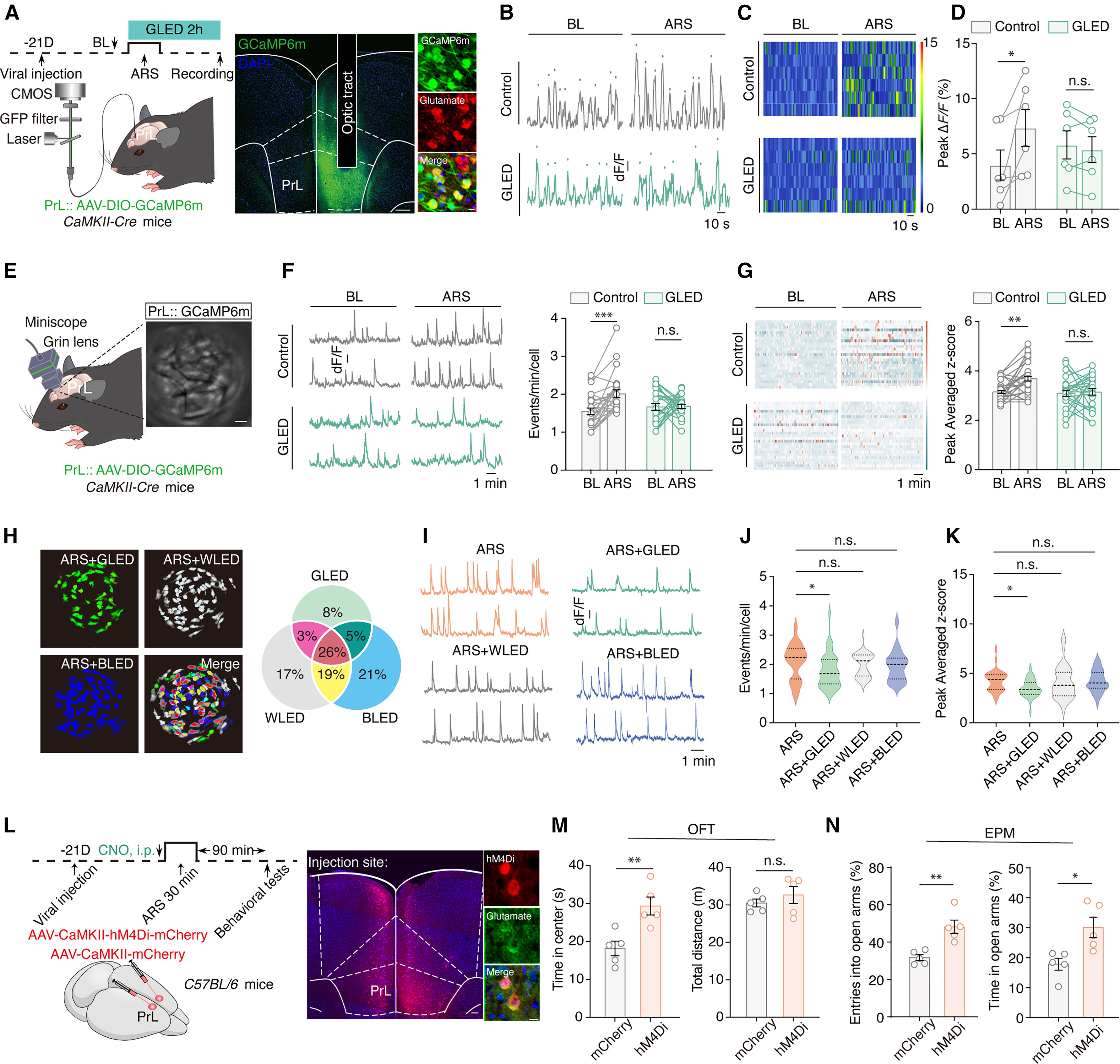
PrL glutamatergic neurons show increased calcium activity after acute stress. Green light attenuates this increase, a pattern also seen with single-cell imaging. White and blue illumination do not produce the same changes under the tested conditions. Without stress, green light has no significant effect on this population or the measured behaviors. Four hours after acute restraint, neither elevated avoidance nor hyperactivity remains detectable, placing the findings within a transient stress response.
Suppressing these neurons likewise increases center and open-arm exploration in stressed mice without significantly changing total distance. This intervention adds a causal link: tempering excessive PrL excitation can reduce stress-related avoidance.
Source: Mingjun Zhang et al. · Cell Reports · CC BY-NC-ND 4.0; © 2026 The Author(s). Published by Elsevier Inc.
Original figure and English caption ↗
A route from vision to a stress-related network
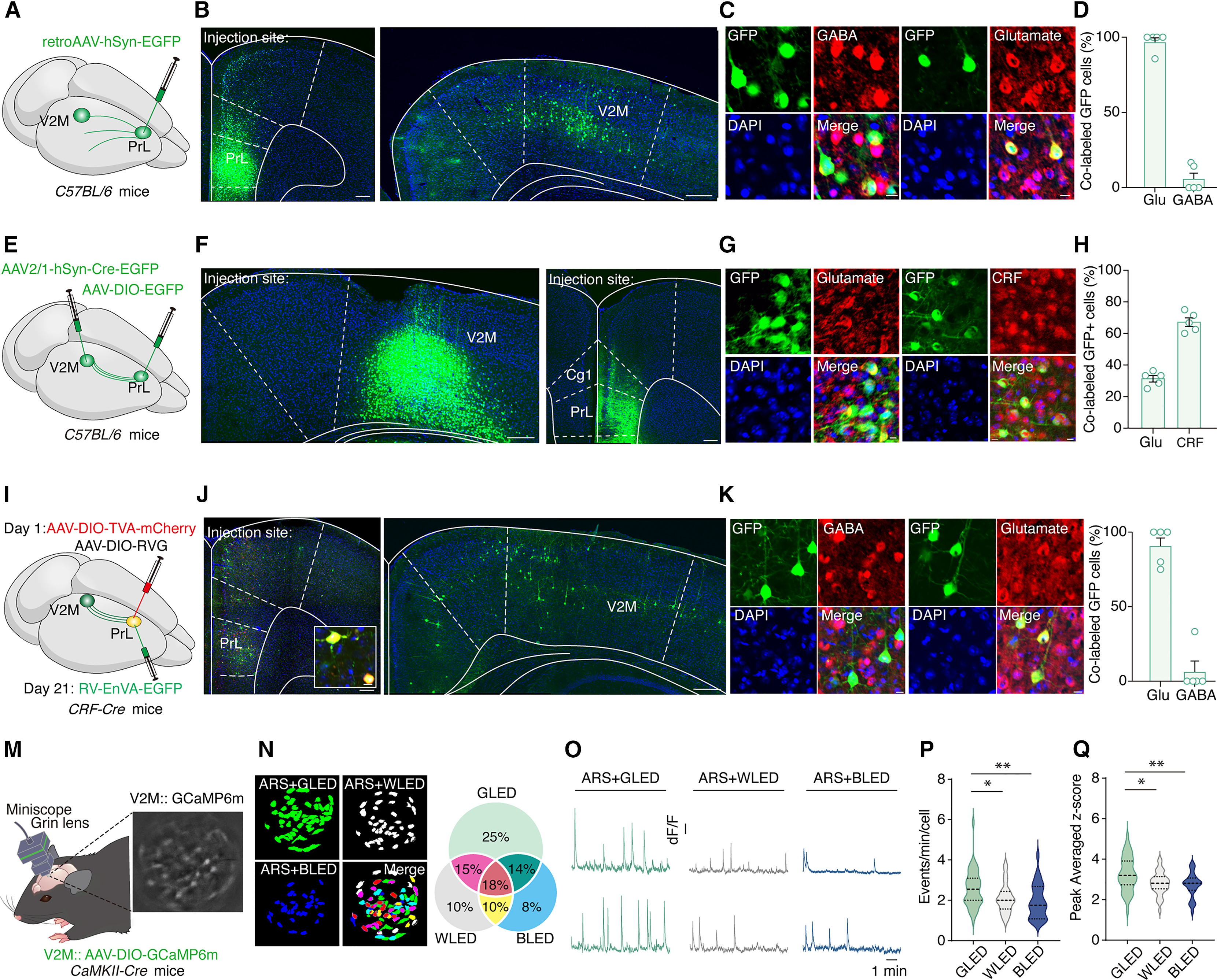
Tracing inputs to PrL identifies many projecting neurons in V2M, most bearing a glutamate marker. Additional tracing shows that both CRF-labeled and glutamatergic PrL neurons receive V2M input, providing an anatomical route for visual information to enter the local network.
V2M activity also differs across illumination conditions. Green light recruits a larger fraction of glutamatergic neurons and produces stronger calcium responses. The population is heterogeneous, with subsets differing in sensitivity to the tested light conditions.
Source: Mingjun Zhang et al. · Cell Reports · CC BY-NC-ND 4.0; © 2026 The Author(s). Published by Elsevier Inc.
Original figure and English caption ↗
How an excitatory input recruits inhibition
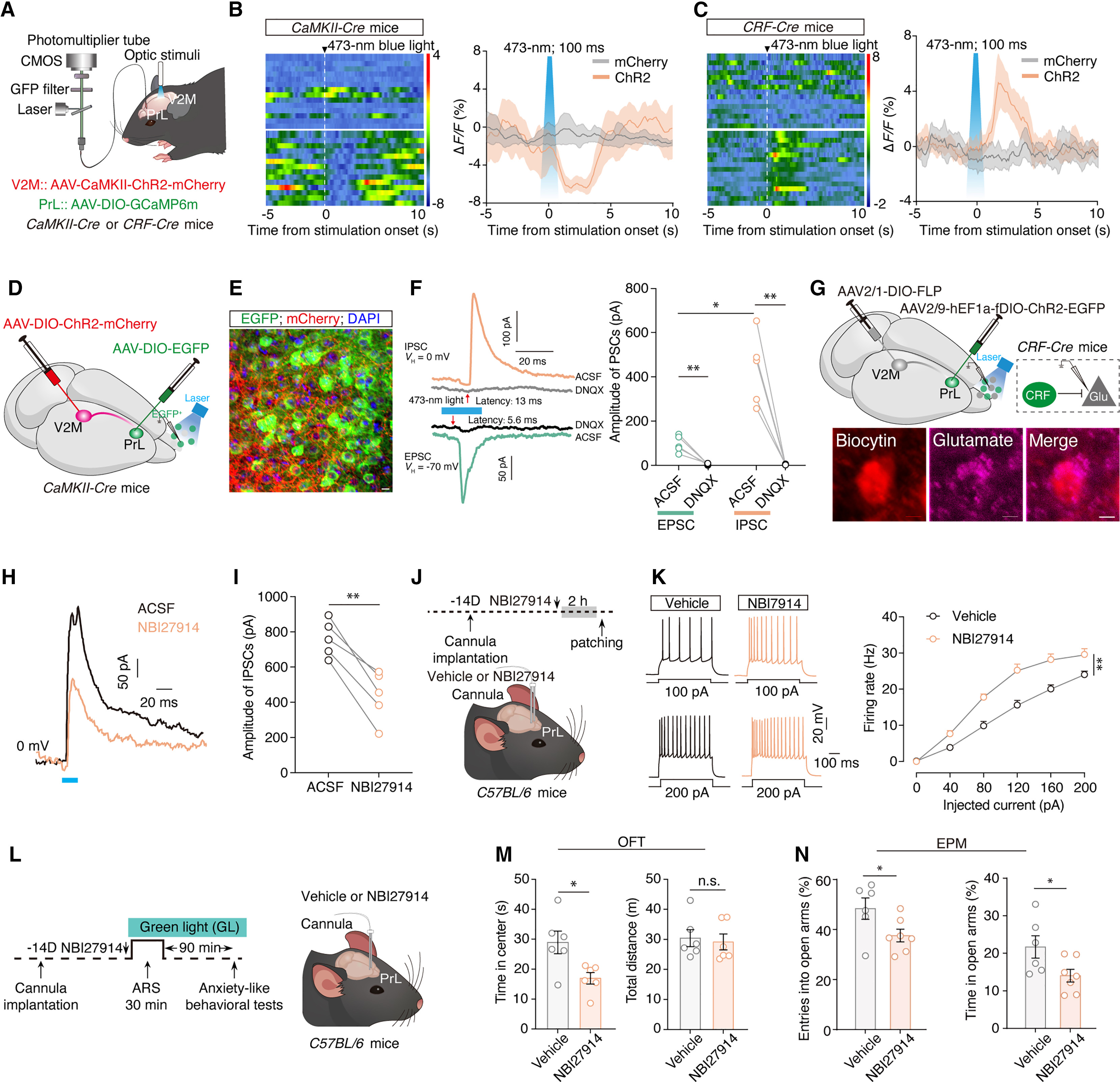
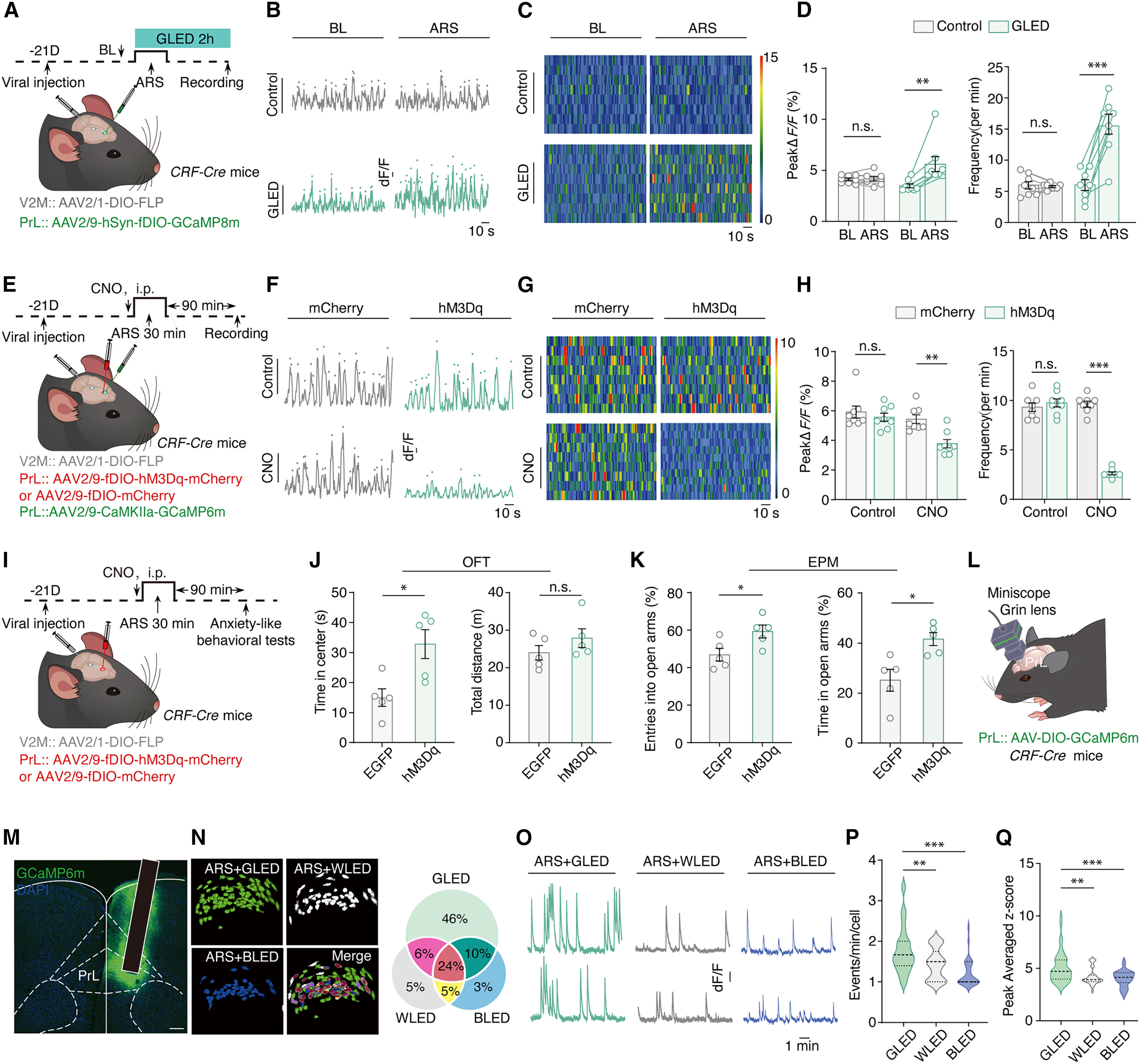
Activating V2M glutamatergic neurons increases calcium signals in locally CRF-labeled PrL cells but decreases signals in PrL glutamatergic neurons. In slices, visual input evokes both faster excitatory currents and later inhibitory currents, consistent with input recruiting a local network that then restrains another population.
CRFR1 signaling contributes to this restraint. Blocking the receptor partially reduces the relevant inhibitory current and increases firing of PrL glutamatergic neurons. During stress with green light, blockade also weakens improved exploration without significantly changing total distance. Residual inhibitory current points to additional components of local regulation.
Source: Mingjun Zhang et al. · Cell Reports · CC BY-NC-ND 4.0; © 2026 The Author(s). Published by Elsevier Inc.
Original figure and English caption ↗
Visual-input recipients can influence behavior
PrL recipient cells associated with V2M input show increased activity during stress with green light. Activating this recipient population reduces PrL glutamatergic activity and increases exposed-space exploration; inhibiting it produces changes in the opposite direction. Neither intervention significantly changes total distance.
Separate imaging of locally CRF-labeled PrL neurons finds that green light recruits more responsive cells than white or blue light. Together, input-recipient experiments and local CRF-cell imaging connect visual recruitment, local inhibition and behavioral choices.
Source: Mingjun Zhang et al. · Cell Reports · CC BY-NC-ND 4.0; © 2026 The Author(s). Published by Elsevier Inc.
Original figure and English caption ↗
A projection connects visual input to exploration
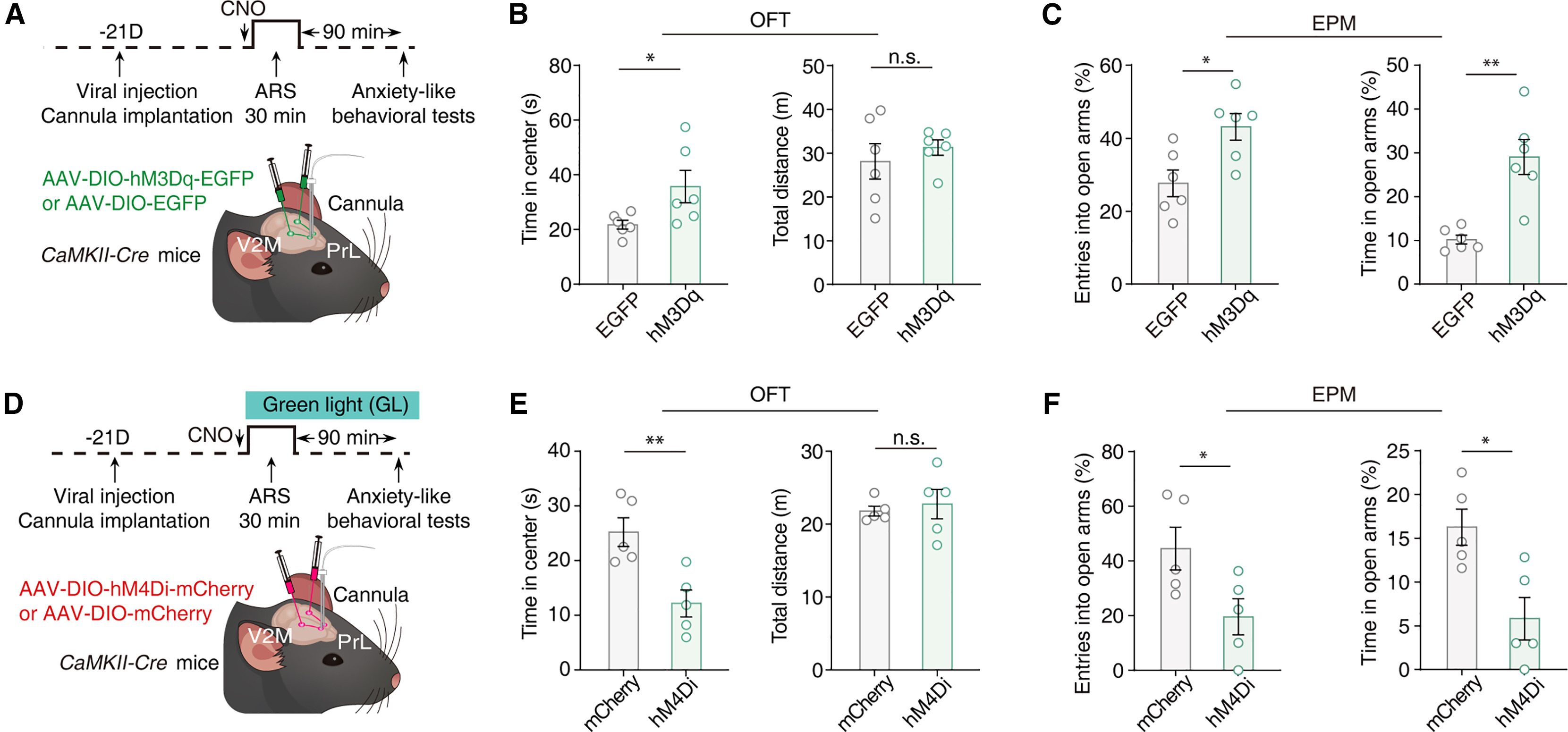
The final experiments target PrL terminals of V2M glutamatergic neurons. Increasing this input promotes exploration after acute stress, whereas reducing it weakens the behavioral improvement under green light. Neither direction significantly alters total open-field distance, providing complementary evidence for the projection’s behavioral contribution. An excitatory visual input can therefore coexist with reduced activity in a prefrontal population: the outcome also depends on which local cells it recruits.
Source: Mingjun Zhang et al. · Cell Reports · CC BY-NC-ND 4.0; © 2026 The Author(s). Published by Elsevier Inc.
Original figure and English caption ↗
Beyond color, the state of the brain matters
In adult male mice, this work shows how illumination can reshape a local network during stress. An optical question remains: the colors were matched in illuminance, but equal brightness to the human eye does not ensure equal photon flux or retinal stimulation in mice. The relationships among spectrum, delivered stimulus and animal state remain to be resolved.