How does chronic stress change the prefrontal cortex? Following the inflammatory mechanisms of compound Ciwujia granules
A rat study connects stress-related behavior, prefrontal neuronal activity and TNF-associated inflammation to examine the mechanisms of compound Ciwujia granules.
What traces does prolonged stress leave in the brain? Alongside changes in neuronal activity, an inflammatory response may reshape how animals engage with their surroundings. Following the effects of compound Ciwujia granules, researchers traced the story into the prefrontal cortex, where neurons and microglia connect stress, inflammation and behavior.
Compound Ciwujia granules improved selected behavioral and endocrine measures in stressed rats. Activity in dorsomedial prefrontal glutamatergic neurons and TNF-associated inflammation emerged as key mechanistic leads.
Contents · 6 sections
Where does stress leave its mark?
The preparation combines Acanthopanax senticosus and Schisandra chinensis. Earlier work suggested stress-related and anti-inflammatory effects, but observing behavioral improvement does not establish how it occurs. The authors ask whether defined brain regions, neuronal populations and inflammatory processes contribute.
In 2026, Tian F and colleagues reported the work in CNS Neuroscience & Therapeutics. Using a six-week chronic unpredictable mild stress model in rats, they compared compound Ciwujia granules, fluoxetine and control conditions. The investigation followed behavioral changes into brain activity and inflammatory signaling to ask why these changes occur together.
More exploration, less immobility: behavior begins to shift
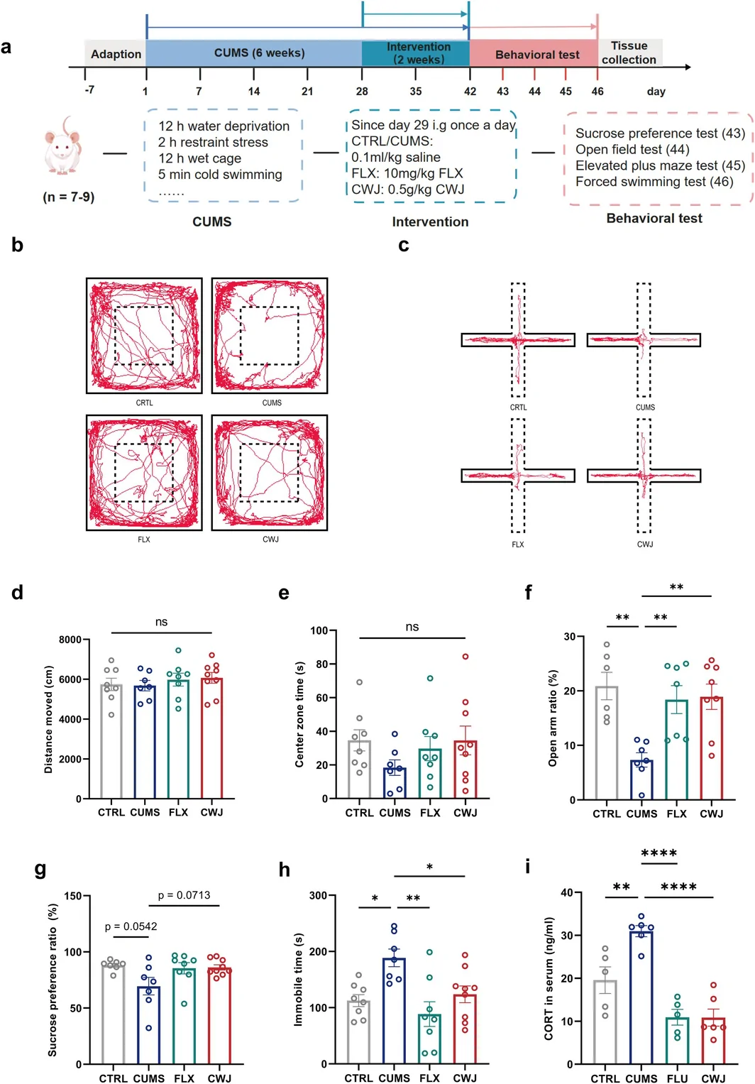
Stressed rats showed reduced open-arm exploration in the elevated plus maze and longer immobility in the forced-swim test. The compound increased the open-arm ratio, reduced immobility and lowered elevated serum corticosterone. These converging measures indicate effects on stress-related behavior and endocrine state in this model.
Sucrose preference followed a different pattern: it fell after stress and rose after treatment, but neither key pairwise comparison reached statistical significance. Exploration, immobility and corticosterone responded more clearly than sucrose preference. The different measures reveal distinct aspects of the stress response and motivate a closer look inside the brain.
Source: Tian F et al. · CNS neuroscience & therapeutics · CC BY
Original figure and English caption ↗
Following brain activity to the prefrontal cortex
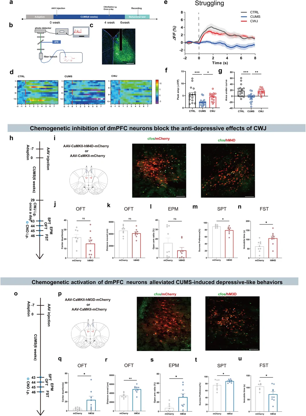
Whole-brain functional measurements mapped activity changes across several regions after treatment. Guided by these observations and earlier work, the team focused on the dorsomedial prefrontal cortex, or dmPFC. Stress reduced c-Fos activity labeling there, while treatment partly restored it. This raised a more specific question: which cells track the changes in behavior?
During struggling in the forced-swim test, dmPFC glutamatergic neurons showed event-related calcium signals. These responses were lower in stressed rats than in controls and increased with compound treatment. The regional observations were thus connected to a defined neuronal population and its activity during behavioral events.
Changing neuronal activity and watching behavior respond
To test their role, the researchers inhibited dmPFC glutamatergic neurons after compound treatment. The rats gave a differentiated behavioral response: sucrose preference decreased, while locomotor distance, open-arm ratio and immobility did not change significantly. The connection between these cells and behavior therefore varied across the measured outcomes.
Activating these neurons improved several stress-related measures and also changed locomotor distance. The difference in open-field center time was no longer significant after adjustment for distance. Neuronal activity, movement and exploration thus contributed together to the observed behavioral pattern.
Source: Tian F et al. · CNS neuroscience & therapeutics · CC BY
Original figure and English caption ↗
An inflammatory clue beside the neurons
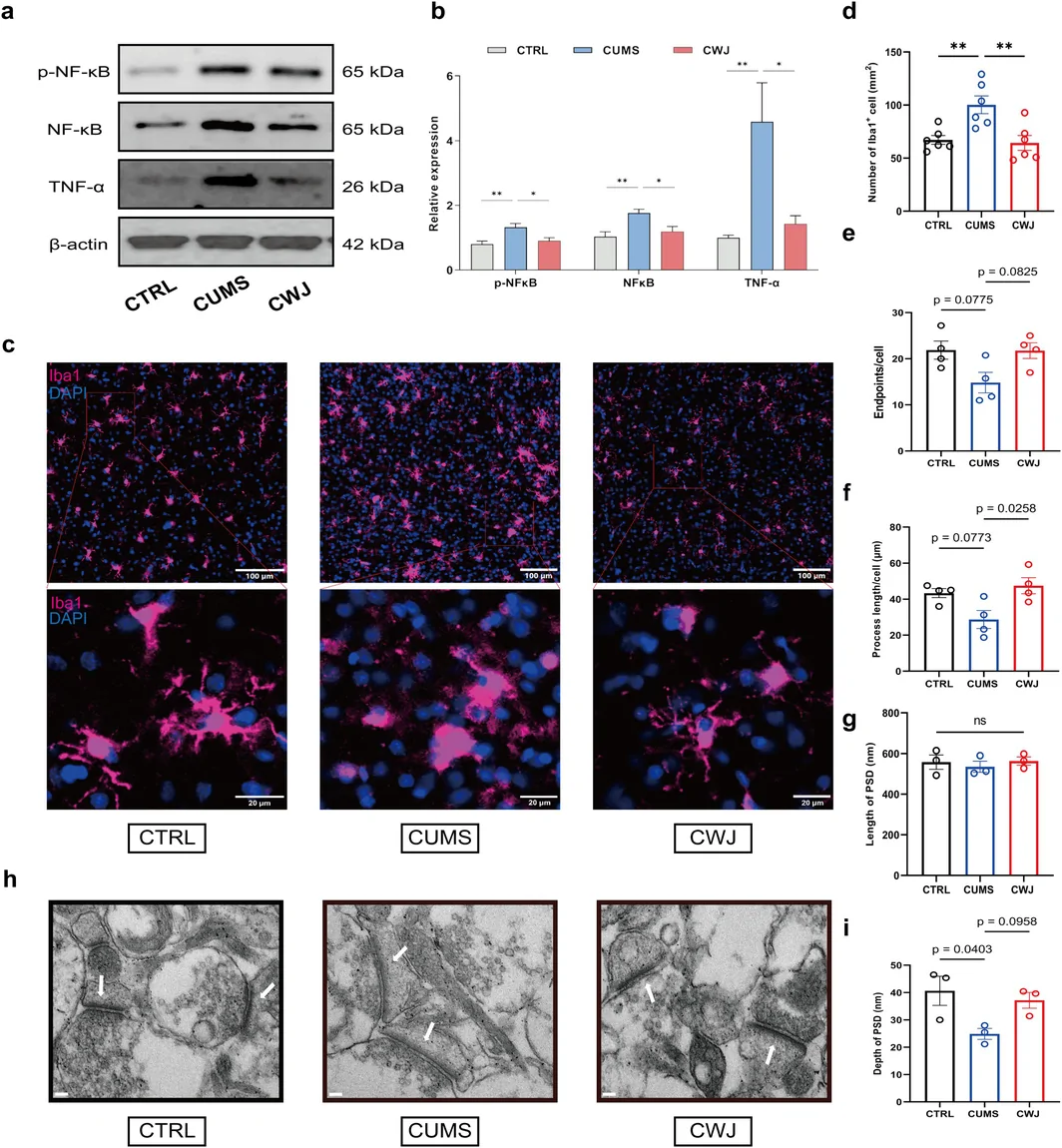
A second clue emerged at the molecular level. Transcriptomic and network-pharmacology analyses highlighted TNF-associated inflammation. Measurements then showed that stress increased TNF-α, NF-κB and phosphorylated NF-κB, while treatment reduced these changes. Selected inflammatory transcripts moved in the same direction, linking the neuronal observations to their local inflammatory environment.
Microglia showed a visible response as well. Treatment reduced the elevated number of Iba1-positive cells and lengthened processes that stress had shortened. Process-endpoint counts did not recover significantly, and synaptic ultrastructural improvement remained unclear. Images and quantitative measurements revealed changes in some inflammatory and morphological features alongside different responses in others.
Source: Tian F et al. · CNS neuroscience & therapeutics · CC BY
Original figure and English caption ↗
A prefrontal conversation opens new questions about stress
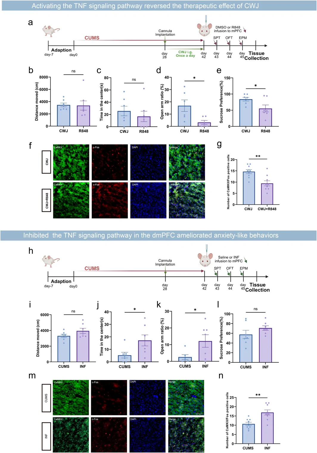
Pharmacological interventions extended the mechanism analysis. Increasing the inflammatory process partly counteracted behavioral benefits and reduced activity-related neuronal labeling. A complementary TNF-inhibition experiment improved selected stress-related measures and increased neuronal labeling. Together, the opposing interventions support a link between local inflammation and dmPFC neuronal state.
Together, the findings connect the compound intervention, prefrontal neurons and local inflammation. During prolonged stress, the immune environment and circuit state may influence one another and ultimately shape behavior. This rat study sets up further questions about the active constituents, the sequence of inflammatory and neuronal changes, and the different responses across stress-related measures.
Source: Tian F et al. · CNS neuroscience & therapeutics · CC BY
Original figure and English caption ↗