How does a context retrieve drug memory? Excitatory and inhibitory coordination in a hippocampal–prefrontal circuit
Following contextual retrieval of morphine reward memory, the study shows that hippocampal inputs shape distinct prefrontal populations rather than simply raising regional excitation.
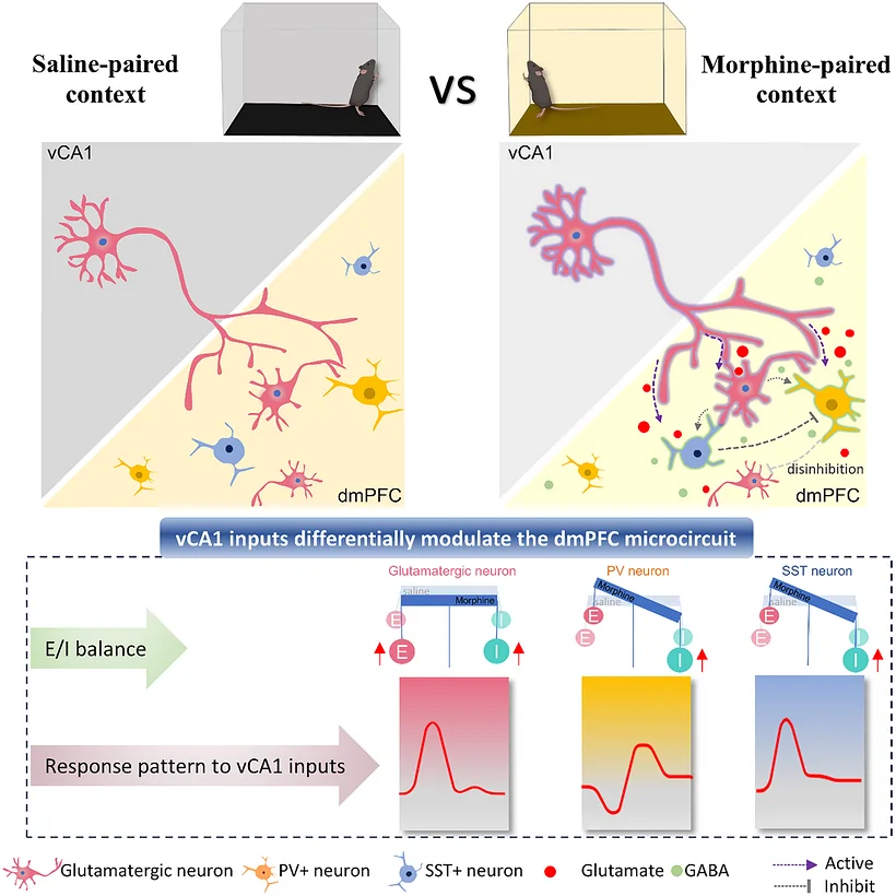
Why can a familiar place bring an earlier experience back to mind? An environment associated with a drug can evoke its memory even when the drug is absent. Following a hippocampal connection to the prefrontal cortex, researchers uncovered a richer story than the transmission of a memory signal: excitatory cells and two inhibitory populations work together to shape contextual memory expression in mice.
A glutamatergic vCA1–dmPFC projection contributes to morphine reward memory retrieval, with different roles for PV and SST inhibitory cells. Local NMDA receptor signaling links the projection to behavioral expression.
Contents · 6 sections
Which neurons become active on returning to a familiar place?
Published in Advanced Science in 2026, the study used morphine-conditioned place preference to associate an environment with drug experience. Re-exposure recruited glutamatergic activity in vCA1 and dmPFC and increased activity labeling in the relevant projection neurons.
Interestingly, a single morphine exposure did not produce the same significant calcium response in these populations. The clearer response appeared when mice returned to an environment already associated with the drug. This distinction shifted attention toward contextual recall and the interaction between the hippocampus and prefrontal cortex.
Turning down the pathway changes memory expression
Inhibiting the vCA1-to-dmPFC projection reduced preference for the drug-associated context during intervention. When inhibition stopped, the behavioral expression returned. The connection thus influenced the memory coming into play at that moment. Locomotor and object-recognition controls helped place the change alongside other aspects of behavior.
The regions also showed functional interdependence. Raising prefrontal glutamatergic activity did not restore retrieval during hippocampal inhibition, and enhancing hippocampal activity did not compensate for prefrontal inhibition. Effective expression thus depends on coordinated circuit state rather than maximal activity in either region alone.
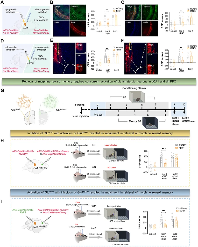
Source: Shi Z et al. · Advanced science (Weinheim, Baden-Wurttemberg, Germany) · CC BY
Original figure and English caption ↗
Two inhibitory populations play different roles
Two prefrontal inhibitory populations, PV and SST interneurons, also entered the picture. During retrieval, PV population calcium activity fell while SST activity showed no significant change. Yet activating PV or inhibiting SST reduced place preference; the opposite interventions had no significant effect. A stable population signal could therefore coexist with a functional contribution to behavior.
SST cells also formed direct inhibitory connections onto PV cells, and this inhibition strengthened after repeated morphine exposure. The authors proposed that SST-to-PV inhibition contributes to local disinhibition, changing output through an inhibitory layer. Hippocampal input and local prefrontal connections together suggest a more detailed model of memory processing.
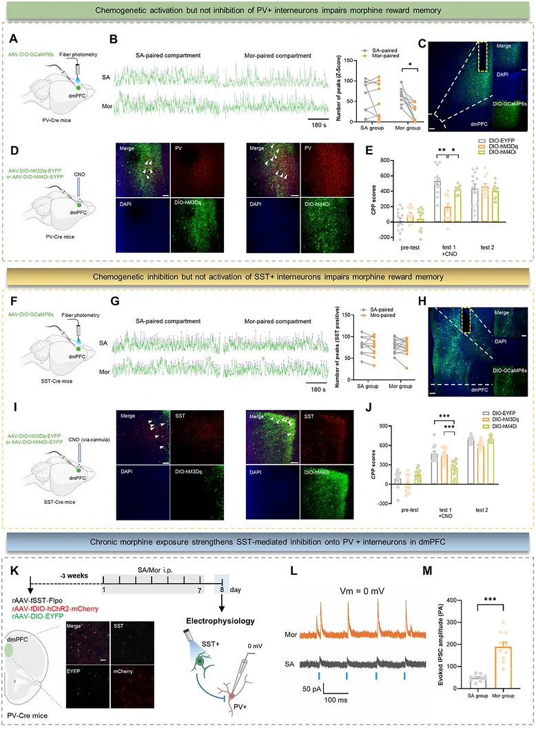
Source: Shi Z et al. · Advanced science (Weinheim, Baden-Wurttemberg, Germany) · CC BY
Original figure and English caption ↗
One hippocampal input, different activity patterns
Stimulating vCA1 terminals produced different temporal responses: sustained increases in glutamatergic cells, a brief SST response and an initial PV suppression followed by enhancement. Synaptic measurements also showed cell-type-dependent changes in excitatory–inhibitory input. Long-range input therefore reorganizes local processing rather than producing one uniform response.
Context re-exposure increased dmPFC glutamate signals, while inhibiting the hippocampal projection reduced the prefrontal calcium response. Local NMDA receptor blockade weakened memory expression during pathway activation, and an NMDA intervention partly restored behavior during projection inhibition. These observations connect transmitter signaling, receptors and behavior.
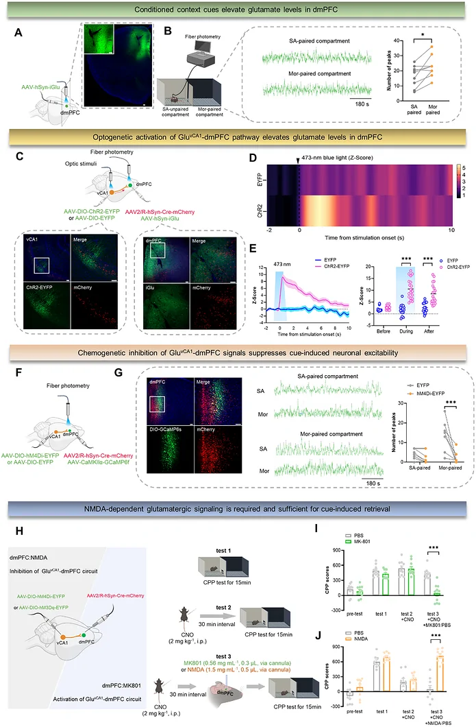
Source: Shi Z et al. · Advanced science (Weinheim, Baden-Wurttemberg, Germany) · CC BY
Original figure and English caption ↗
Over a longer timescale: withdrawal and analgesic tolerance
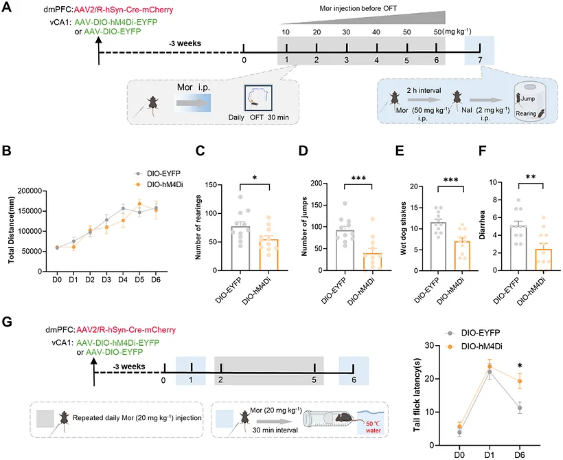
The team followed the pathway over the longer timescale of repeated morphine exposure. Sustained inhibition reduced selected physical withdrawal signs and the development of analgesic tolerance, while acute morphine analgesia was unaffected under the tested conditions. The connection thus participated in some adaptations beyond contextual memory.
Why does one projection affect memory expression and longer-term adaptation while showing a different relationship to acute analgesia? These observations open questions about how hippocampal–prefrontal communication contributes to different processes as experience accumulates, linking contextual memory with adaptation and pain modulation.
Source: Shi Z et al. · Advanced science (Weinheim, Baden-Wurttemberg, Germany) · CC BY
Original figure and English caption ↗
A memory echoes through the cooperation of cells
The study connects hippocampal contextual input with processing in distinct prefrontal populations. Different roles for PV, SST and glutamatergic cells make the broad idea of hippocampal activation of prefrontal cortex more specific: one input can produce different temporal responses and interact with local inhibitory organization.
Further work can resolve how cells coordinate during natural retrieval, which cells carry the relevant NMDA receptors, and how local changes in movement influence place preference. The study turns the moment of contextual recall into a partnership between long-range input and local excitation and inhibition, revealing a cellular division of labor in memory expression.
Source: Shi Z et al. · Advanced science (Weinheim, Baden-Wurttemberg, Germany) · CC BY
Original figure and English caption ↗