How can reduced hippocampal inhibition influence depression-related behavior?
A mouse study links 5-HT2C receptors with hippocampal GABA signaling. Receptor blockade weakens inhibitory transmission, with nNOS–CAPON interaction and vesicle-release proteins providing mechanistic clues.
Serotonin is often discussed in terms of more or less, yet different receptors can produce different cellular effects. A mouse study published online in Brain on May 3, 2024 examined 5-HT2C receptors and hippocampal excitation–inhibition balance. Receptor blockade reduced CA3 GABA signals and inhibitory-event frequency in pyramidal cells alongside changes in depression-related behavioral readouts. Calcium signaling, nNOS–CAPON interactions and vesicle-release processes supplied further molecular detail.
The relative excitation bias after 5-HT2C blockade accompanied lower GABA and inhibitory-event frequency. Disrupting nNOS–CAPON interaction improved related inhibitory transmission and behavioral readouts, linking receptor signaling with network regulation.
Contents · 7 sections
The same transmitter can act through different receptors
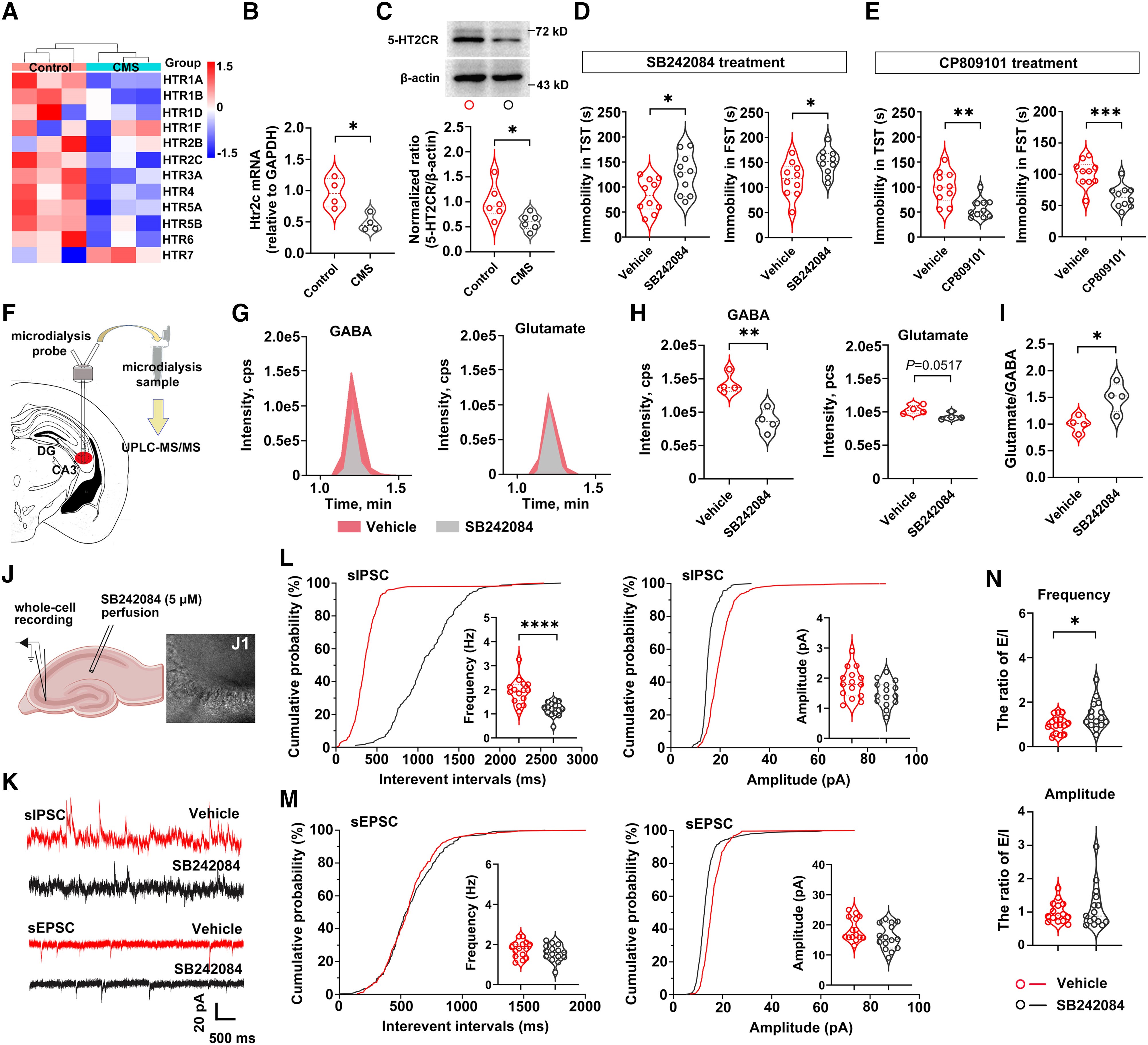
Serotonin acts through multiple receptors across cells and brain regions, and previous work on 5-HT2C and emotion-related behavior has produced differing findings. Chronic mild stress in this study lowered hippocampal receptor RNA and protein, motivating an analysis of local network effects.
Receptor antagonism increased immobility in two stress-coping tests, whereas agonism reduced it. These animal behavioral measures concern responses under particular tasks and do not directly measure clinical mood or treatment efficacy in humans.
A relative excitation bias accompanies reduced inhibition
CA3 extracellular GABA was lower after receptor blockade, while the corresponding glutamate comparison was not significant. The glutamate-to-GABA ratio increased, indicating a relative shift with the clearest change on the inhibitory side.
Pyramidal-cell spontaneous inhibitory-current frequency decreased without a significant amplitude change. Excitatory-current frequency and amplitude were not significantly altered. The excitation-to-inhibition frequency ratio changed, whereas the amplitude ratio did not, defining the measured balance shift.
Source: Hu-Jiang Shi et al. · Brain · © The Author(s) 2024. Published by Oxford University Press on behalf of the Guarantors of Brain.
Original figure and English caption ↗
Calcium and protein interactions change together
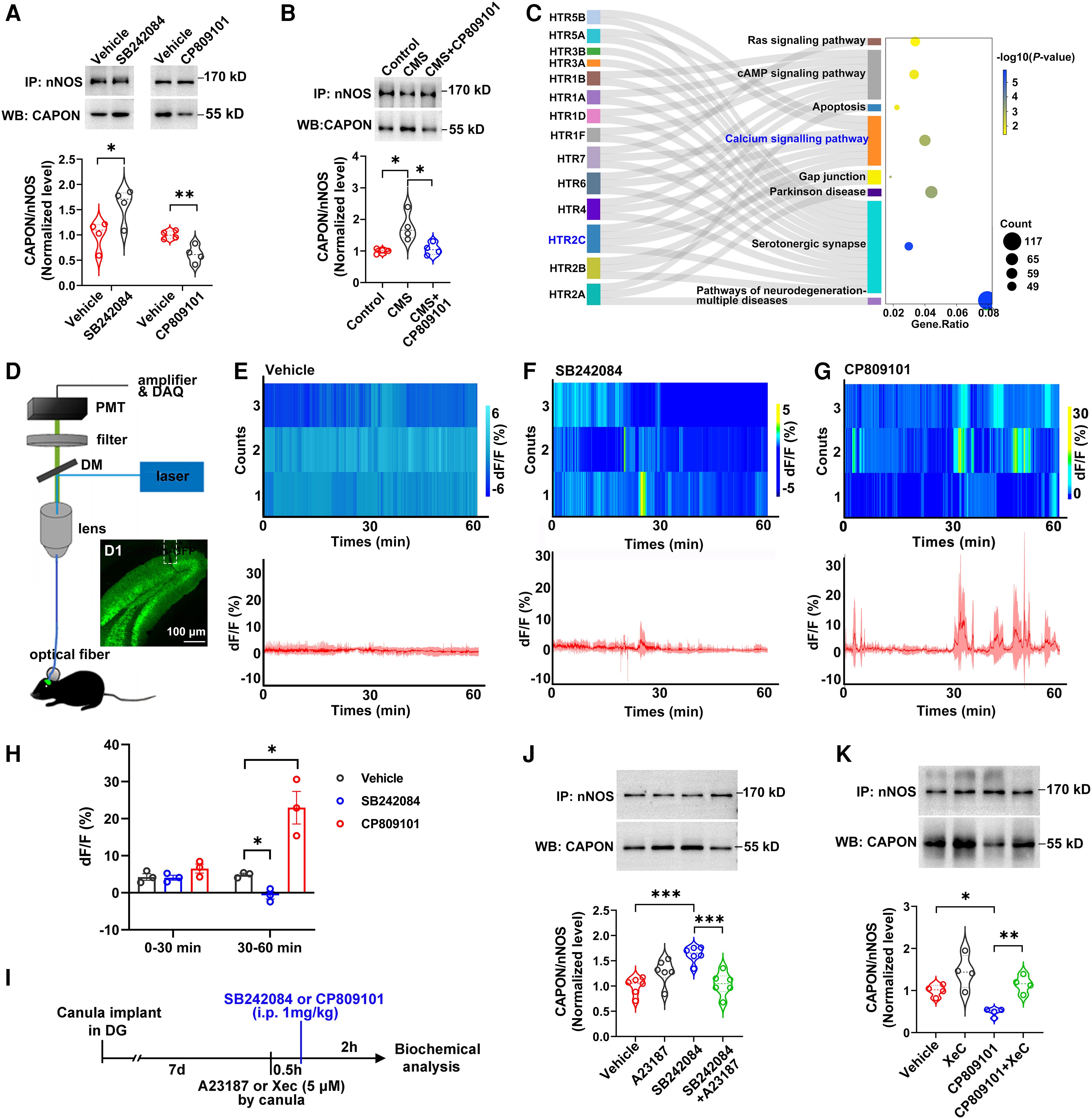
Receptor antagonism increased hippocampal nNOS–CAPON complexes, while agonism reduced their association. The analysis concerned interaction between nNOS and its partner, rather than equating total protein abundance with function.
Dentate-gyrus population calcium showed corresponding changes in the later observation window without significant early-window differences. Further calcium-related comparisons supported a role in regulating protein association, without specifying every cell’s spike count or the complete cellular source.
Source: Hu-Jiang Shi et al. · Brain · © The Author(s) 2024. Published by Oxford University Press on behalf of the Guarantors of Brain.
Original figure and English caption ↗
Disrupting the interaction improves related behavioral readouts
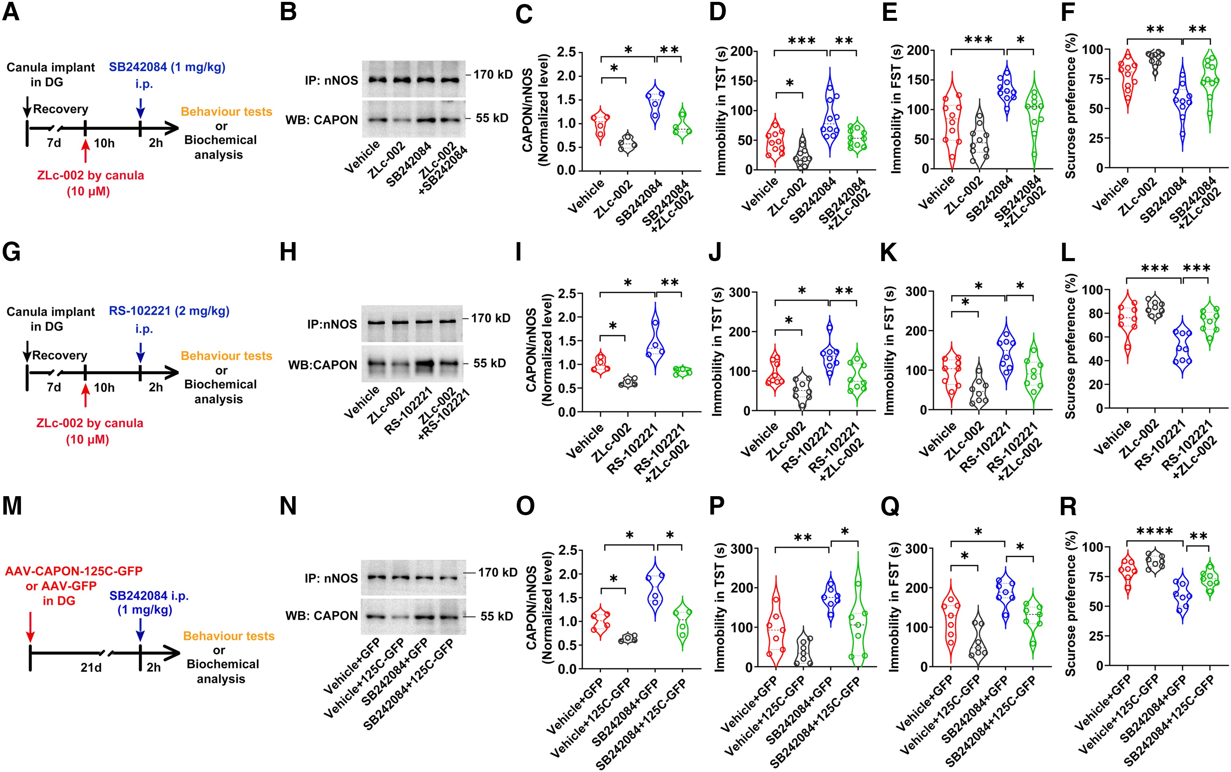
If nNOS–CAPON association merely followed receptor changes, disrupting it might not affect behavior. Different approaches to reducing the interaction lowered immobility and improved sucrose preference under receptor antagonism, with convergent comparisons across two antagonist conditions.
The findings provide a functional link between protein association and behavior in the tested pharmacological conditions. Contributions from different stress contexts, other regions and additional transmitter changes remain to be resolved.
Source: Hu-Jiang Shi et al. · Brain · © The Author(s) 2024. Published by Oxford University Press on behalf of the Guarantors of Brain.
Original figure and English caption ↗
Vesicle-release proteins provide a link to reduced inhibition
Neurotransmitter release depends on coordinated vesicle fusion involving SNARE protein complexes. Receptor blockade reduced complex assembly, while disruption of nNOS–CAPON association improved the assembly readout, providing a functional link between receptor signaling and release.
Release-related functional comparisons further supported a contribution to behavioral improvement. The hippocampal protein measurements did not isolate changes in every cell type or synapse, leaving finer cellular sources unresolved.
GABA transmission reconnects molecules with the network
Disrupting nNOS–CAPON association improved antagonist-related GABA reduction and inhibitory-event frequency, without significant corresponding excitatory-current changes. Frequency and amplitude ratios remained distinct, focusing the evidence on specific inhibitory transmission.
Reducing CA3 inhibitory-neuron activity weakened the behavioral benefit of interaction disruption, supporting local GABA-related involvement. The critical inhibitory subtypes and coordination between dentate gyrus and CA3 remain questions for further study.
An emotion-related network change can begin on the inhibitory side
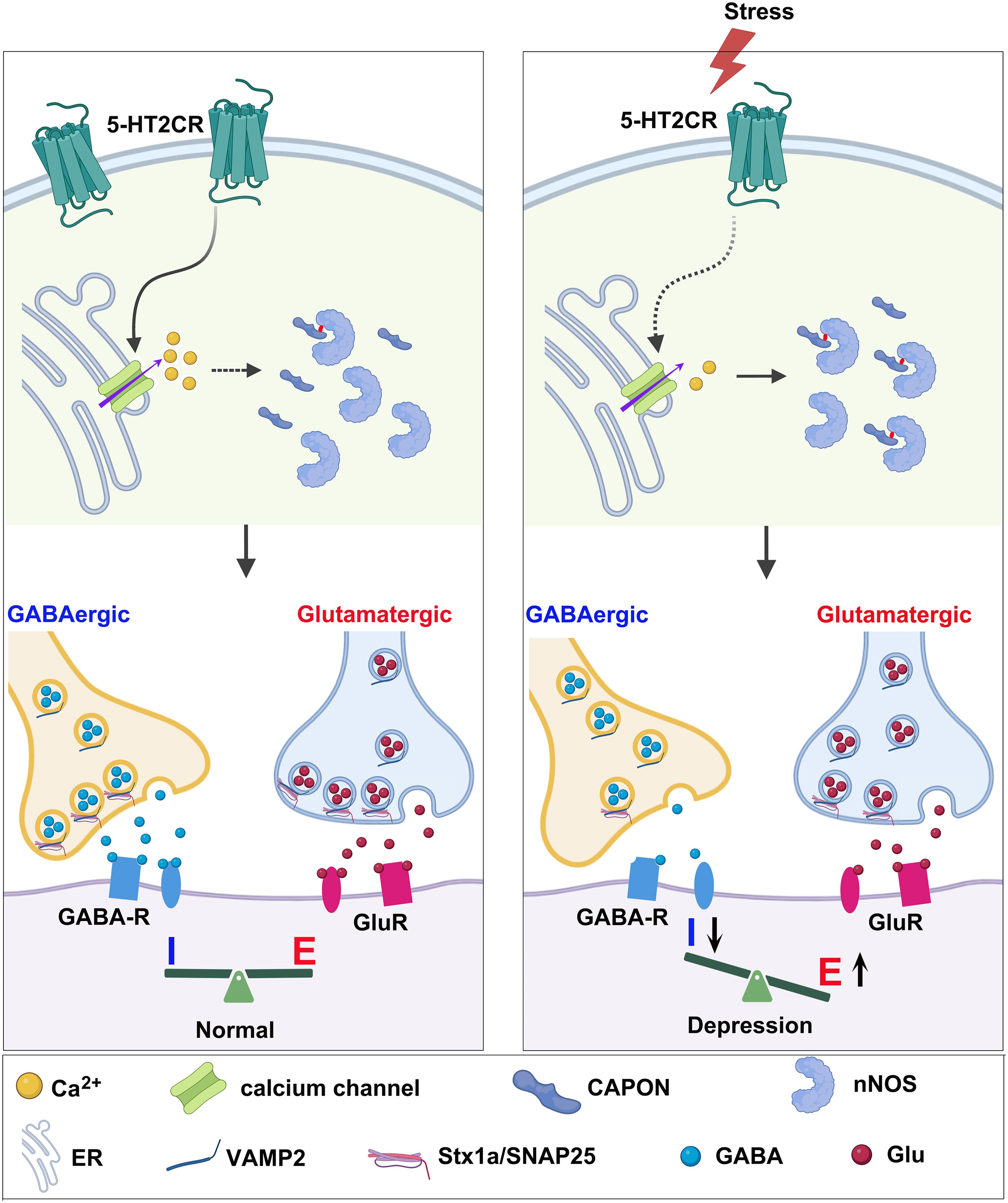
The study connects receptor signaling, calcium-related regulation, protein association, release and behavior. The authors’ schematic summarizes these comparisons, with the relative excitation bias particularly associated with weakened inhibition.
The mechanism does not encompass all depression biology. Calcium sources, cell-type contributions and other transmitters such as dopamine remain relevant questions. The findings explain depression-related behavior under specific mouse conditions, while human therapeutic relevance requires further evidence.
Source: Hu-Jiang Shi et al. · Brain · © The Author(s) 2024. Published by Oxford University Press on behalf of the Guarantors of Brain.
Original figure and English caption ↗