After stress, an amygdala inhibitory network takes a turn
In adult male mice, increasing basolateral-amygdala CCK-interneuron activity reduces anxiety-like behavior after acute stress. Slice experiments reveal that activity patterns can also strengthen their inhibitory effect.
Stress can end while changes in behavior remain. After acute stress, mice explore exposed spaces less, while activity-marker expression rises in basolateral amygdala. Could its local circuitry help quiet the network? A 2024 study in Neurobiology of Stress examines inhibitory interneurons expressing cholecystokinin (CCK). These cells suppress local network responses, and particular activity patterns strengthen that suppression. Their activation also changes behavior after stress.
Activating basolateral-amygdala CCK interneurons after stress increases time in exposed areas and reduces local c-Fos labeling without significantly changing total distance traveled. Unstressed mice show no corresponding significant behavioral change.
Contents · 7 sections
Excitatory cells have neighbors that regulate them
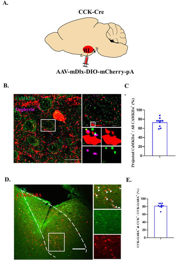
Basolateral amygdala (BLA) processes emotion-related information. Excitatory cells transmit signals outward, while inhibitory interneurons act locally. Together they shape the size of the network response to incoming signals. CCK interneurons belong to this local regulatory system.
Tissue images show CCK-cell terminals surrounding excitatory cells and apposed to an inhibitory-synapse marker. About seven in ten sampled excitatory cells show this relationship. These spatial associations motivate activating CCK cells and examining the local network response.
Source: Fang W et al. · Neurobiology of stress · CC BY-NC
Original figure and English caption ↗
Reducing the response while the network remains active
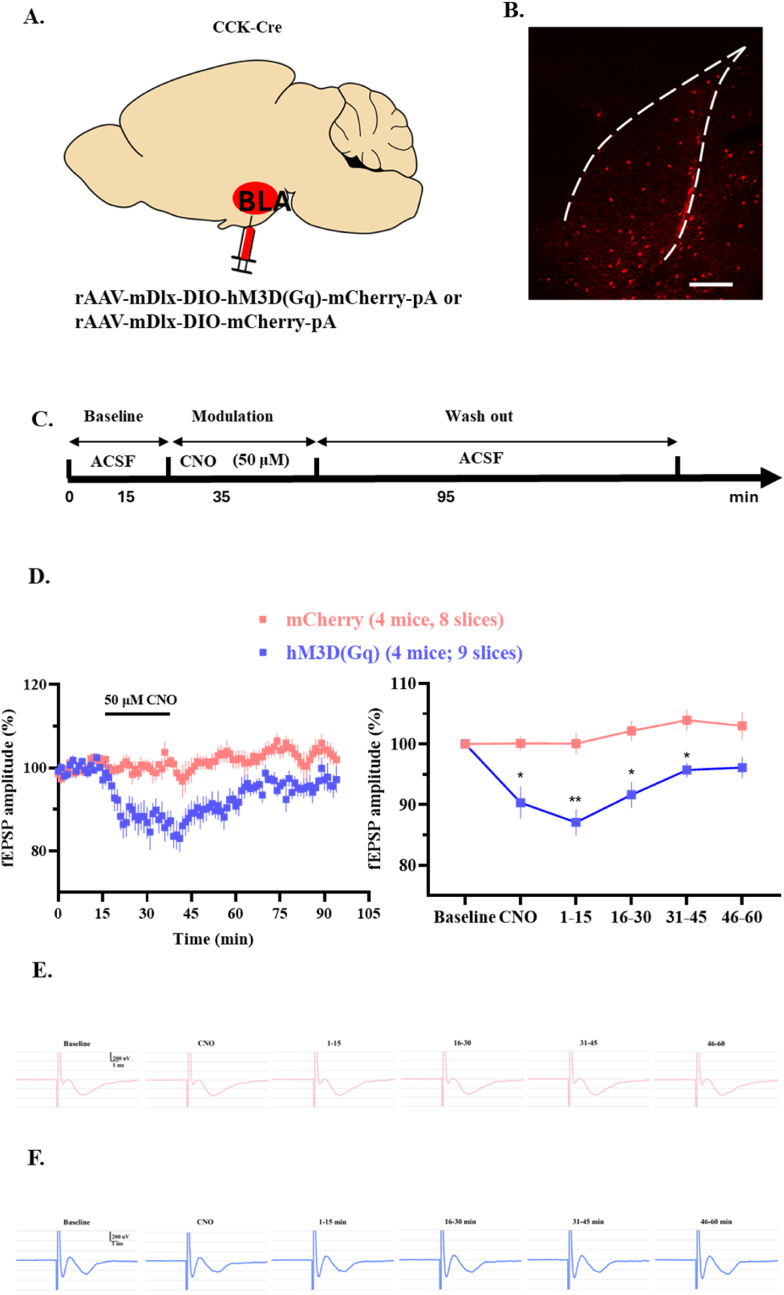
In slices, increasing CCK-interneuron activity reduces the amplitude of field excitatory postsynaptic potentials evoked by electrical stimulation. The same external input therefore produces a smaller local response. This population signal captures how local inhibition influences the network response.
The reduction is not confined to the intervention itself. Suppression persists for a period after the activating drug is washed out, then recordings approach baseline. In this slice experiment, the change in network responsiveness therefore outlasts the activating condition.
Source: Fang W et al. · Neurobiology of stress · CC BY-NC
Original figure and English caption ↗
Mice return to exposed spaces
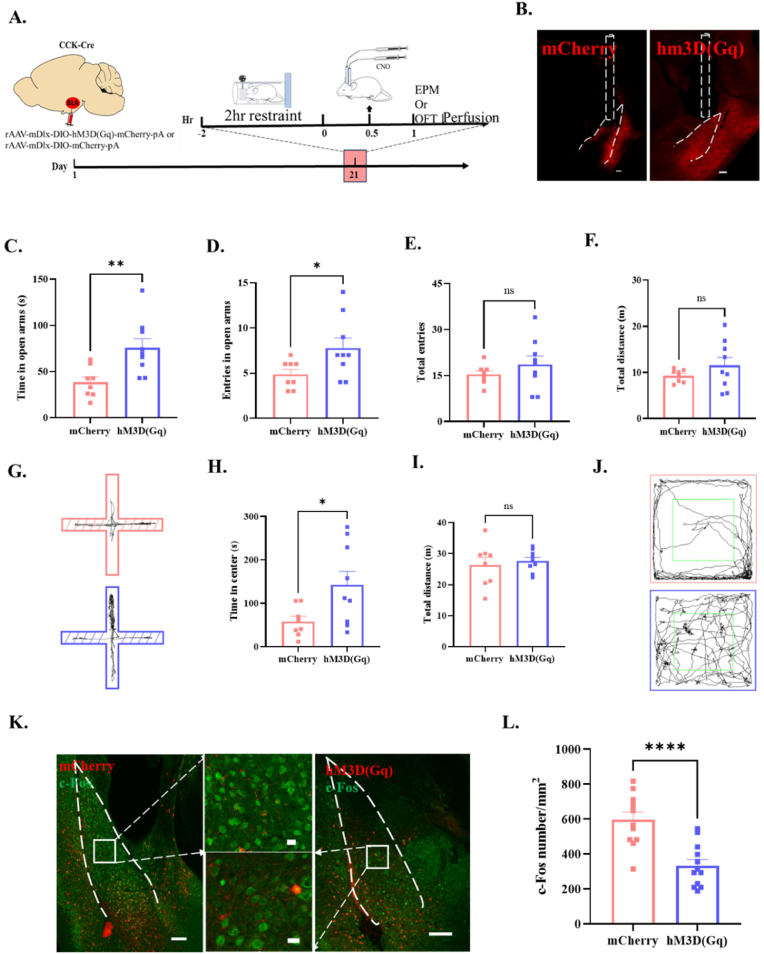
After acute restraint stress, mice spend less time in an open-field center and on elevated-maze open arms, without significantly changing total distance traveled. Exploration shifts toward more sheltered areas. Increased BLA c-Fos labeling also associates stress with a change in local activity-related signals.
Activating CCK interneurons after stress increases open-arm entries and time, as well as open-field center time, relative to matched controls. Total distance and total maze-arm entries remain statistically unchanged. BLA c-Fos labeling also declines, connecting the behavioral shift with a local activity-marker change.
Source: Fang W et al. · Neurobiology of stress · CC BY-NC
Original figure and English caption ↗
Different activity patterns leave different network changes
Can cellular activity itself strengthen suppression? After high-frequency activation in slices, recruiting CCK cells again suppresses field potentials more strongly. The study describes this persistent enhancement as inhibitory long-term potentiation. Plasticity can thus occur on the inhibitory side of the network.
Low-frequency activation does not produce the same potentiation. Both conditions initially cause comparable immediate suppression; the difference emerges after the activity patterns. The recordings distinguish the ability to inhibit the network from the ability to strengthen that inhibition.
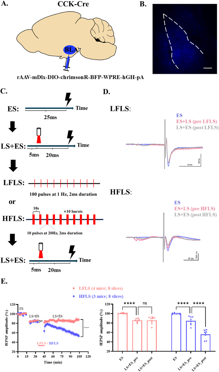
Source: Fang W et al. · Neurobiology of stress · CC BY-NC
Original figure and English caption ↗
A CCK fragment supports potentiation under low-frequency conditions
CCK is both a marker of these cells and a neuropeptide. In supplementary experiments, low-frequency activity alone does not potentiate suppression, but adding the exogenous CCK-8S fragment enables potentiation under that condition. This links local inhibitory plasticity with peptide signaling.
The team proposes that stronger cellular activity may recruit peptide signaling to potentiate inhibition. The evidence combines activity-pattern comparisons, peptide supplementation and pharmacological observations. Actual CCK release remains a proposed step in this explanation.
Exploration changes after a brief activation period ends
The team next briefly activates CCK cells after stress, allows mice to rest, and then examines behavior. High-frequency activation increases open-arm and open-field center time and open-arm entries, without significant overall movement changes. BLA c-Fos labeling declines. Behavioral differences are therefore detectable after activation ends.
Slice potentiation and post-stress behavioral improvement provide two complementary observations. The former shows a change in network responsiveness; the latter shows that this population can influence exploration after stress. Together they support a contribution of local inhibition to BLA regulation of stress responses.
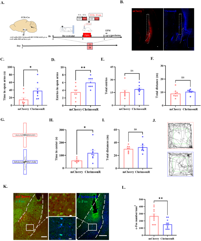
Source: Fang W et al. · Neurobiology of stress · CC BY-NC
Original figure and English caption ↗
The same activation has no matching effect without stress
In unstressed mice, neither activation approach significantly changes the tested exploratory measures or BLA c-Fos labeling. The effect is therefore state-dependent in these experiments: increasing local inhibition after stress accompanies a behavioral shift, while baseline measures do not show the same response.
This adult male mouse study gives cellular detail to the idea of quieting the amygdala: a defined population, two activation approaches and an inhibitory effect that can strengthen. Regulation of emotion-related networks depends on which cells are recruited, their activity patterns and the animal’s preceding state.