How can a small group of stress-responsive neurons tip the habenula toward depression-like behavior?
A mouse study identifies a small subset of stress-responsive neurons linking the hypothalamus and lateral habenula, tracing how local plasticity expands circuit recruitment during chronic stress.
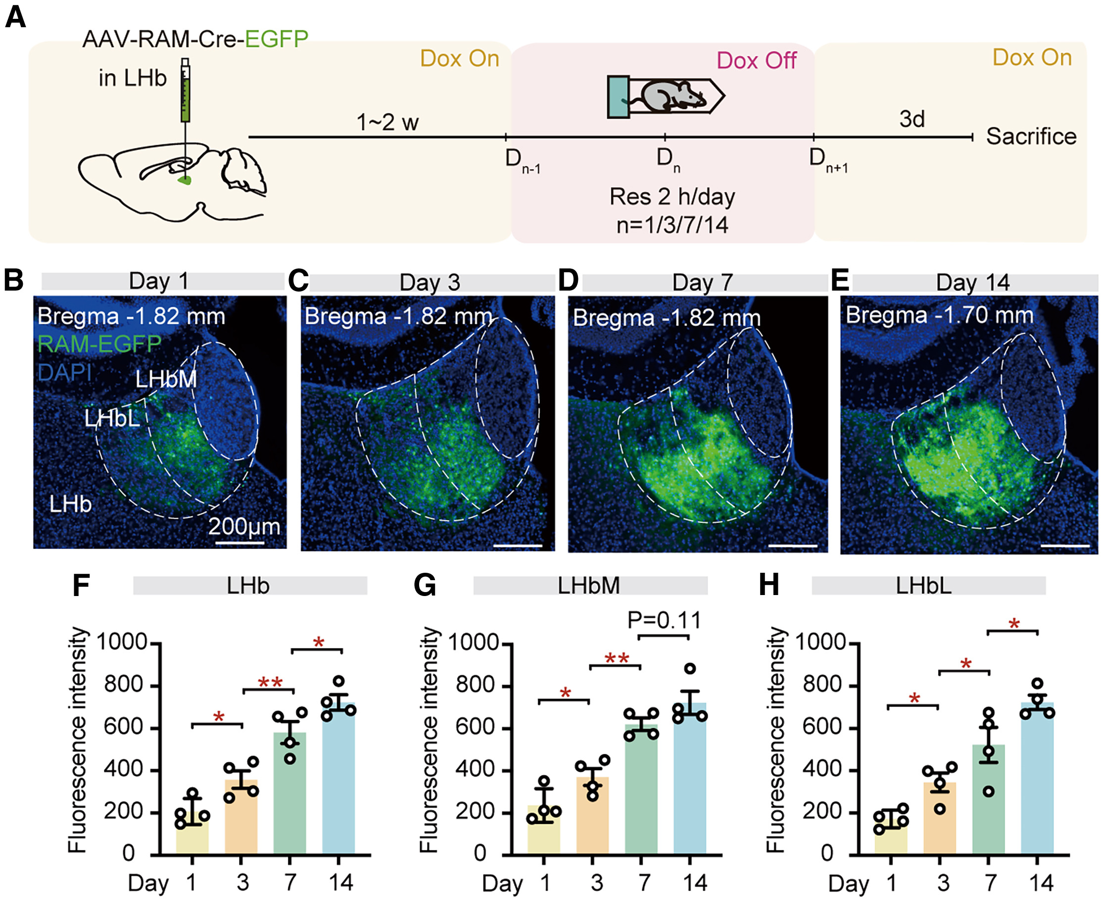
A single acute stress episode activates only a small fraction of neurons in the lateral hypothalamus (LH) and lateral habenula (LHb), yet repeated chronic stress leads to widespread LHb hyperactivity and depression-like behaviors. How this small initial ensemble of stress-responsive "starter" cells amplifies stress signals across days to remodel the entire habenular network has remained poorly understood. Published in Neuron on October 9, 2024, a study led by Dr. Yiyan Dong and Prof. Yihui Cui at Zhejiang University School of Medicine (in collaboration with Prof. Shuang Qiu; "A small population of stress-responsive neurons in the hypothalamus-habenula circuit mediates development of depression-like behavior in mice") combined Robust Activity Marking (RAM) ensemble tagging, Cre-on/Cre-off optogenetic slice electrophysiology, EPSP-burst-dependent plasticity (EBDP) induction, fiber photometry, and synaptic tracing to show that stress-responsive starter neurons in the middle LH (mLH+) preferentially innervate starter neurons in the medial LHb (LHbM+), undergo Hebbian EBDP, and recruit neighboring non-starter neurons across the medial and lateral LHb (LHbM- and LHbL-) via local intra-LHb excitatory connections during 14-day chronic restraint stress (CRS).
Acute stress recruits ~7%–8% of cells in the mLH and LHb as "starter" neurons (mLH+ and LHbM+); mLH+ inputs form preferential, high-weight synapses onto LHbM+ neurons that undergo 40-Hz phasic EPSP-burst-dependent plasticity (EBDP), which subsequently recruits surrounding LHbM- and LHbL- non-starter neurons through local intra-LHb excitatory synapses to expand the active LHb ensemble from the medial to the lateral division over 14 days of chronic restraint stress.
Contents · 5 sections
RAM Tagging Identifies mLH+ and LHbM+ Stress Starter Neurons Required for CRS-Induced Depression-like Behavior
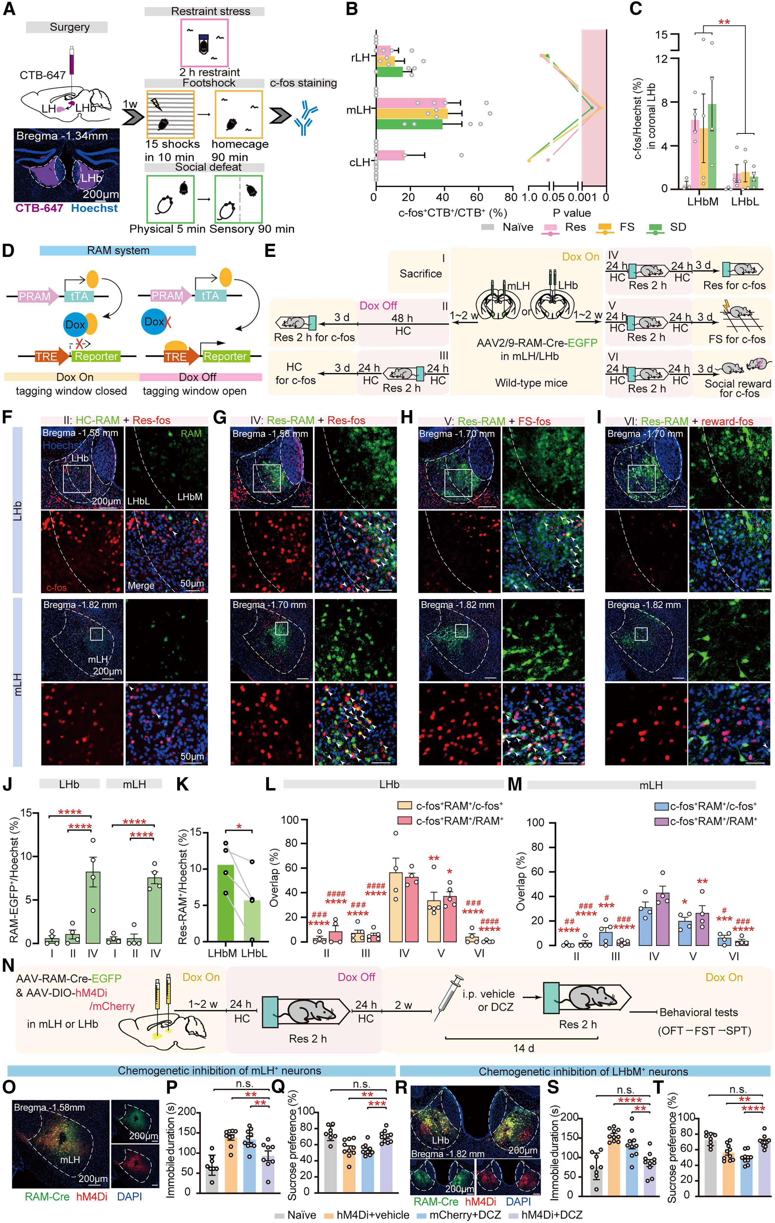
Acute restraint (Res), footshock (FS), and social defeat (SD) each activated ~5% of cells in the LH and LHb. Retrograde CTB-647 tracing from the LHb revealed that stress-activated LHb-projecting neurons were enriched in the middle LH (mLH, ~40% c-fos+CTB+/CTB+), while c-fos+ LHb neurons were concentrated in the medial division (LHbM) over the lateral division (LHbL). Using the doxycycline-off RAM system (AAV-RAM-Cre-EGFP) after a 2-h restraint episode, the authors tagged 7%–8% of total cells (~10% of NeuN+ neurons in the LHb, mainly in LHbM, and ~5% in the mLH), which showed high reactivation efficiency (LHb: 56.22% ± 11.93%; mLH: 30.91% ± 4.60%) and specificity (LHb: 52.57% ± 3.39%; mLH: 42.82% ± 5.64%) upon restraint re-exposure, strong overlap with footshock, and only baseline overlap with female social reward.
To test whether these stress-responsive "starter" cells (mLH+ and LHbM+) are required for chronic stress-induced depression-like behaviors, the authors expressed hM4Di via RAM-Cre in either mLH+ or LHbM+ neurons and administered DCZ (10 μg/kg, i.p.) prior to each daily restraint session during 14-day chronic restraint stress (CRS). Daily chemogenetic silencing of either mLH+ or LHbM+ starter cells completely prevented CRS-induced immobility in the forced swim test (FST) and anhedonia in the sucrose preference test (SPT).
Source: Zhiwei Zheng et al. · Neuron · © Elsevier Inc.
Original figure and English caption ↗
mLH+ Starter Neurons Form Preferential, High-Weight Excitatory Synapses Onto Postsynaptic LHbM+ Starter Neurons
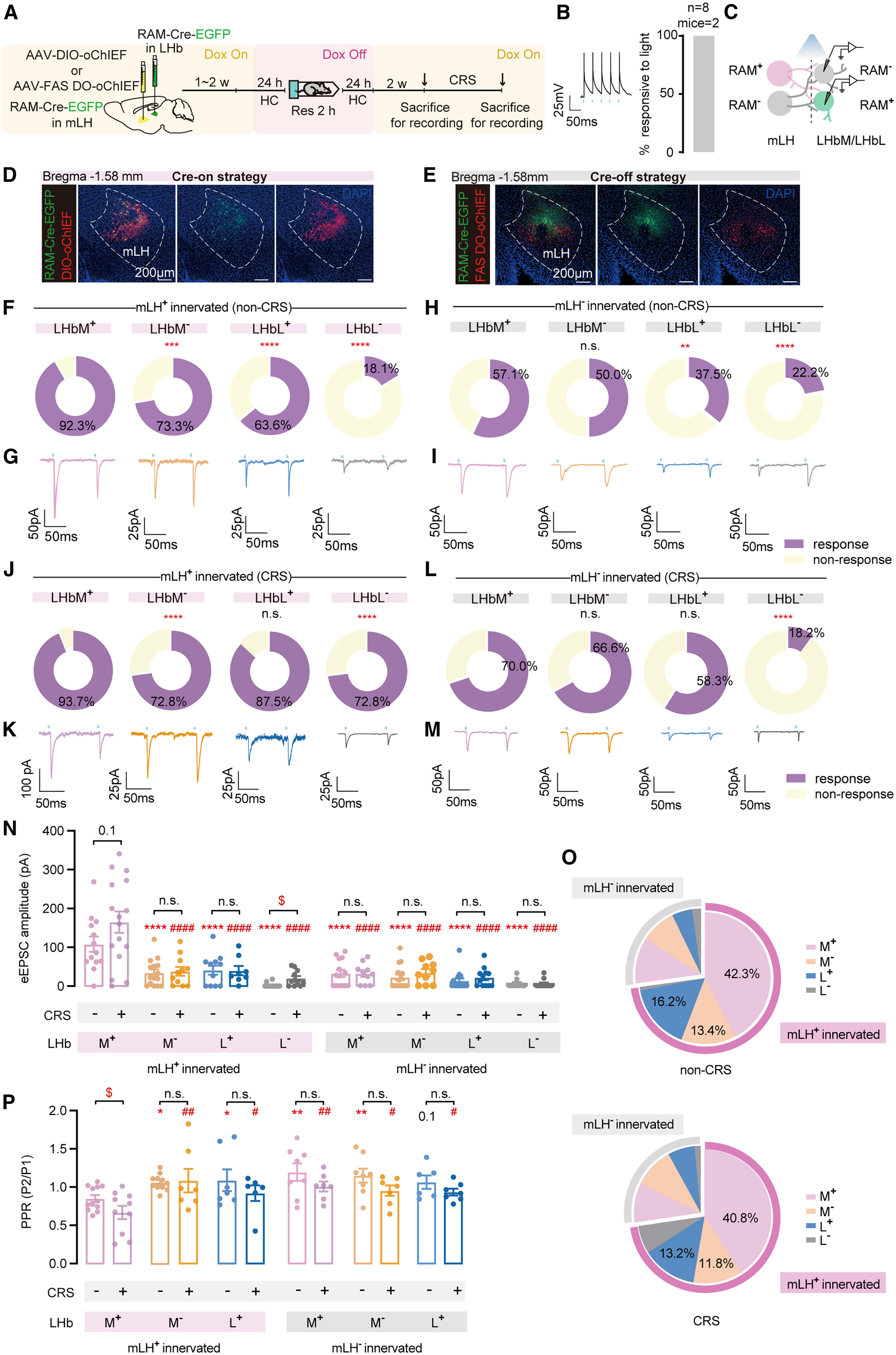
To dissect the functional connectivity between mLH starter/non-starter inputs (mLH+ vs mLH-) and four postsynaptic LHb subpopulations (LHbM+, LHbM-, LHbL+, and LHbL-), the authors combined RAM tagging with Cre-on (DIO-oChIEF) or Cre-off (FAS DO-oChIEF) optogenetic slice recordings. Under non-CRS conditions, photostimulation of mLH+ terminals evoked excitatory postsynaptic currents (eEPSCs) in 92.3% of LHbM+ neurons (compared with 73.3% in LHbM-, 63.6% in LHbL+, and 18.1% in LHbL-), whereas mLH- inputs showed substantially weaker responsiveness across all four groups (57.1%, 50.0%, 37.5%, and 22.2%).
Moreover, the mLH+→LHbM+ subcircuit exhibited the largest eEPSC amplitude, accounted for the predominant share of total synaptic weight, and displayed lower paired-pulse ratios (PPRs, indicating higher presynaptic release probability). Following 14-day CRS, mLH+→LHbM+ synapses retained the largest eEPSC amplitude and synaptic weight and selectively exhibited a significant further reduction in PPR.
Source: Zhiwei Zheng et al. · Neuron · © Elsevier Inc.
Original figure and English caption ↗
40-Hz Phasic Stimulation Selectively Induces EPSP-Burst-Dependent Plasticity at mLH+→LHbM+ Synapses and Drives Depression-like Behavior
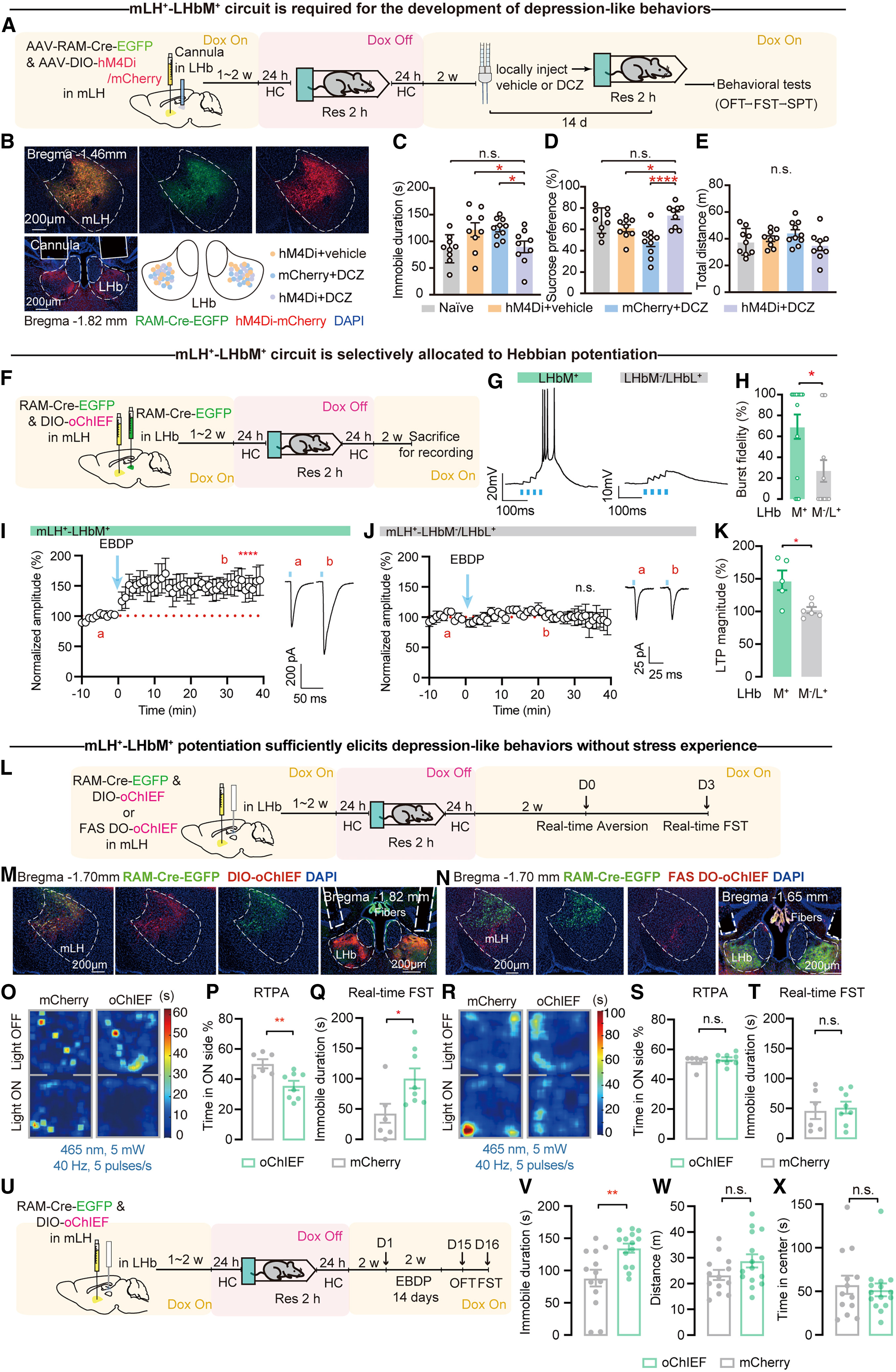
Local intra-LHb microinfusion of DCZ to silence hM4Di-expressing mLH+ terminals during daily restraint sessions of 14-day CRS prevented FST immobility and SPT anhedonia without altering open-field locomotion (whereas silencing mLH- terminals in Figure S3 had no protective effect). In acute brain slices, delivering 40-Hz phasic photostimulation (4 pulses/s) to mLH+ terminals reliably evoked EPSP-burst pairings and induced robust Hebbian EPSP-burst-dependent potentiation (EBDP) selectively in postsynaptic LHbM+ neurons, but not in LHbM- or LHbL+ neurons.
In stress-naive mice, in vivo 40-Hz phasic photostimulation of the mLH+→LHb pathway—but not the mLH-→LHb pathway—acutely drove real-time place aversion (RTPA) and increased immobility during real-time FST, and 14 days of chronic daily EBDP photostimulation produced persistent FST immobility measured 2 days after stimulation ceased.
Source: Zhiwei Zheng et al. · Neuron · © Elsevier Inc.
Original figure and English caption ↗
LHbM+ Starter Neurons Recruit Neighboring LHbM- and LHbL- Neurons via Local Intra-LHb Excitatory Connections
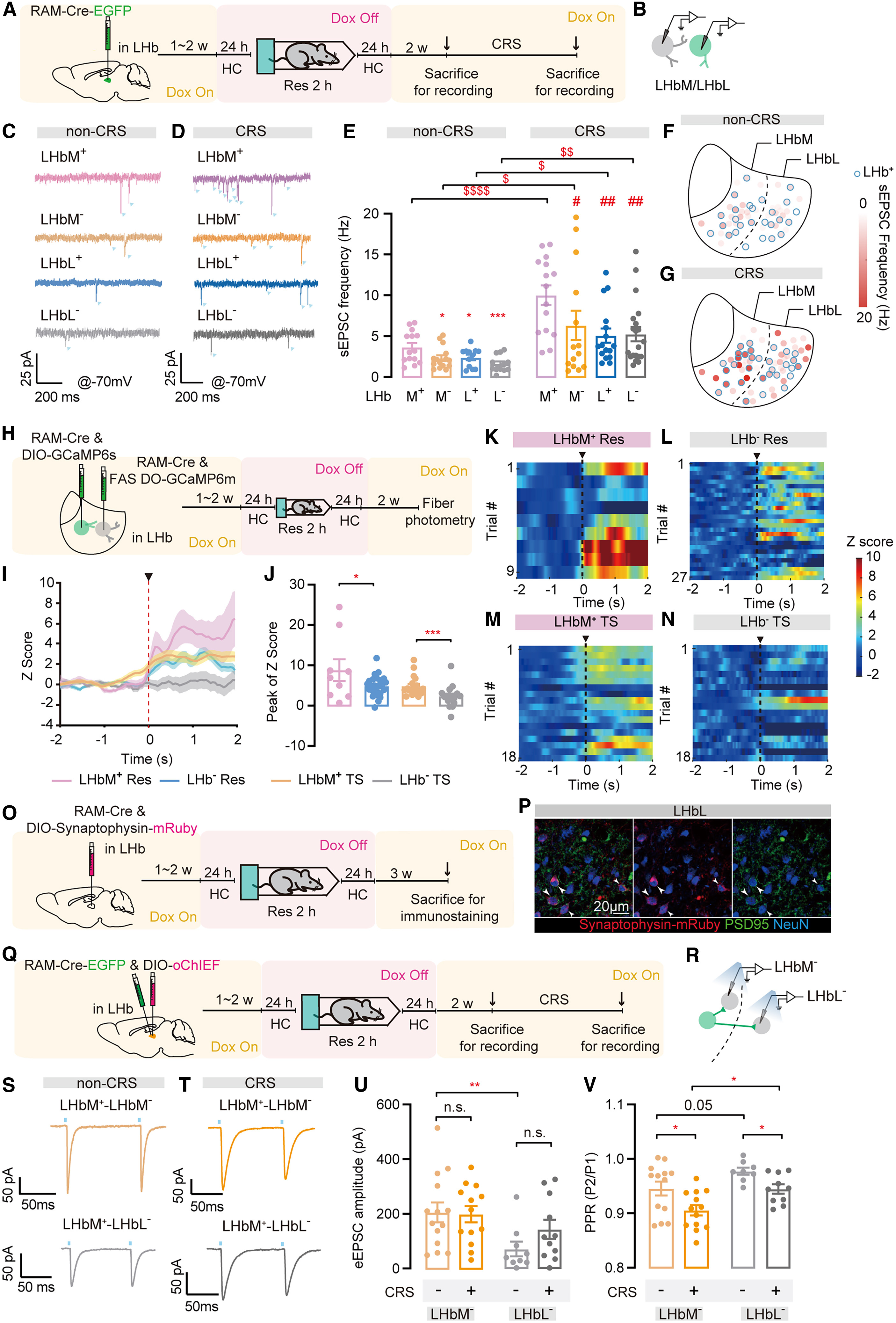
Under non-CRS baseline conditions, LHbM+ starter cells exhibited the highest spontaneous EPSC (sEPSC) frequency among all four LHb subpopulations and showed stronger in vivo GCaMP6s calcium responses to restraint (Res) and tail suspension (TS) than non-starter (LHb-) neurons. After 14-day CRS, however, sEPSC frequency increased 2- to 3-fold across all four LHb subpopulations (LHbM+, LHbM-, LHbL+, and LHbL-), accompanied by heightened in vivo stress-evoked calcium responses in LHb- neurons.
Expressing Cre-on synaptophysin-mRuby in LHbM+ starter neurons revealed abundant presynaptic puncta colocalized with PSD95 and NeuN in the lateral LHb (LHbL). Whole-cell slice recordings with Cre-on oChIEF in LHbM+ neurons demonstrated direct local intra-LHb monosynaptic excitatory connections from LHbM+ starter cells onto both LHbM- (3.5-fold larger basal eEPSC amplitude) and LHbL- non-starter neurons, and both local pathways (LHbM+→LHbM- and LHbM+→LHbL-) exhibited significantly decreased PPRs after 14-day CRS.
Source: Zhiwei Zheng et al. · Neuron · © Elsevier Inc.
Original figure and English caption ↗
Stage-Specific RAM Labeling Reveals Progressive Spatial Expansion of Recruited LHb Neurons From LHbM to LHbL Across Chronic Stress
Finally, to directly visualize how the stress-recruited LHb ensemble evolves over the course of chronic stress, the authors performed stage-specific RAM labeling (AAV-RAM-Cre-EGFP in the LHb) at Day 1, Day 3, Day 7, or Day 14 of the 14-day CRS paradigm.
Quantitative mapping demonstrated that recruited LHb+ fluorescence progressively increased across CRS and spread spatially from the medial division (LHbM, where recruitment rose steeply from Day 1 to Day 7) into the lateral division (LHbL, which expanded markedly through Day 14). Together, these findings establish a two-stage circuit mechanism whereby preferential mLH+→LHbM+ connectivity and EBDP plasticity initiate stress encoding in medial starter cells, which then progressively recruit lateral LHb neurons via local intra-LHb excitatory connections to drive depression-like behavior.
Source: Zhiwei Zheng et al. · Neuron · © Elsevier Inc.
Original figure and English caption ↗