How a sugar modification on a protein buffers dopamine changes under stress
A male-mouse study identifies a molecular tug of war between glycosylation and STAT3 phosphorylation in VTA dopamine cells, shaping exploration and reward-related behavior after acute stress.
Why can a brief stressful episode reduce exploration and the appeal of normally attractive experiences? A study published on July 21, 2025 in Advanced Science follows this question into dopamine cells of the ventral tegmental area (VTA). Acute restraint changes chemical marks on STAT3, alongside reduced neuronal activity and accumbal dopamine signals. At the same time, a sugar modification on the protein increases. These opposing changes help shape how mice subsequently approach exposed spaces, unfamiliar conspecifics, and environments associated with sweet rewards.
Enhancing O-GlcNAc glycosylation suppresses stress-induced STAT3 Ser727 phosphorylation and buffers associated behavioral changes; mimicking phosphorylation at that site weakens the buffering effect.
Contents · 7 sections
One stressor activates two kinds of modification
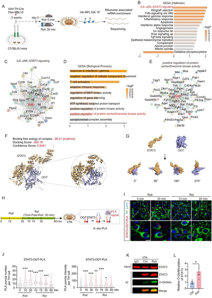
Dopamine cells receive electrical signals from other brain regions, while their internal machinery also adjusts to the environment. Examining gene activity after stress highlights signaling associated with STAT3. Because STAT3 can regulate gene expression, it offers a route from a brief stressful event to changes inside the cell.
An intriguing pairing then emerges: proximity signals between STAT3 and OGT, the enzyme that adds O-GlcNAc, increase under stress, as does STAT3 glycosylation. This sugar is a chemical modification of a protein that alters its molecular state, distinct from the sweet reward experienced when an animal consumes sugar.
Source: Shao M et al. · Advanced science (Weinheim, Baden-Wurttemberg, Germany) · CC BY
Original figure and English caption ↗
Buffering targets one phosphorylation site
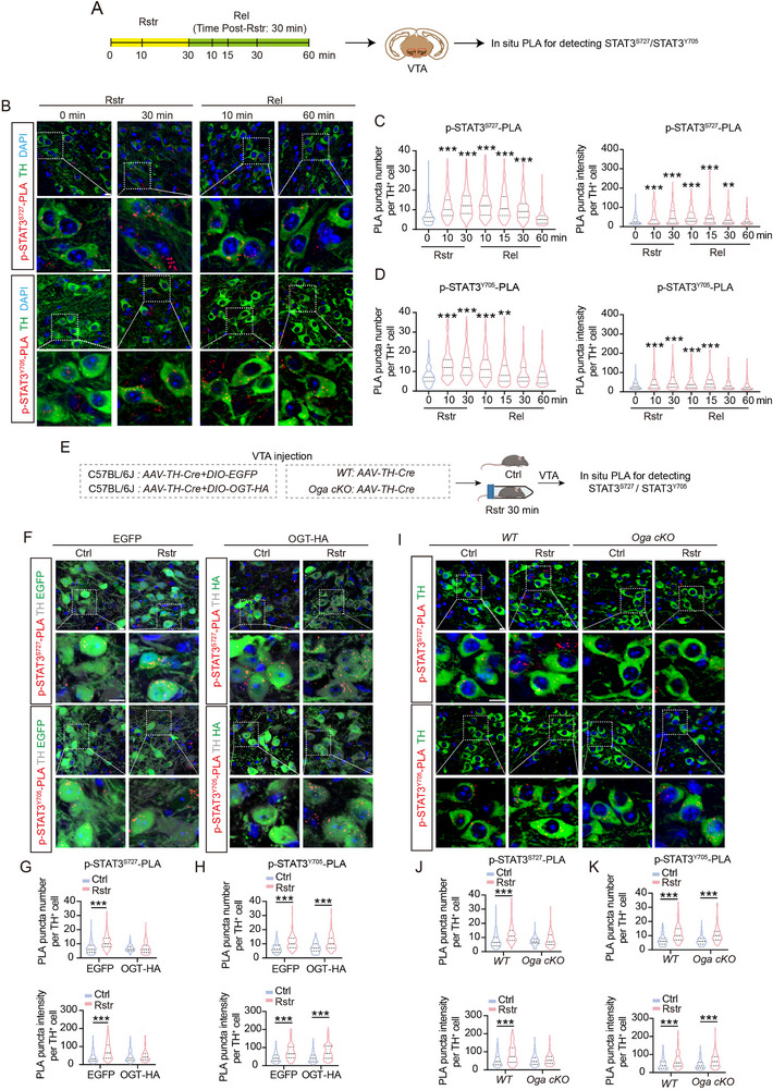
STAT3 also carries another important class of mark: phosphorylation. Under stress, phosphorylation at Ser727 and Tyr705 rises and subsequently returns toward baseline after release. Total protein abundance does not rise alongside these changes; the state of existing molecules is changing.
Increasing OGT or reducing OGA, the enzyme that removes the sugar mark, enhances glycosylation and prevents the stress-induced increase at Ser727. The Tyr705 response remains. Glycosylation therefore counterbalances a particular STAT3 site rather than shutting down the entire stress response.
Source: Shao M et al. · Advanced science (Weinheim, Baden-Wurttemberg, Germany) · CC BY
Original figure and English caption ↗
The dopamine decrease depends on STAT3
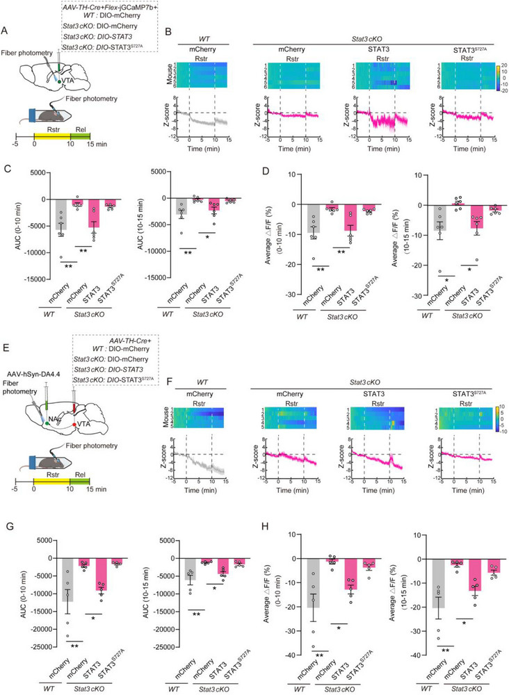
How does this molecular tug of war reach neural activity? In control mice, VTA dopamine-cell calcium signals fall during restraint, and dopamine signals detected in the nucleus accumbens also decrease, remaining depressed for a period after release. Removing STAT3 from these cells prevents the corresponding stress-induced decreases.
Restoring normal STAT3 brings the decreases back, whereas restoring a version that cannot be phosphorylated at Ser727 does not. Natural responses during sucrose licking or conspecific sniffing remain broadly similar between controls and the deletion group: loss of STAT3 does not abolish all dopamine-cell responses to rewarding experiences.
Source: Shao M et al. · Advanced science (Weinheim, Baden-Wurttemberg, Germany) · CC BY
Original figure and English caption ↗
Mice approach exposed spaces less
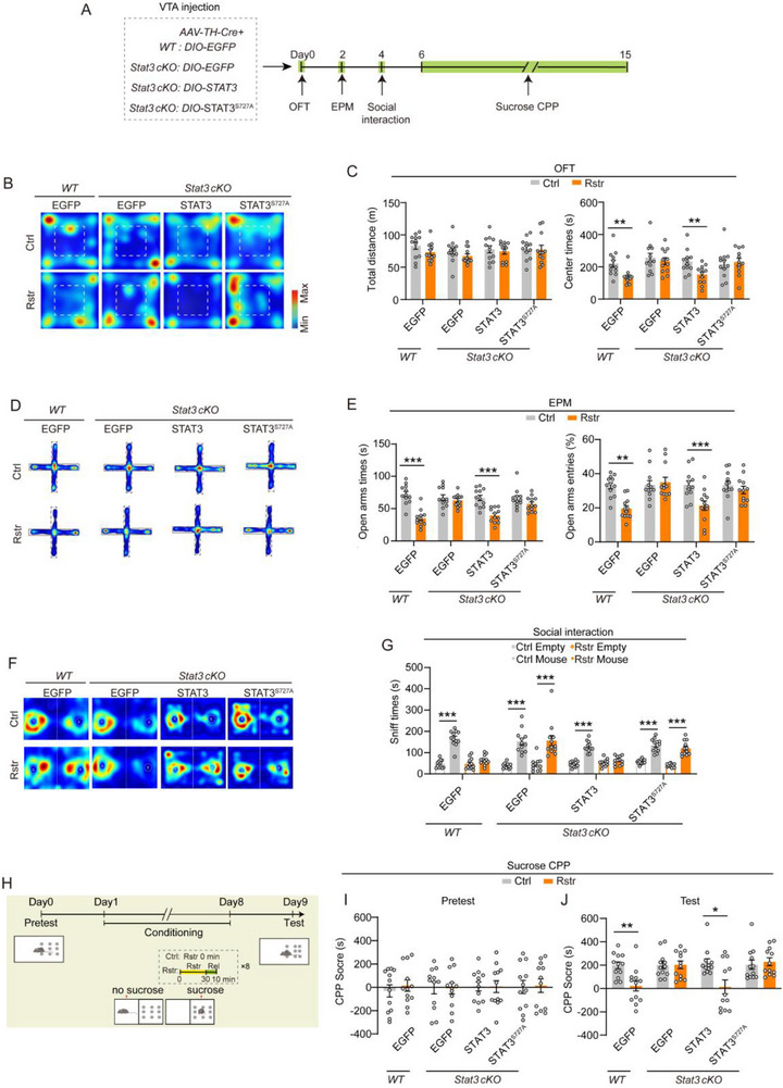
Behavioral changes accompany these signals. After stress, control mice spend less time in the center of an open field and reduce time in, and entries into, the open arms of an elevated maze. Deleting STAT3 from VTA dopamine cells abolishes these stress-induced increases in avoidance.
Restoring normal STAT3 reinstates these changes, but the Ser727 phosphorylation-deficient version does not. The state of a single intracellular site is thus linked to how mice allocate exploration time. Concordant effects across settings connect the behavioral observations to a molecular mechanism.
Source: Shao M et al. · Advanced science (Weinheim, Baden-Wurttemberg, Germany) · CC BY
Original figure and English caption ↗
Glycosylation preserves reward-related preferences
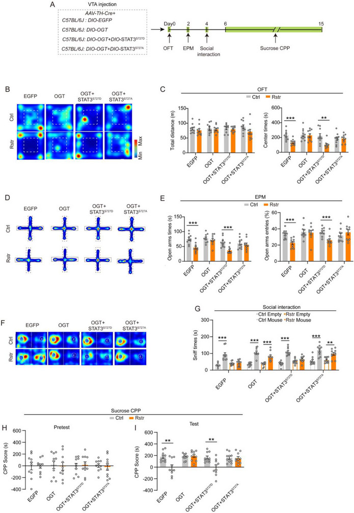
The researchers next increase OGT. After stress, these mice maintain greater exploration of exposed areas and retain preferences for an unfamiliar conspecific and a sucrose-associated environment. Reducing OGA produces similar buffering: making the sugar mark easier to add or harder to remove shifts behavior in the same direction.
Replacing STAT3 with a version that mimics Ser727 phosphorylation weakens the OGT-associated buffer, reinstating increased avoidance and reduced reward preference after stress. This result connects the relationship between increased glycosylation and behavioral buffering more specifically to the phosphorylation state of STAT3.
Source: Shao M et al. · Advanced science (Weinheim, Baden-Wurttemberg, Germany) · CC BY
Original figure and English caption ↗
Gene regulation points toward inhibitory receptors
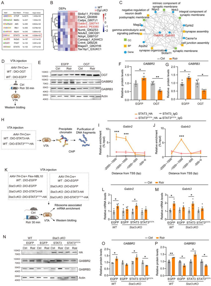
How can a modification at one STAT3 site influence the cell’s working state? Stress increases expression of GABBR2 and GABRB3, two GABA-receptor subunits. Normal STAT3 binds upstream regions of the corresponding genes; preventing Ser727 phosphorylation alters this binding and the stress-induced increase in expression.
Increasing OGT also suppresses the rise in receptor-subunit abundance. Because GABA commonly conveys inhibition to neurons, these expression changes offer a possible downstream explanation for reduced dopamine activity. Direct measurements are still needed to determine how receptor currents change and how much each subunit contributes.
Source: Shao M et al. · Advanced science (Weinheim, Baden-Wurttemberg, Germany) · CC BY
Original figure and English caption ↗
The stress response contains its own buffer
The study reveals more than a one-way pathway that amplifies stress. Acute stress increases STAT3 phosphorylation while recruiting glycosylation that counterbalances it. Existing proteins inside dopamine cells thereby become a point of control over short-term adaptation.
These findings come from acute restraint stress in male mice. They connect changes in exploration and reward-related behavior to intracellular chemical marks that can counterbalance one another. Whether prolonged or repeated stress changes this balance is a specific question arising from the mechanism.