Facing acute stress, how do midbrain astrocytes help buffer mood-related circuits?
A mouse study shows that ventral tegmental area astrocytes release adenosine during stress, dampening local GABA transmission and limiting anxiety- and depression-like behaviors.
Dopaminergic (DA) neurons in the ventral tegmental area (VTA) are central regulators of reward processing, motivation, and stress-related mood disorders. Although astrocytes are increasingly recognized as active partners in synaptic modulation via intracellular calcium signaling and gliotransmitter release, how VTA astrocytes respond to aversive stress and shape local circuit inhibition to buffer against anxiety and depression has remained unclear. On June 11, 2025, Yizheng Wang, Jiani Li, and colleagues at the Beijing Institute of Basic Medical Sciences and Fudan University (with Jun Du and Huapeng Diao as co-first authors) published a study titled "Astrocytic modulation of GABA transmission in the VTA limits progression of anxiety- and depression-like behaviors in adult mice" in Cell Reports. Combining in vivo fiber photometry (GCaMP6f, GRAB_DA1h, and GRAB_Ado), optogenetic manipulations, whole-cell patch-clamp recordings, and cell-type-specific shRNA knockdowns, the authors demonstrate that acute aversive stressors selectively trigger IP3R2-dependent calcium elevations and adenosine release from VTA astrocytes; astrocytic adenosine activates presynaptic A1 receptors on VTA GABAergic neurons to suppress inhibitory transmission onto DA neurons, enhance nucleus accumbens (NAc) dopamine release, and protect adult mice against stress-induced anxiety- and depression-like behaviors.
Acute aversive stressors (tail suspension, restraint, and foot shock), but not natural rewards, selectively activate IP3R2-dependent Ca2+ signaling and vesicular adenosine release in VTA astrocytes, which acts on adenosine A1 receptors (Adora1) on VTA GABAergic neurons to reduce sIPSC frequency, disinhibit VTA dopamine neurons, elevate NAc dopamine release, and limit stress-induced anxiety- and depression-like behaviors.
Contents · 5 sections
VTA Astrocytes Exhibit Robust Calcium Transients to Acute Aversive Stressors Rather Than Natural Rewards
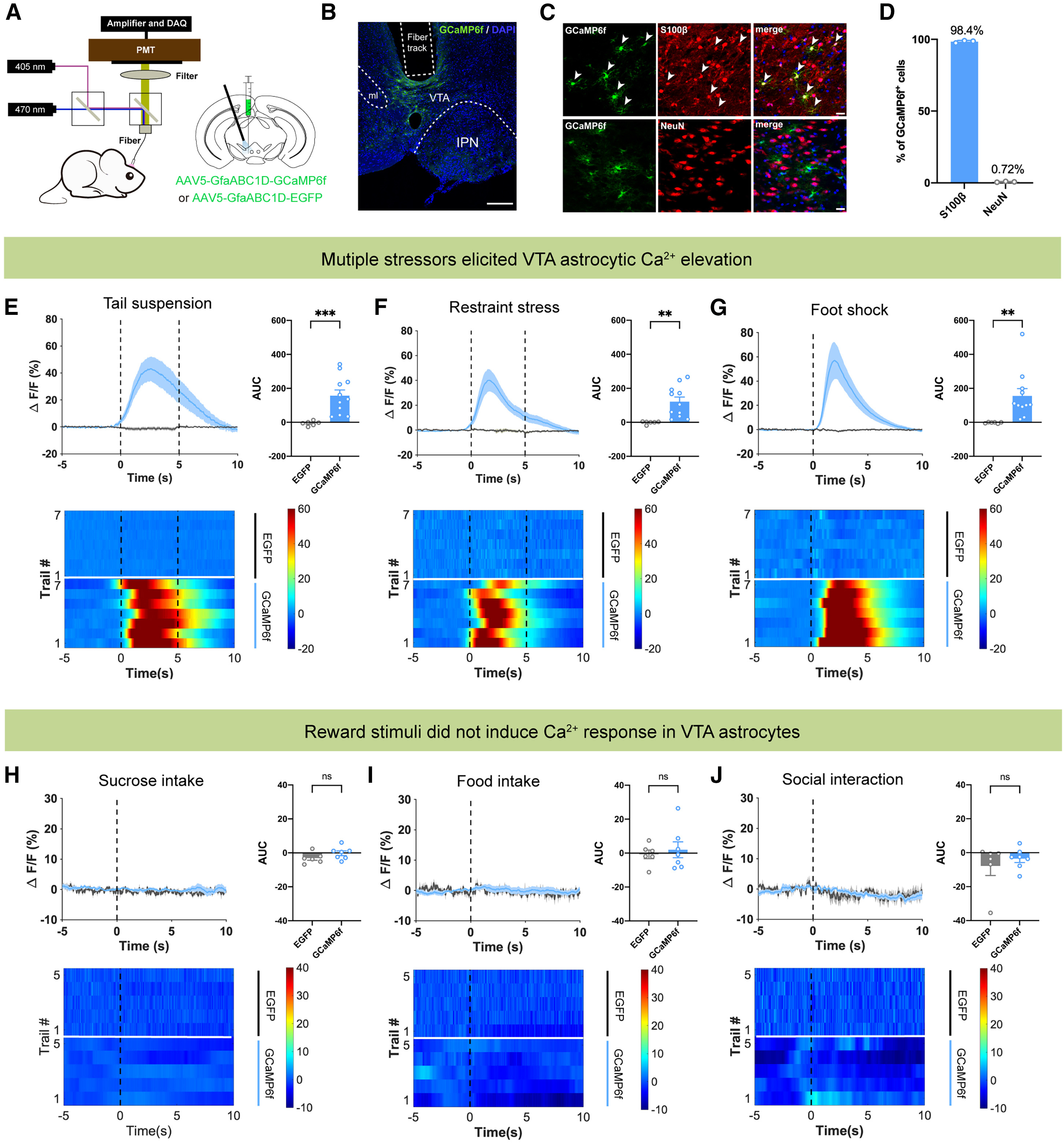
To monitor VTA astrocyte activity in freely behaving adult mice, the researchers injected the astrocyte-specific calcium indicator AAV5-GfaABC1D-GCaMP6f into the VTA—where 98.4% of GCaMP6f-expressing cells co-localized with the astrocytic marker S100β and only 0.72% overlapped with the neuronal marker NeuN—and implanted an optical fiber above the VTA for in vivo fiber photometry.
Comparing aversive and rewarding paradigms revealed striking selectivity: acute tail suspension (5 s), physical restraint (5 s), and electric foot shock (0.3 mA, 1 s) each evoked rapid, robust increases in VTA astrocytic GCaMP6f fluorescence (ΔF/F and AUC), whereas natural reward behaviors—including sucrose licking, food pellet consumption, and social interaction with a female mouse—elicited no significant calcium responses, demonstrating that VTA astrocytes selectively respond to acute aversive stressors.
Source: Jun Du et al. · Cell Reports · © Elsevier Inc.
Original figure and English caption ↗
Optogenetic Activation of VTA Astrocytes Elevates NAc Dopamine Release and Drives D1R-Dependent Anxiolytic and Antidepressant Effects
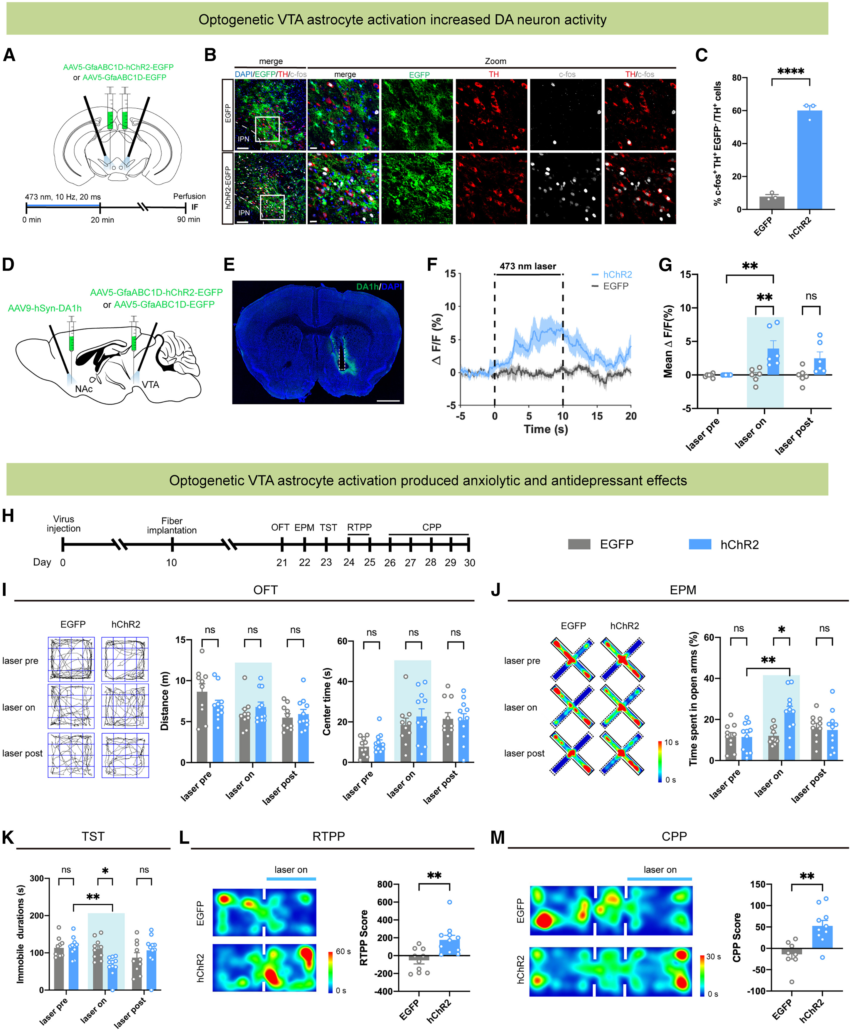
To determine the functional consequences of VTA astrocyte activation, the authors bilaterally expressed AAV5-GfaABC1D-hChR2-EGFP in VTA astrocytes and delivered blue light stimulation (473 nm, 10 Hz, 20 ms pulses). Optogenetic activation of VTA astrocytes significantly increased the percentage of c-Fos+/TH+ dopaminergic neurons in the VTA and triggered robust dopamine release in the downstream nucleus accumbens (NAc) as measured by AAV9-hSyn-GRAB_DA1h fiber photometry.
Behaviorally, photoactivation of VTA astrocytes did not alter baseline locomotion in the OFT, but significantly increased open-arm exploration time in the EPM, reduced immobility duration in the TST, and induced both real-time place preference (RTPP) and conditioned place preference (CPP). Crucially, systemic administration of the dopamine D1 receptor antagonist SCH23390 (0.5 mg/kg) completely abolished the anxiolytic, antidepressant, and rewarding effects of VTA astrocyte photoactivation, demonstrating that these behavioral benefits depend on downstream dopaminergic signaling.
Source: Jun Du et al. · Cell Reports · © Elsevier Inc.
Original figure and English caption ↗
VTA Astrocytes Suppress Presynaptic GABA Release onto Dopamine Neurons via Adenosine A1 Receptors
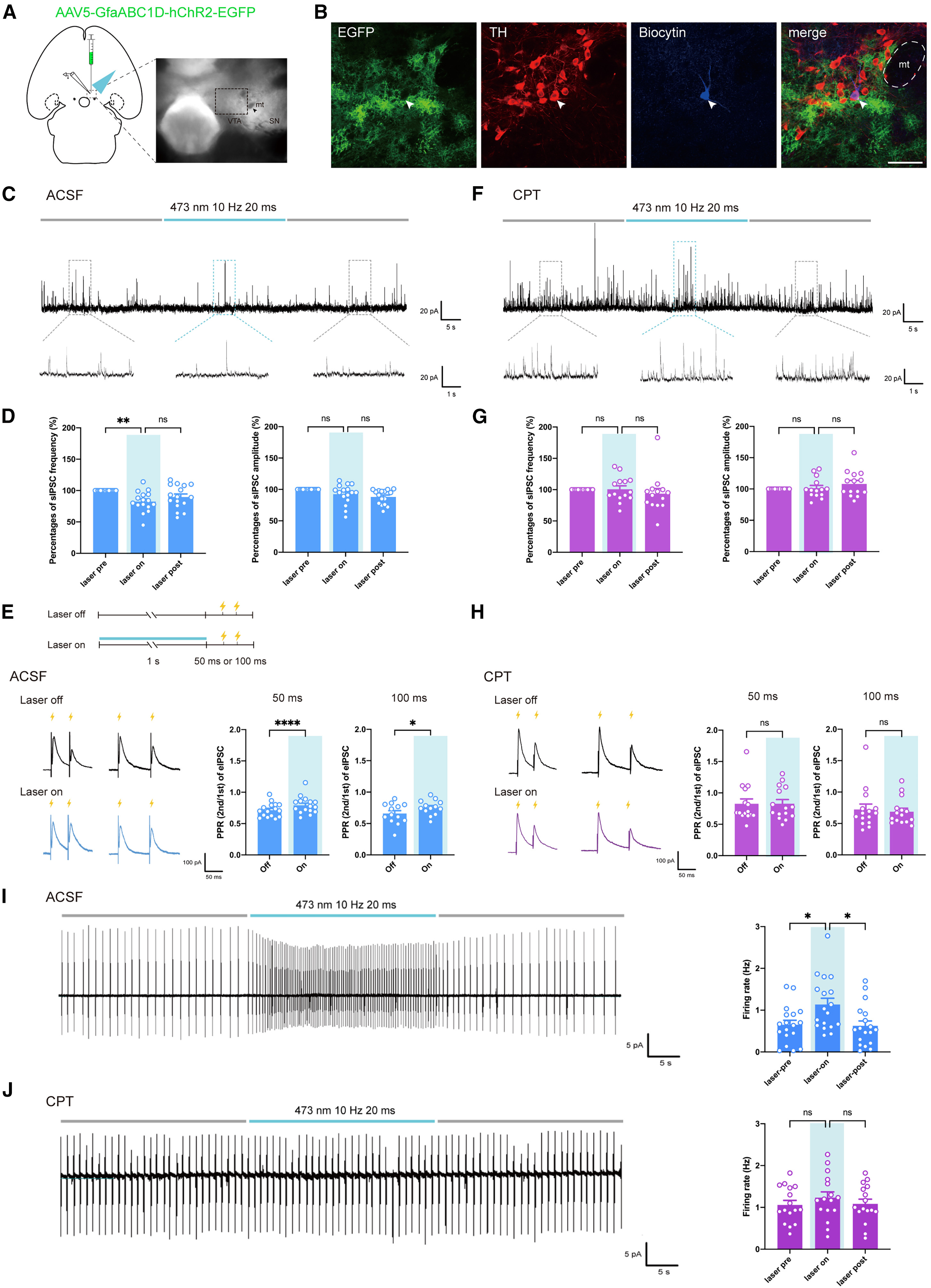
To dissect the synaptic mechanism by which VTA astrocytes excite DA neurons, the authors performed whole-cell patch-clamp recordings from biocytin-filled, TH-verified VTA DA neurons in acute brain slices. Photostimulation of hChR2-expressing VTA astrocytes had no effect on spontaneous EPSCs (sEPSCs), but significantly decreased the frequency—without altering the amplitude—of spontaneous IPSCs (sIPSCs), increased the paired-pulse ratio (PPR at 50 ms and 100 ms intervals) of electrically evoked eIPSCs, and elevated the spontaneous action potential firing rate of VTA DA neurons, indicating presynaptic suppression of GABA release and consequent disinhibition of DA neurons.
Pharmacological dissection revealed that bath application of the selective adenosine A1 receptor (A1R) antagonist CPT (10 μM) completely blocked the astrocyte-stimulation-induced reduction in sIPSC frequency, increase in eIPSC PPR, and elevation in DA neuronal firing rate, whereas the selective adenosine A2A receptor antagonist SCH58261 (1 μM) had no effect—pinpointing adenosine signaling through presynaptic A1Rs as the mediator of GABAergic suppression.
Source: Jun Du et al. · Cell Reports · © Elsevier Inc.
Original figure and English caption ↗
Knockdown of Adenosine A1 Receptors in VTA GABAergic Neurons Abolishes Astrocytic Modulation and Behavioral Rescue
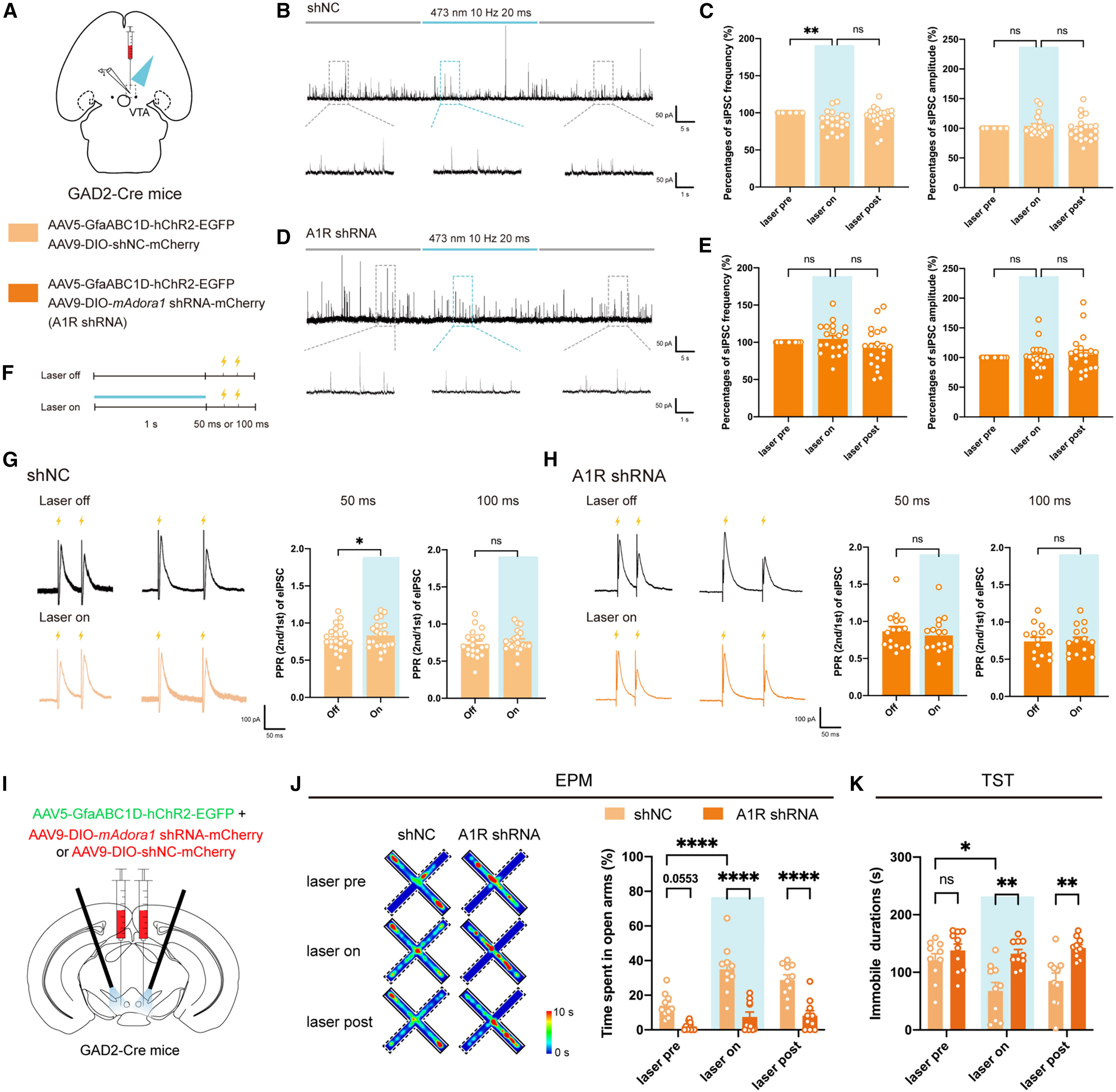
RNAscope in situ hybridization combined with immunostaining confirmed abundant Adora1 expression in VTA GABAergic neurons. To test the causal necessity of GABAergic A1Rs, the authors injected Cre-dependent AAV9-EF1a-DIO-mAdora1-shRNA-mCherry into the VTA of GAD2-Cre mice to selectively knock down A1Rs in VTA GABAergic interneurons. Selective A1R knockdown induced baseline anxiety-like behavior in the OFT and EPM and markedly increased susceptibility to a 10-day subthreshold chronic mild stress (SCMS) paradigm across the OFT, EPM, SPT, TST, and FST.
Co-expressing astrocyte-specific hChR2 with DIO-mAdora1-shRNA in GAD2-Cre mice demonstrated that while optogenetic VTA astrocyte activation decreased sIPSC frequency, increased 50 ms eIPSC PPR, increased EPM open-arm exploration, and reduced TST immobility in shNC controls, all of these electrophysiological and behavioral effects were completely abolished in mice lacking A1Rs in VTA GABAergic neurons.
Source: Jun Du et al. · Cell Reports · © Elsevier Inc.
Original figure and English caption ↗
Disrupting Astrocytic IP3R2 Ca2+ Signaling or Vesicular Gliotransmission Blunts Adenosine Surges and Increases Stress Vulnerability
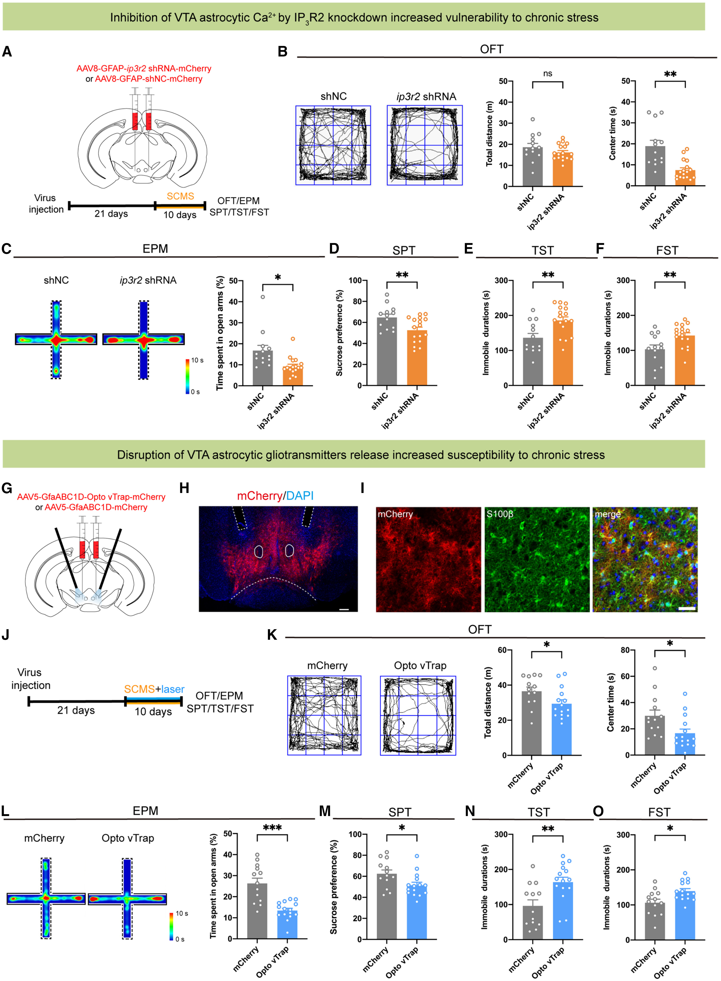
Finally, the authors examined whether endogenous VTA astrocytic calcium signaling and vesicular gliotransmitter release protect against stress vulnerability. Type 2 inositol 1,4,5-trisphosphate receptors (IP3R2) mediate endoplasmic reticulum calcium release in astrocytes. Knocking down IP3R2 in VTA astrocytes via AAV8-GFAP-ip3r2-shRNA-mCherry (90.22% co-localized with S100β+) markedly blunted tail-suspension- and foot-shock-evoked astrocytic Ca2+ transients as well as stress-induced extracellular adenosine surges measured by the GRAB_Ado1.0 sensor, while decreasing 50 ms eIPSC PPR and increasing evoked eIPSC amplitudes onto VTA DA neurons (Original Figure 5).
Behavioral testing demonstrated that either knocking down astrocytic IP3R2 or optogenetically trapping astrocytic vesicles with AAV5-GfaABC1D-Opto-vTrap-mCherry during 10-day subthreshold chronic mild stress (SCMS) precipitated pronounced anxiety- and depression-like phenotypes—including reduced OFT center time, decreased EPM open-arm exploration, lower sucrose preference in the SPT, and prolonged immobility in both the TST and FST—establishing VTA astrocytic IP3R2-dependent adenosine release and GABAergic A1R-mediated disinhibition of dopamine neurons as an endogenous resilience mechanism against stress-induced mood disorders.
Source: Jun Du et al. · Cell Reports · © Elsevier Inc.
Original figure and English caption ↗