Delayed changes after stress: a prefrontal receptor and vulnerability to avoidance
A male-mouse study finds increased PrL GHS-R1a expression and weaker excitatory input six days after repeated stress; raising receptor expression worsens post-stress behavioral and synaptic changes.
Stress ends while brain changes continue to unfold. A study published on March 13, 2025 in Communications Biology finds that shortly after repeated restraint stress, male mice show no clear reduction in open-arm exploration, although activity markers in prefrontal PrL cells increase. Six days later, those cells are less active and avoidance-related behavior is stronger. GHS-R1a, a receptor usually associated with ghrelin signaling, also increases in this region after a delay. Altering receptor expression connects this later molecular change to excitatory input and post-stress vulnerability.
Increasing GHS-R1a expression in PrL excitatory cells strengthens post-stress avoidance and synaptic changes, while effects of reducing expression differ across tasks and states.
Contents · 7 sections
Cells activate first, behavior changes later
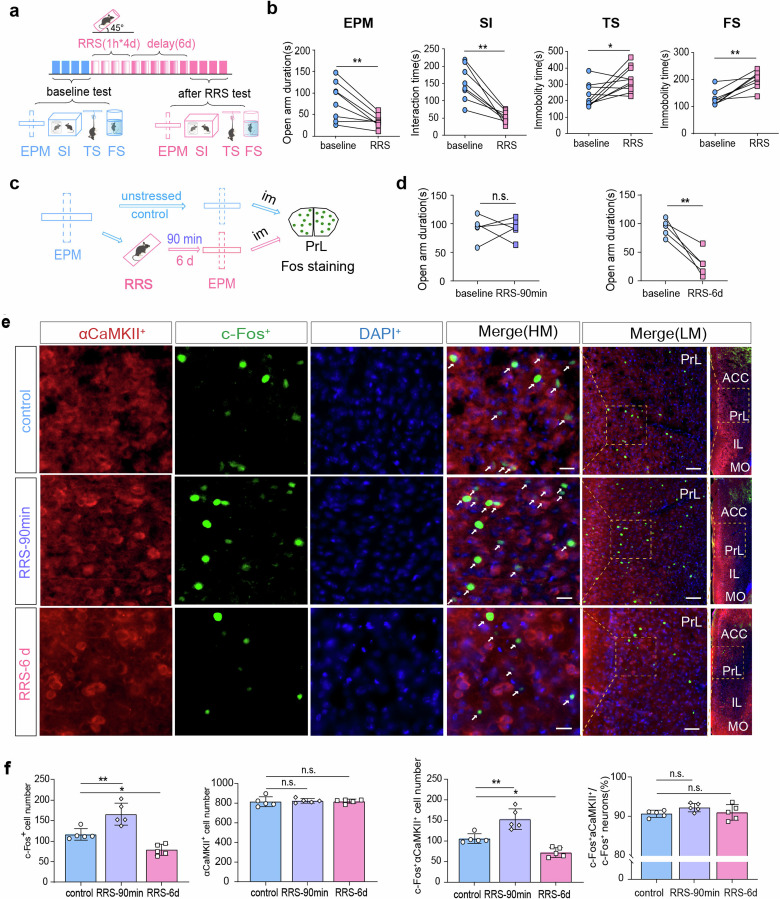
Shortly after repeated stress ends, open-arm time remains similar to baseline. Six days later, open-arm exploration and social interaction decrease while tail-suspension and swimming immobility increase. An observable delay separates the stress event from later behavioral changes.
Activity markers in PrL αCaMKII-positive excitatory cells rise early but fall later. In neighboring IL, markers rise at both time points. Even within prefrontal cortex, stress-related time courses differ, directing the study toward PrL.
Source: Yang L et al. · Communications biology · CC BY-NC-ND
Original figure and English caption ↗
A response to entering open arms weakens after stress
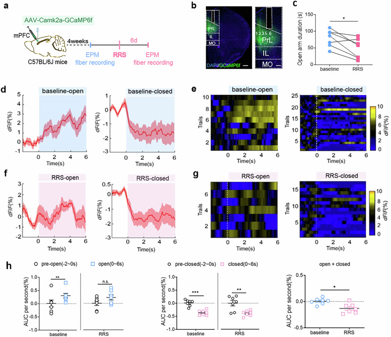
At baseline, entering an open arm increases PrL calcium signals. After repeated stress, that clear entry-associated increase is absent. Entering a closed arm lowers calcium in both states, while overall recorded activity is lower after stress.
Electrical recordings show lower frequency and amplitude of miniature excitatory events at the later stage, while inhibitory events remain broadly unchanged. The weaker response has an input-level counterpart: reduced excitatory drive accompanies altered dynamics during environmental events.
Source: Yang L et al. · Communications biology · CC BY-NC-ND
Original figure and English caption ↗
Increasing PrL activity after stress increases exploration
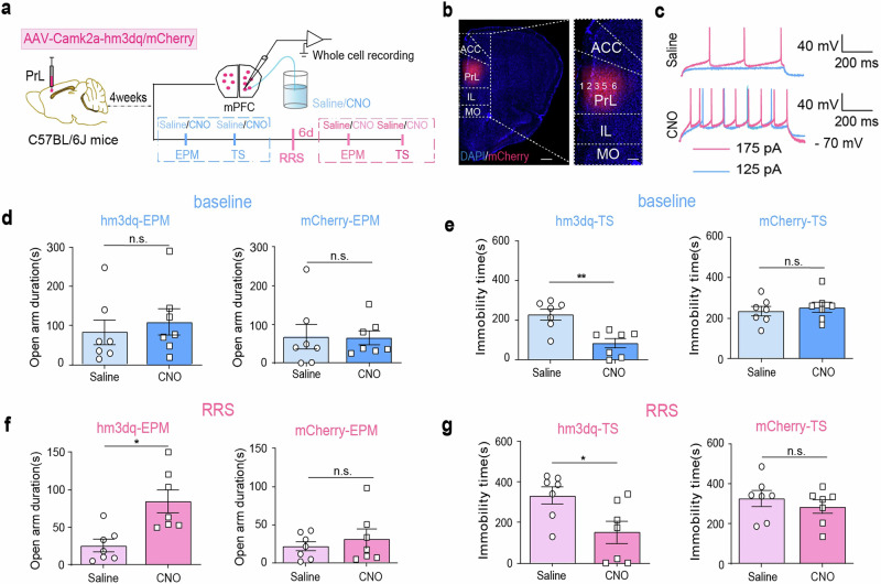
Directly increasing activity in these PrL cells raises open-arm time and reduces tail-suspension immobility in stressed mice. In unstressed mice, open-arm exploration does not clearly change, although immobility decreases. Effects depend on both task and state.
This gives functional meaning to the early activation and later low activity: increasing these cells’ activity changes some post-stress measures. The study next searches for a delayed cellular change that could contribute to weakened inputs and responses.
Source: Yang L et al. · Communications biology · CC BY-NC-ND
Original figure and English caption ↗
More cells carry GHS-R1a transcripts
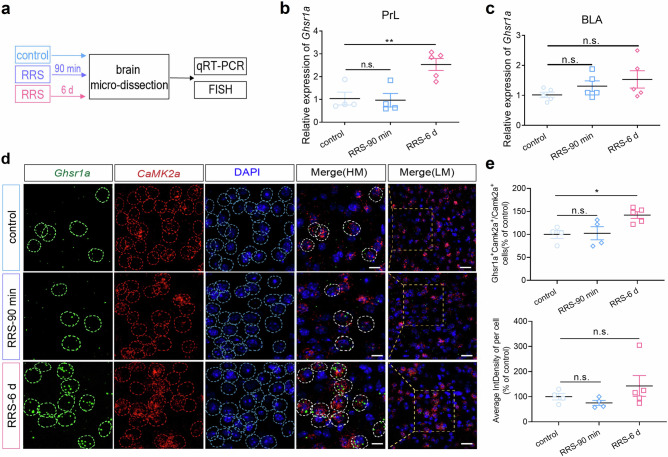
GHS-R1a is usually studied as a ghrelin receptor. Here, its PrL transcripts increase six days after stress without a clear early change; the basolateral amygdala lacks the same alteration. The expression time course accompanies behavioral and cellular-activity changes.
In situ detection shows a larger fraction of αCaMKII-positive cells carrying receptor transcripts, without a clear increase in average signal intensity within each positive cell. A broader receptor-positive population and uniform upregulation in every cell represent different observational scales.
Source: Yang L et al. · Communications biology · CC BY-NC-ND
Original figure and English caption ↗
Behavioral improvement differs by task
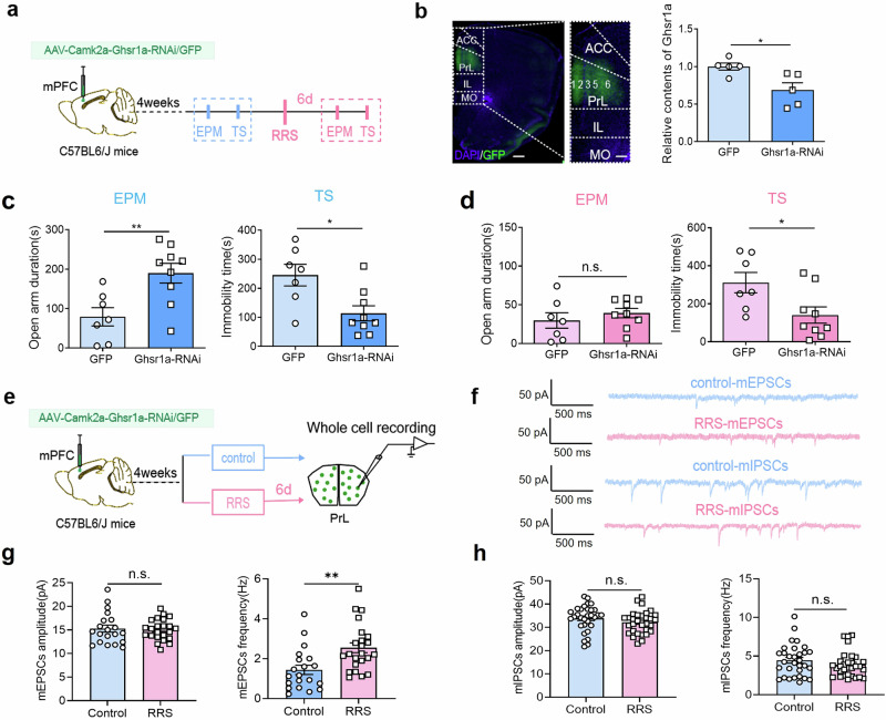
Reducing GHS-R1a in these PrL cells increases open-arm exploration and reduces immobility before stress. After stress, immobility remains lower but open-arm time does not clearly differ. The effects on different stress-associated measures are therefore not identical.
In receptor-reduced mice, excitatory-event frequency rises after stress, while amplitude and inhibitory events remain broadly unchanged. This differs from the declining excitatory input observed in ordinary stressed mice, supporting a contribution of receptor expression to stress-related synaptic responses.
Source: Yang L et al. · Communications biology · CC BY-NC-ND
Original figure and English caption ↗
Little baseline change, greater avoidance after stress
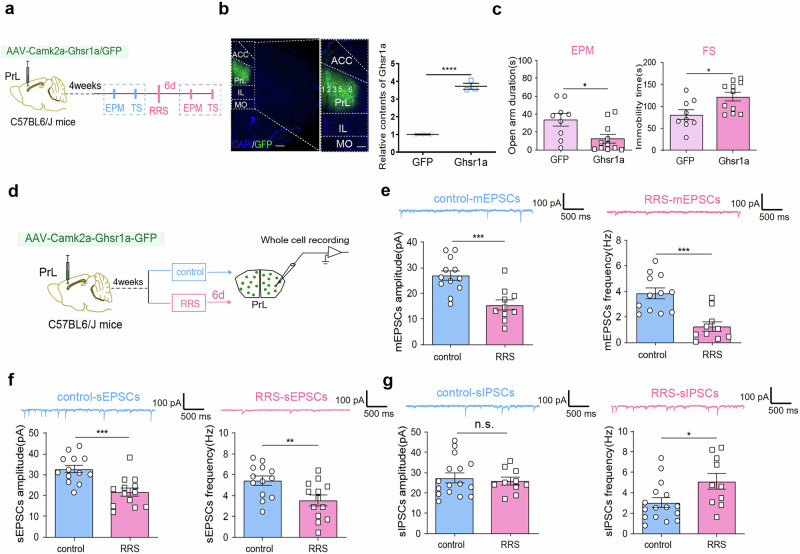
Increasing GHS-R1a expression does not clearly alter tested behavior before stress. After repeated stress, open-arm exploration falls and swimming immobility increases. Higher expression thus changes vulnerability in a stressful context rather than producing an identical effect in every condition.
After stress, receptor-overexpressing cells show lower excitatory-event frequency and amplitude and higher spontaneous inhibitory-event frequency. The input changes jointly shift toward reduced excitatory drive, accompanying stronger avoidance.
Source: Yang L et al. · Communications biology · CC BY-NC-ND
Original figure and English caption ↗
Receptor effects depend on cells and states
Increasing the same receptor in corresponding basolateral-amygdala cells does not produce the same behavioral changes. Region, cellular population and stress history jointly shape the effect, explaining why receptor identity or overall abundance alone cannot predict behavioral direction.
This adult male-mouse study describes delayed adjustment after stress. How GHS-R1a alters excitatory and inhibitory inputs, and how much peripheral ghrelin contributes, remain specific questions. The findings connect a metabolism-associated receptor to delayed prefrontal responses to stress.