Chronic stress shifts a molecular switch, making hippocampal cells more excitable
A mouse study connects Rap1 activation to modification of the Kv4.2 channel: protein abundance stays broadly unchanged while cellular electrical properties and anxiety-related exploration change.
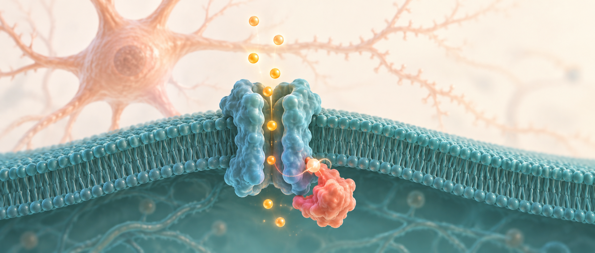
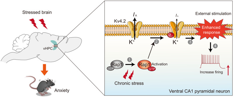
Counting proteins is not enough to understand a cell: their activation state also matters. A study published on May 23, 2025 in Science Advances finds that chronic restraint stress increases hippocampal Rap1 activity in mice without clearly increasing its total abundance. This small GTPase acts as a molecular switch. Its activation in ventral-hippocampal pyramidal cells is linked to greater excitability and reduced exploration of exposed areas. Following the mechanism to a phosphorylation site on the potassium channel Kv4.2 reveals how stress can alter the working state of existing proteins.
Reducing Rap1 activity in ventral-hippocampal pyramidal cells or providing a Kv4.2 version that cannot be phosphorylated at Thr607 buffers associated excitability and exploration changes.
Contents · 7 sections
Protein abundance stays stable while activity rises
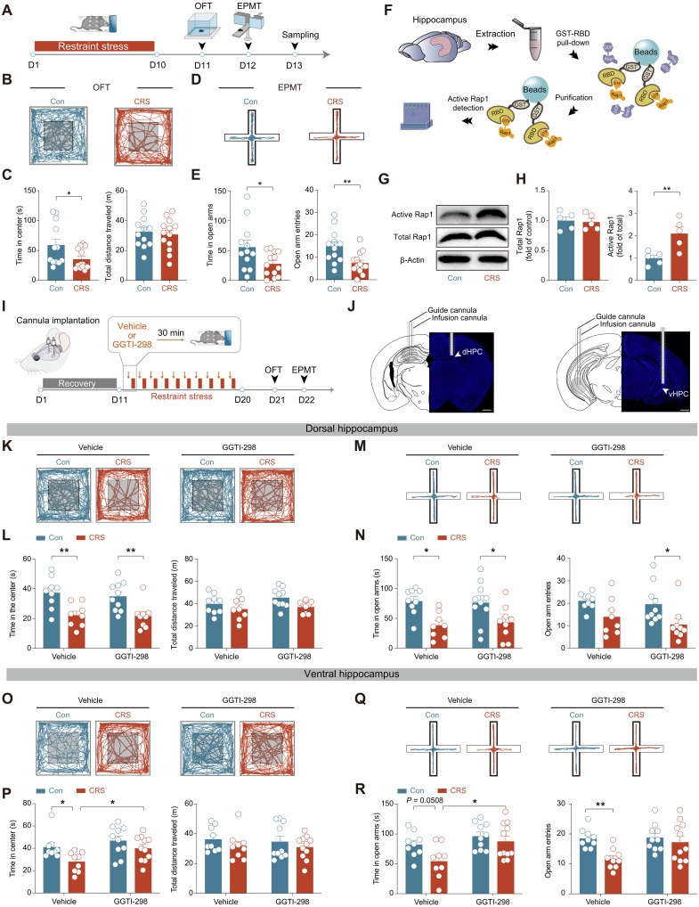
After chronic stress, mice spend less time in the open-field center and explore open maze arms less, while total distance traveled remains broadly unchanged. Hippocampal measurements show no clear rise in total Rap1 protein; the increase concerns its active signaling form. Rap1 switches between states bound to different nucleotides and is more active when bound to GTP. The same amount of Rap1 can therefore convey different levels of intracellular signaling.
A single acute restraint does not produce the same Rap1 activation or subsequent behavioral changes, whereas chronic unpredictable stress produces similar findings. Increased activity remains measurable days after the final restraint, indicating a molecular-switch change that persists for a period beyond the stress episode.
Source: Pan HQ et al. · Science advances · CC BY-NC
Original figure and English caption ↗
The effect centers on ventral-hippocampal pyramidal cells
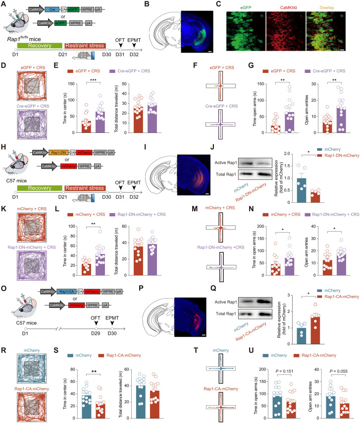
The study narrows its focus from the whole hippocampus to particular cells. Reducing Rap1 or its activity in ventral-hippocampal pyramidal cells increases center and open-arm exploration after chronic stress. The examined astrocyte and inhibitory-interneuron manipulations do not produce the same result.
Conversely, increasing pyramidal-cell Rap1 activity in mice without chronic stress reduces open-field center time. Differences in the two elevated-maze measures do not reach significance. The molecular manipulation can alter exploration in one setting, while effects vary across tasks.
Source: Pan HQ et al. · Science advances · CC BY-NC
Original figure and English caption ↗
Cells respond more strongly to the same setting
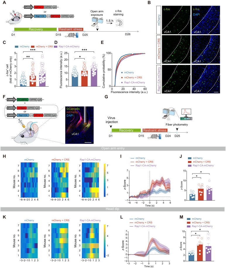
Calcium signals in ventral CA1 pyramidal cells rise when mice move from closed to open arms. After chronic stress or enhanced Rap1 activity, the response is larger. Looking beyond the edge of an open arm also evokes a stronger calcium response.
Activity-associated protein markers change in a similar direction. Suppressing this population’s activity buffers behavioral changes in both stressed mice and the Rap1-activation group. Altered molecular state amplifies the connection between an external setting and the cellular response.
Source: Pan HQ et al. · Science advances · CC BY-NC
Original figure and English caption ↗
The change centers on the cell’s electrical response
The researchers initially examine dendrites, spines and synaptic transmission. Branching and spine density show no clear change, and miniature excitatory and inhibitory events remain broadly similar in frequency and amplitude. Greater activity does not accompany a wholesale change in the measured structural and synaptic features.
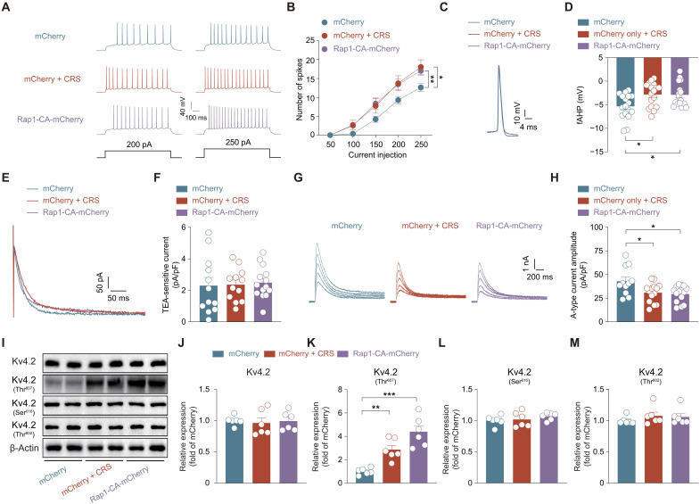
When given the same electrical input directly, cells from stressed and Rap1-activation groups produce more spikes. Fast afterhyperpolarization following an action potential is weakened, and A-type potassium current is reduced. Altered responsiveness makes the same input more readily produce repeated firing. Resting membrane potential, action-potential threshold and BK-associated potassium current remain broadly similar. Stable basic measures focus the change more closely on fast afterhyperpolarization and A-type current.
Source: Pan HQ et al. · Science advances · CC BY-NC
Original figure and English caption ↗
One Kv4.2 site is linked to the change
Kv4.2 provides a molecular lead for the A-type current. Like Rap1, total channel abundance does not clearly change. Phosphorylation increases at Thr607, while two other examined sites show no corresponding change. Once again, similar abundance accompanies a different molecular state.
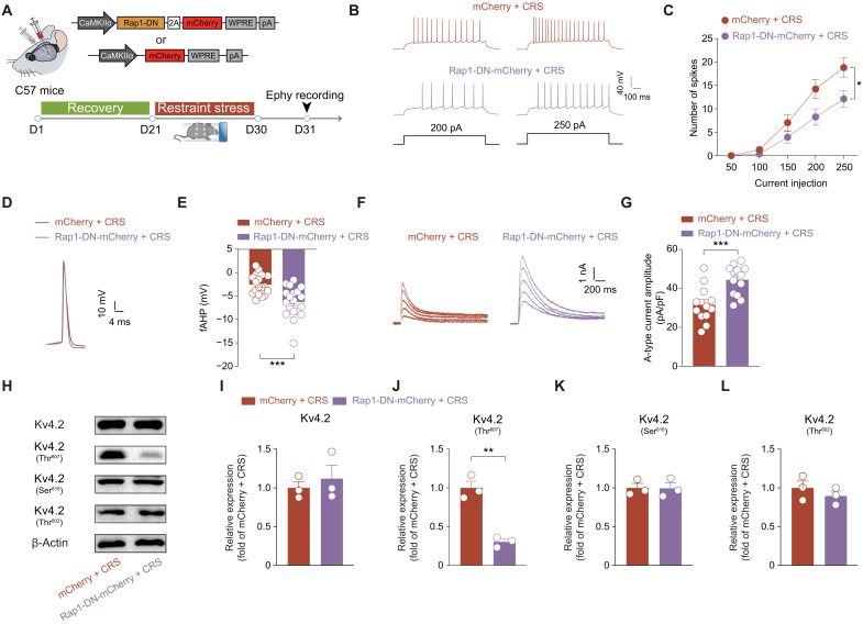
Reducing Rap1 activity weakens the Thr607 change, strengthens A-type current and fast afterhyperpolarization, and reduces firing. Corresponding changes link the molecular switch, channel state and cellular electrical response.
Source: Pan HQ et al. · Science advances · CC BY-NC
Original figure and English caption ↗
A different channel version buffers cellular and behavioral changes
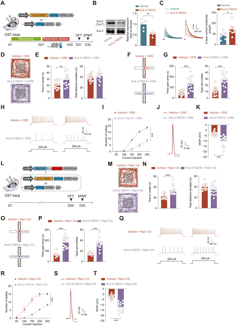
The study next provides a Kv4.2 version that cannot be phosphorylated at Thr607. In chronically stressed mice, A-type current increases, cells become less prone to repeated firing, and center and open-arm exploration rise. This further connects a chemical mark on the channel with behavioral change.
The same version buffers hyperexcitability and avoidance in Rap1-activation mice. Concordant effects under two upstream conditions support Kv4.2-associated regulation as a downstream contributor to Rap1’s effects.
Source: Pan HQ et al. · Science advances · CC BY-NC
Original figure and English caption ↗
Stress can change function without clearly changing abundance
This male-mouse study connects activation after chronic stress, channel modification, cellular excitability and exploration. Unchanged total abundance at two points illustrates how measuring only protein quantity can miss altered function in molecules already present.
How stress persistently shifts Rap1, and which kinase links Rap1 to the channel, remain specific open questions. The findings focus attention on intracellular molecular states that alter electrical responsiveness, alongside broader questions about structural change.
Source: Pan HQ et al. · Science advances · CC BY-NC
Original figure and English caption ↗