Beyond recycling: how astrocyte lysosomes respond to stress
A male mouse study links astrocytic TFEB, the lysosomal channel TRPML1 and ATP release to social avoidance and other behaviors after stress.
Lysosomes are often described as cellular recycling stations that break down and reuse materials. In astrocytes, they also participate in releasing signals outside the cell. A study published in Advanced Science in 2024 focuses on medial prefrontal cortex (mPFC), where susceptible mice show altered astrocytic lysosomes and lower channel protein TRPML1 after prolonged stress. Following this change connects the transcription factor TFEB with ATP release, bringing intracellular organelles into the story of stress-related behavior.
The astrocytic TFEB–TRPML1 axis supports lysosomal exocytosis and ATP release. Disrupting it increases selected depression-like behaviors in male mice; restoring mPFC TRPML1 improves tested behaviors.
Contents · 7 sections
Intracellular traces of stress
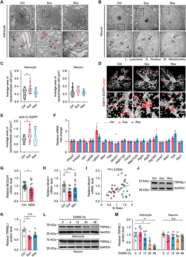
Some mice show marked social avoidance after chronic social defeat. Compared with control and resilient mice, susceptible animals have larger mPFC astrocytic lysosomes, while neuronal lysosomal morphology in the same region does not clearly change. The alteration first emerges in a particular cell type.
Susceptible mice also show lower brain Mcoln1 expression and its encoded protein, TRPML1. Lower MCOLN1 expression in peripheral blood from patients with major depressive disorder provides a separate clinical association. Larger lysosomes do not necessarily function more efficiently, prompting examination of their channel.
Source: Mo JW et al. · Advanced science (Weinheim, Baden-Wurttemberg, Germany) · CC BY
Original figure and English caption ↗
One channel, different cellular consequences
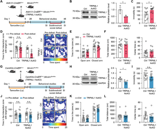
Deleting Mcoln1 in adult astrocytes increases immobility in forced swimming. After a milder social defeat challenge, mice show more social avoidance and lower sucrose preference. A relatively mild stressor thus becomes more likely to accompany behavioral changes.
Exposed-space exploration, total distance and tested motor abilities remain largely unchanged. Deleting the same gene in CaMKIIα-marked neurons does not produce the corresponding swimming and interaction effects. TRPML1 function depends on its cellular location, without a general motor deficit accompanying these behavioral differences.
Source: Mo JW et al. · Advanced science (Weinheim, Baden-Wurttemberg, Germany) · CC BY
Original figure and English caption ↗
Narrowing the effect to prefrontal cortex
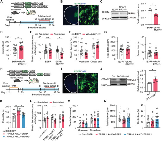
Deleting Mcoln1 only in mPFC astrocytes reproduces increased immobility and reduced social interaction after stress. Restoring the protein specifically in mPFC astrocytes in animals with widespread astrocytic TRPML1 deletion improves both behaviors.
Local deletion and restoration converge on the same region. The question moves from whether glia matter to how lysosomes in prefrontal astrocytes shape the signaling environment around neighboring cells.
Source: Mo JW et al. · Advanced science (Weinheim, Baden-Wurttemberg, Germany) · CC BY
Original figure and English caption ↗
ATP: from intracellular vesicles to extracellular signals
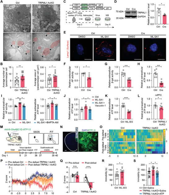
TRPML1 is a calcium channel on lysosomal membranes. Cultured astrocytes lacking it contain more and larger lysosomes but have impaired exocytotic responses, with lower lysosomal enzyme activities and ATP in the medium. Channel activation increases ATP release, and blocking exocytosis weakens that increase.
ATP serves as an extracellular signal as well as an energy molecule. Across the cell membrane, its role extends from intracellular energy supply to communication among neighboring cells. Extracellular ATP is lower in knockout brain slices. In living mice, attack-related ATP responses are similar between groups before defeat, but become markedly lower in the knockout group afterward. Stress separates signals that had initially been similar.
Systemic ATP administration reduces swimming immobility in knockout mice. Together with the cellular and slice findings, this places ATP in an important position in the behavioral mechanism, with supplementation delivered systemically in this experiment.
Source: Mo JW et al. · Advanced science (Weinheim, Baden-Wurttemberg, Germany) · CC BY
Original figure and English caption ↗
TFEB changes unfold over time
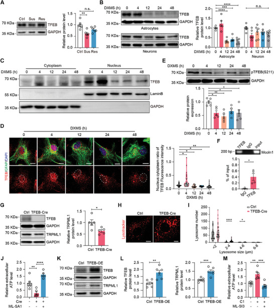
TFEB, which regulates lysosome-related genes, supplies an upstream clue. Its protein abundance is lower in susceptible mouse mPFC. In cultured astrocytes exposed to a glucocorticoid analogue, TFEB initially enters the nucleus more readily, whereas longer exposure reduces nuclear localization and total protein. Early and later responses differ.
TFEB binds the Mcoln1 promoter in astrocytes. Reducing TFEB lowers TRPML1 and ATP release, while activating TRPML1 can still increase ATP release. This downstream intervention bypasses an upstream loss to restore part of the signaling process.
Source: Mo JW et al. · Advanced science (Weinheim, Baden-Wurttemberg, Germany) · CC BY
Original figure and English caption ↗
Downstream restoration connects the molecular chain to behavior
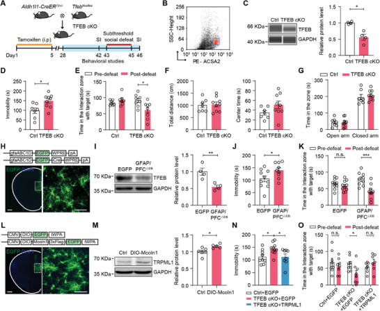
Astrocytic TFEB deletion also increases swimming immobility and avoidance after mild social defeat, with local mPFC deletion reproducing these changes. Increasing TRPML1 in mPFC astrocytes against a TFEB-deficient background improves swimming and social interaction.
Restoring a downstream protein alleviates behavioral consequences of upstream loss, linking TFEB and TRPML1 to stress responses. Exposed-space and movement measures do not change broadly after deletion, giving this molecular chain a selective behavioral contribution.
Source: Mo JW et al. · Advanced science (Weinheim, Baden-Wurttemberg, Germany) · CC BY
Original figure and English caption ↗
A recycling system also supports communication
The study extends lysosomes from intracellular recycling to communication in the brain. Behavioral causal evidence comes from adult male mice, release mechanisms are mainly resolved in cultured cells and slices, and patient evidence is peripheral-blood expression association. Across these levels, changes in a cell’s internal organization may also change its capacity to send signals outward.