Astrocytes also shape the coordination of brain networks
An astrocytic-calcium-related mouse model shows connectivity changes partly paralleling a large depression dataset. Cellular and terminal manipulations further link imaging changes with behavior.
Resting-state functional connectivity compares how closely signals from different regions fluctuate over time. Behind this large-scale coordination, which cells participate besides neurons? Astrocytes around neurons can regulate synaptic activity and communication across regions. A study published in Science Advances on November 16, 2022 combines an astrocytic-signaling-related genetic mouse model, mouse imaging and a large depression dataset. A model with reduced common astrocytic calcium responses shows connectivity changes partly resembling patient patterns. Subsequent cell-population and local-terminal stimulation further examines relationships between networks and behavior.
Astrocyte-targeted stimulation partially alters imaging connectivity. Stimulation of prefrontal neurons and their striatal terminals also accompanies improved immobility and sucrose-preference measures in model mice.
Contents · 6 sections
A calcium-signaling-related deletion shifts network coordination
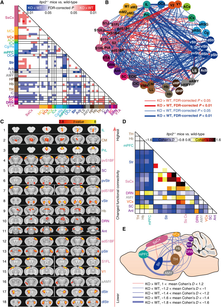
The study uses germline Itpr2-knockout mice. Itpr2 encodes IP3R2, and its loss markedly reduces a common class of astrocytic calcium responses. Several resting-state connections decrease and others increase relative to wild type, while overall hippocampal and ventral-tegmental connectivity changes are less apparent.
Prefrontal, striatal, amygdalar and thalamic networks stand out. Selected weaker connections correlate with greater tail-suspension immobility. The network does not uniformly disconnect: distinct links change differently, motivating more specific cellular and regional tests.
Source: Liu J et al. · Science advances · CC BY-NC
Original figure and English caption ↗
Human data show both shared patterns and differences
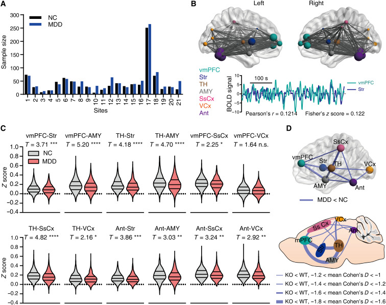
The team analyzes 1080 participants with major depression and 931 controls from 21 sites. Ventromedial-prefrontal correlations with striatum and amygdala are lower in patients, alongside selected thalamic and other connections, paralleling several mouse changes. Prefrontal-related results are comparatively stable across analyses.
Similarity does not extend to every connection. The patient prefrontal–visual comparison is nonsignificant, and some other links change in different directions across species. The human atlas omits some smaller nuclei analyzed in mice, restricting comparisons to regions available in both datasets. Human observations provide large-scale patterns, while mice permit cellular manipulation; the evidence addresses different levels of the question.
Source: Liu J et al. · Science advances · CC BY-NC
Original figure and English caption ↗
Astrocyte-targeted stimulation raises selected connections
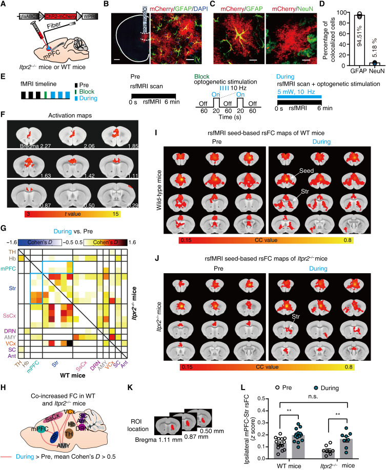
The team expresses a light-sensitive protein chiefly in mPFC astrocytes, with most labeled cells carrying GFAP. During stimulation, local BOLD signals rise and selected connections, including mPFC–striatum, increase in both wild-type and knockout mice.
Some initially reduced model connections shift in the opposite direction, producing partial rather than network-wide restoration. This experiment examines cellularly targeted effects on imaging connectivity; subsequent work tests neuronal activity and behavior.
Source: Liu J et al. · Science advances · CC BY-NC
Original figure and English caption ↗
A different imaging approach highlights prefrontal–striatal changes
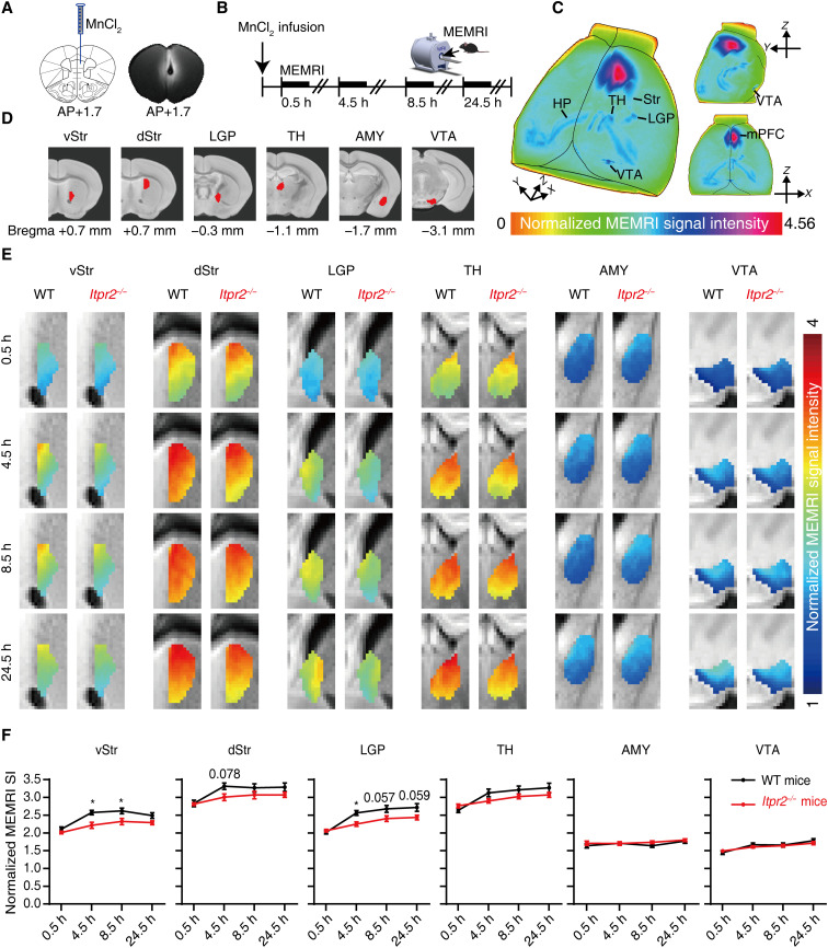
Manganese-enhanced imaging offers another view: active neurons can take up manganese and transport it along axons. Following mPFC delivery, model mice show lower ventral-striatal accumulation at selected earlier points and an early decrease in lateral globus pallidus. Differences are not significant in every subdivision.
Thalamic, amygdalar and ventral-tegmental manganese signals show no clear group differences. Notably, prefrontal–amygdalar resting correlation changes without a corresponding accumulation difference, showing that functional correlation and activity-dependent transport do not map one-to-one. The team consequently focuses on prefrontal–striatal circuitry.
Source: Liu J et al. · Science advances · CC BY-NC
Original figure and English caption ↗
Prefrontal neuronal activation improves imaging and behavioral measures
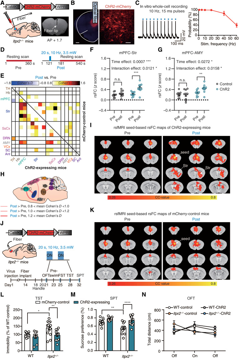
After prefrontal neuronal activation, post-stimulation resting-state recordings show increased mPFC–striatal and mPFC–amygdalar connectivity, with selected changes approaching wild-type levels. Not all links change, including selected thalamic and habenular comparisons.
In separate behavioral experiments, knockout mice show less tail-suspension and forced-swim immobility and greater sucrose preference. Wild-type tail-suspension and sucrose measures do not change significantly, and overall locomotion remains largely unchanged. Imaging and behavioral improvements provide convergent functional evidence relating network changes to model phenotypes.
Source: Liu J et al. · Science advances · CC BY-NC
Original figure and English caption ↗
Local terminals can influence a wider network
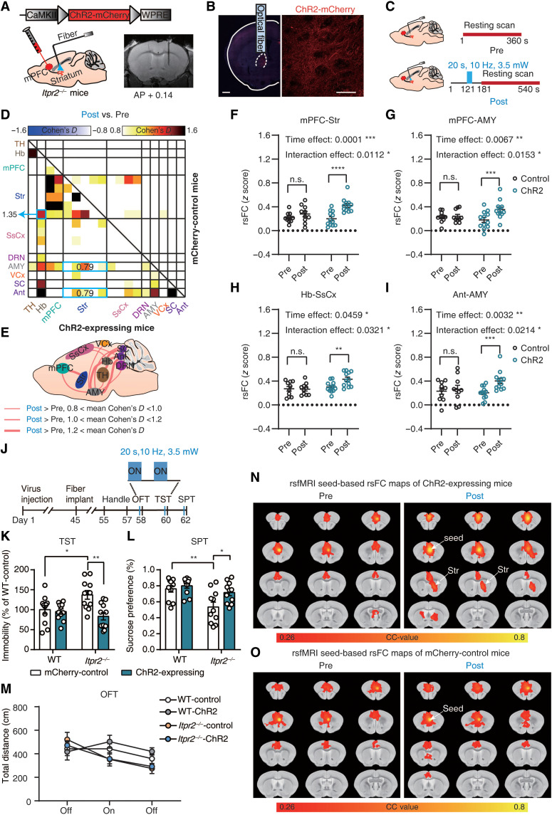
Stimulating mPFC terminals within striatum increases selected model connectivity measures, including prefrontal–striatal and amygdalar-related links, reduces tail-suspension immobility and increases sucrose preference. Both wild-type behaviors remain largely unchanged, with no significant locomotor group difference. A local input manipulation can accompany wider network changes.
The evidence links supporting cells, neurons and large-scale imaging, while BOLD also reflects metabolic and vascular processes and terminal stimulation may recruit upstream circuitry. Astrocytic states are not directly measured in the same human participants. The study offers a testable cellular explanation for shared imaging patterns while retaining distinctions among measurements.
Source: Liu J et al. · Science advances · CC BY-NC
Original figure and English caption ↗