Why does the same sugar evoke different responses before and after feeding? Internal energy signals in flies and mice
Glucose-responsive cells, neuropeptides and early sweet-sensory processing connect in a study of how nutritional state changes sugar responses.

The same sugar can attract a hungry animal differently from one that has just fed. That difference may begin in sensory responses, before the final amount eaten. A 2026 eLife study follows internal glucose, neuropeptides and sweet-sensory cells in flies, then examines related regulation in male mice. The work links post-feeding state with early sensory processing: changes inside the body influence the response to the next mouthful.
Glucose-responsive hugin populations influence fly Gr5a sweet cells through AstA. Mouse NMU-related findings also link nutritional state, sugar preference and brainstem responses.
Contents · 7 sections
Following post-feeding changes from sensation to action
Hungry flies more readily extended their proboscis to sugar and showed stronger calcium responses in Gr5a sweet cells; both weakened after feeding. Parallel behavioral and sensory changes drew attention to an earlier stage than total intake: could internal nutrition alter the response to incoming sugar?
Sweet but nonnutritive sugar also produced some suppression, suggesting sensory adaptation. Some feeding effects persisted after changes to dopamine-related conditions. Post-feeding reduction therefore had multiple sources, motivating a search for an inhibitory route alongside established hunger-promoting mechanisms.
Glucose recruits a responsive subset
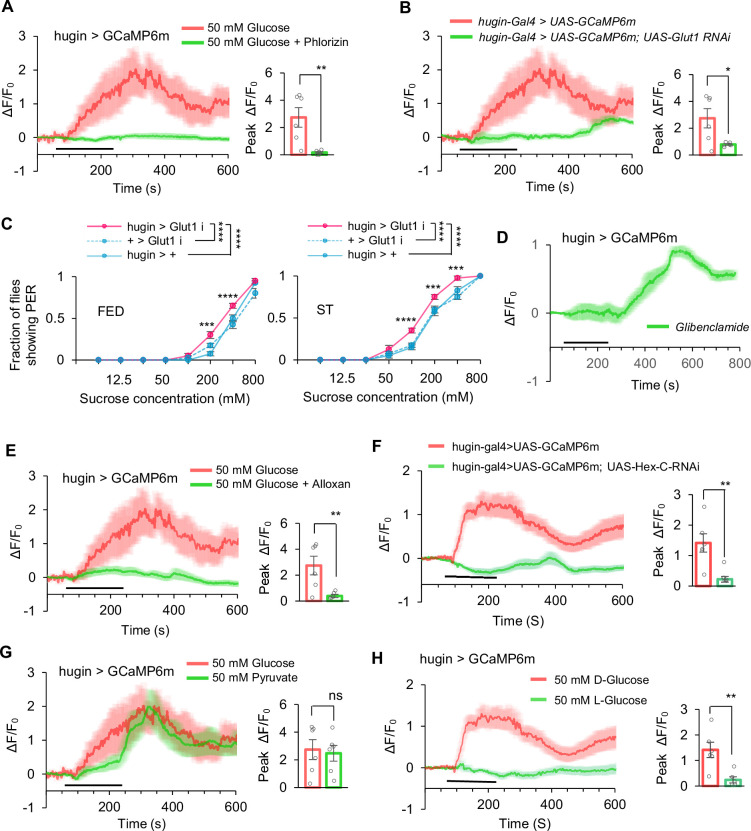
Among screened peptide populations, hugin and AstA responded at higher glucose. Blocking action-potential-dependent transmission preserved hugin but not AstA responses. Only particular anatomical subsets participated, locating glucose sensitivity within more specific cell groups.
Changes affecting glucose transport and metabolism weakened hugin calcium responses, while KATP-related conditions also affected activity. Internal nutrition connected with cellular excitability. The pyruvate comparison was nonsignificant, showing that metabolic conditions did not all yield the same response.
Source: Qin W et al. · eLife · CC BY
Original figure and English caption ↗
A peptide relay passes the signal onward
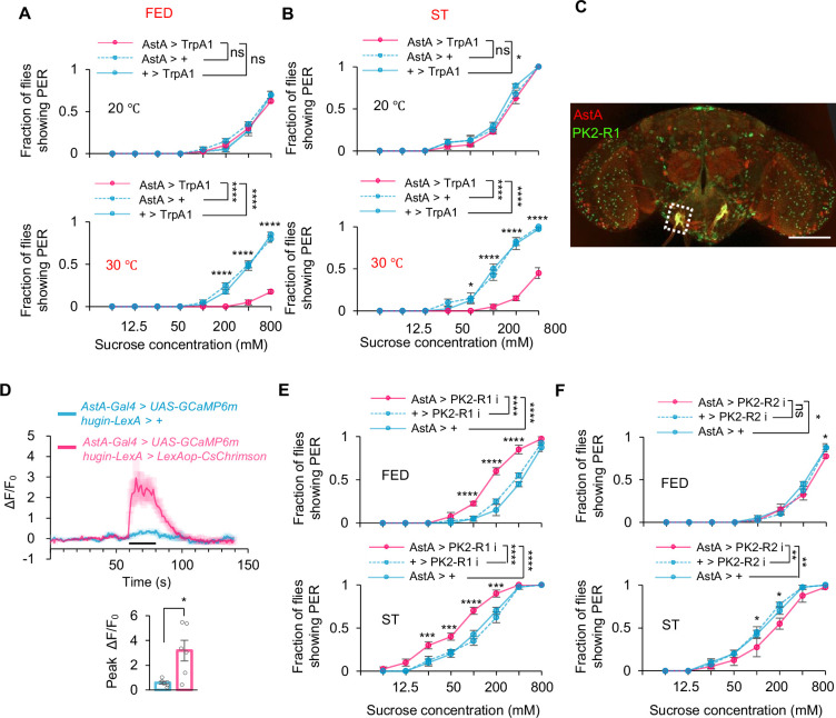
Hugin peptide reduced proboscis and Gr5a responses, but its corresponding receptor was not found on the proboscis sensory cells. AstA provided a relay: it expressed PK2-R1 and responded to increased hugin activity. Reducing this receptor in AstA enhanced sugar responses.
PK2-R2 had different effects, including smaller opposite changes under some hungry conditions. The brain route remained effective after separation from the ventral nerve cord, while intact preparations responded more strongly. A principal relay operated alongside additional neural contributions.
Source: Qin W et al. · eLife · CC BY
Original figure and English caption ↗
Internal state reaches sweet-sensory processing
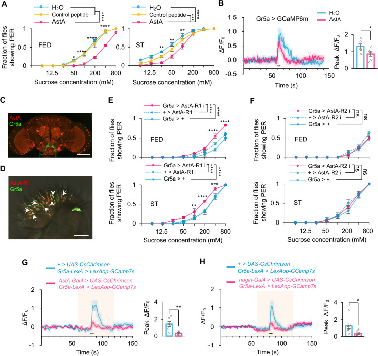
AstA then reached the sensory level. Peptide or increased AstA activity reduced sugar-evoked Gr5a calcium responses, and Gr5a carried AstA-R1. Reducing this receptor enhanced proboscis responses. Internal-glucose information thus passed through a peptide relay into early sweet processing.
Reducing hugin or AstA transmission also increased intake, less than the overall effect of hunger. Regulation remained possible in both hungry and fed conditions. The route formed part of a network involving other nutritional, adaptive and action-related signals.
Source: Qin W et al. · eLife · CC BY
Original figure and English caption ↗
Related regulatory clues appear in mice
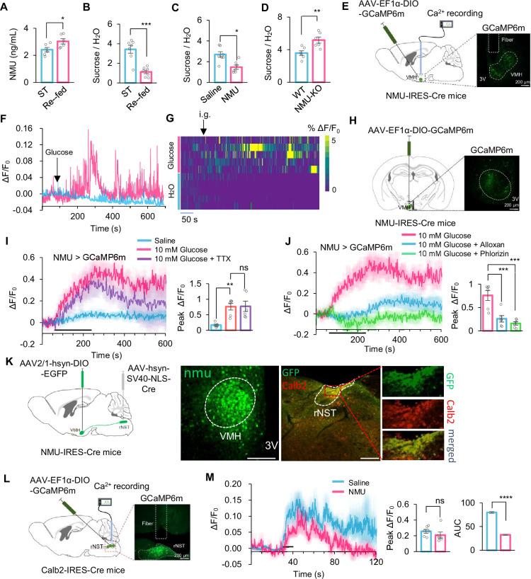
After sugar refeeding in male mice, circulating NMU increased. Exogenous NMU reduced sucrose preference, while gene loss increased it. Ventromedial-hypothalamic NMU cells responded to gastric sugar, and slice glucose responses showed transport- and metabolism-related features.
Mouse and fly results point toward regulation of sugar-related behavior by nutritional peptide signals. Two-bottle preference also includes motivation and post-ingestive feedback, offering a whole-choice view alongside the fly sensory-cell observations.
Brainstem response area and peak differ
Tracing linked hypothalamic NMU populations with rostral-nucleus-of-the-solitary-tract Calb2 cells involved in sweet transmission. Under NMU, calcium-response area during sugar licking decreased, while the peak comparison was nonsignificant. The change appeared in one response dimension.
Glucose-responsive cells, local connections and circulating-peptide effects offer different views. The fly receptor relay is more specifically defined, while mice provide related energy-sensing and behavioral clues. The species share a question while retaining distinct network structures.
Source: Qin W et al. · eLife · CC BY
Original figure and English caption ↗
Bodily state helps shape the next response
Feeding regulation involves more than the decision to eat: internal state can reach sensory processing and alter food-evoked responses. Connecting nutritional information with peptide relays extends hungry–fed differences from behavioral choice to cellular response.
The mouse evidence concerns males and tested sugar conditions. The broader insight is that food responses depend on both the external stimulus and the animal’s internal state. Sensory processing is an adjustable entry point shaped by bodily information.