Similar food intake, increasing weight: Phf6 links weight gain to activity
An estrogen-sensitive preoptic population contributes to movement and energy expenditure; adult local Phf6 deletion reduces activity and enlarges fat cells in female mice.
Weight gain often draws attention to intake, but energy balance also depends on daily movement and expenditure. A Nature Communications study follows cells marked by the chromatin-associated protein Phf6 in medial preoptic area, or MPOA. They strongly overlap with the estrogen receptor Esr1. Adult female mice lacking local Phf6 gain weight without significantly increased feeding, while activity and expenditure fall. The work connects these changes to excitability, hormonal responsiveness and a hypothalamic circuit.
Local MPOA Phf6 loss reduces female-mouse activity and expenditure and weakens cellular estradiol responses; connectivity with VMHvl contributes to activity promotion.
Contents · 7 sections
Which estrogen-sensitive cells carry Phf6?
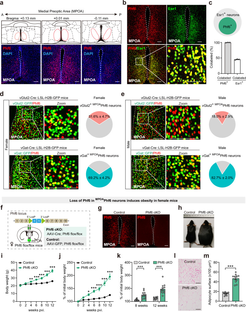
Nearly all Phf6-positive MPOA cells in adult females express Esr1, while accounting for only part of the Esr1 population. They include glutamatergic and GABAergic cells, in sex-dependent proportions. The marker identifies a hormone-sensitive mixed population rather than a purely excitatory class.
Local deletion at eight weeks increases female body weight from week four, with much greater gain by week twelve and enlarged white adipocytes. The adult, region-specific change allows a focused question: what aspect of everyday energy balance falls when these cells change?
Source: Wang J et al. · Nature communications · CC BY-NC-ND
Original figure and English caption ↗
Similar intake, less running and expenditure
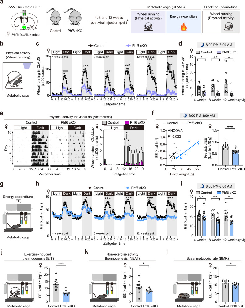
Food intake, feeding frequency and duration do not significantly change, nor do brown-fat surface temperature or measured reproductive hormones. Nighttime wheel running falls, as does later active-period expenditure after body-weight adjustment. The mice move less and expend less rather than clearly eating more.
Exercise-induced thermogenesis shows the largest decline, with smaller significant reductions in nonexercise thermogenesis and basal metabolic rate. Males show some activity and expenditure reductions, but later, weaker weight effects. Expenditure components and sex differ.
Running declines particularly at night, when mice are ordinarily more active, whereas weight gain develops over weeks. Daily activity records and weight trajectories connect reduced expenditure with longer-term energy accumulation, giving the weight difference a behavioral context.
Source: Wang J et al. · Nature communications · CC BY-NC-ND
Original figure and English caption ↗
Cells follow movement and can change it
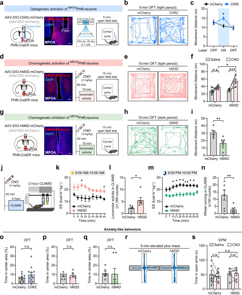
Calcium in MPOA Phf6 cells rises after female mice begin walking or climbing; running and nighttime activity also increase c-Fos labeling. The population tracks activity, prompting a test of whether it can influence movement rather than merely accompany it.
Activation during the less active daytime increases movement and expenditure, while nighttime inhibition decreases movement, running and expenditure. Open-field center and elevated-plus-maze open-arm duration do not significantly change. Activity shifts without corresponding changes in these anxiety-like measures.
Source: Wang J et al. · Nature communications · CC BY-NC-ND
Original figure and English caption ↗
Hormone levels persist, but cellular responses weaken
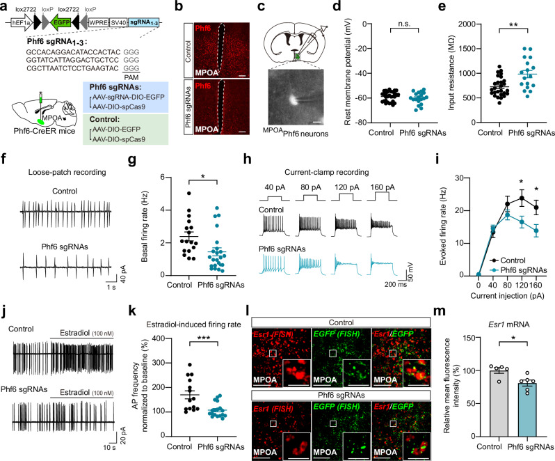
Phf6-deficient cells show reduced spontaneous and stronger-current-evoked firing in slices, unchanged resting potential and increased input resistance. Their output capacity changes. Estradiol normally increases firing, but this response weakens after Phf6 loss.
Esr1 mRNA falls while plasma estradiol does not. The clue lies in hormone-responsive cells rather than reduced circulating hormone. Receptor expression and firing responses connect Phf6 loss with reduced activity at the cellular level.
Source: Wang J et al. · Nature communications · CC BY-NC-ND
Original figure and English caption ↗
VMHvl is an important movement-promoting output
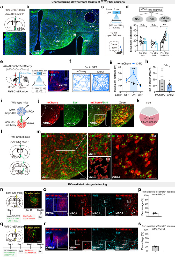
MPOA Phf6 cells project to several regions. Accumbens or paraventricular terminal activation does not significantly increase movement, unlike activation in VMHvl and the adjacent tuberal region. Restricting stimulation to ventrolateral ventromedial hypothalamus, or VMHvl, still increases movement without changing center duration.
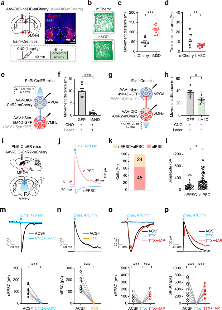
Tracing reveals reciprocal connectivity between MPOA Phf6 and VMHvl Esr1-associated populations. VMHvl inhibition strongly reduces MPOA-evoked movement, while MPOA inhibition partially attenuates VMHvl Esr1-evoked movement. Activity promotion involves cooperation between regions.
Source: Wang J et al. · Nature communications · CC BY-NC-ND
Original figure and English caption ↗
Excitatory and inhibitory inputs converge
MPOA Phf6-terminal stimulation evokes direct excitatory or inhibitory currents in VMHvl cells; nearby SST inhibitory cells also receive input. The mixed population supplies mixed synaptic effects. Direct VMHvl Esr1 activation reduces center duration, unlike MPOA Phf6 activation, showing distinct overall behavioral effects.
Source: Wang J et al. · Nature communications · CC BY-NC-ND
Original figure and English caption ↗
Activity expenditure explains part of the weight change
The mainly adult female-mouse study links a chromatin-associated factor, cellular hormonal responses, activity and expenditure. Altered neural states can lower bodily expenditure, allowing gradual weight gain without clearly increased intake. Understanding energy balance also requires understanding the neural organization of daily activity.