Why does blood pressure stay high after stress? A chloride clue in amygdala cells
In susceptible male rats, chronic stress increases CRF-labeled cell activity; NKCC1-associated chloride changes weaken GABA inhibition and accompany increased sympathetic output.
Stress can raise heart rate and blood pressure. Why do some animals remain hypertensive after it ends? A Journal of Clinical Investigation study compares normotensive rats with borderline-hypertensive rats, following CRF-expressing cells in central amygdala, or CeA. Sustained stress increases their activity and changes the direction of their response to GABA. Chloride distribution and its transporter NKCC1 provide a mechanistic clue.
Stressed susceptible rats show depolarized GABA reversal potential and increased NKCC1 in CeA CRF cells. NKCC1 inhibition restores slice GABA inhibition, while central administration reduces sympathetic activity and blood pressure.
Contents · 7 sections
The same stress produces different recovery
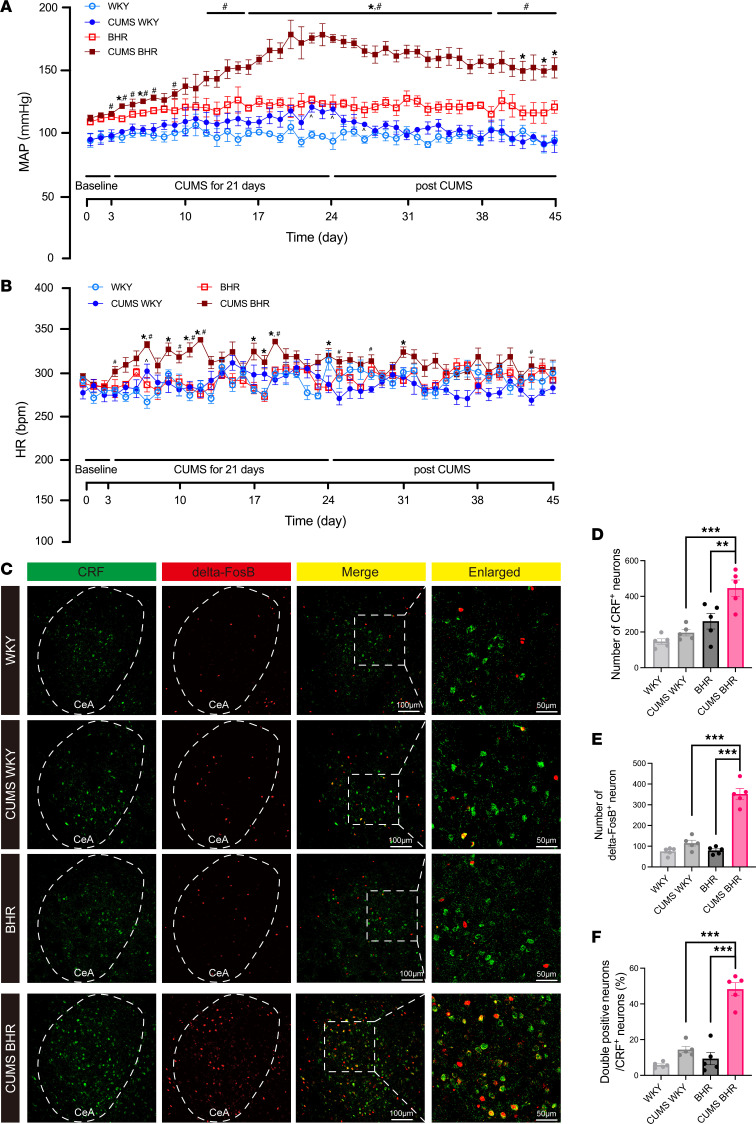
Male borderline-hypertensive rats, or BHRs, are compared with normotensive WKY rats. BHRs are offspring of hypertensive and WKY strains, with higher baseline pressure and susceptibility. Both groups show increased mean arterial pressure during twenty-one days of stress, but WKY recovers rapidly while BHR elevation persists for at least twenty-one days afterward.
Heart rate and serum corticosterone increase in BHRs without significant corresponding WKY changes. CRF colabeling with the sustained-activity marker ΔFosB increases in CeA, prompting a question about this population’s role in persistent cardiovascular changes.
Source: Ma H et al. · The Journal of clinical investigation · CC BY
Original figure and English caption ↗
The cells fire more with comparable input
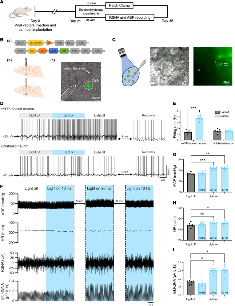
In stressed BHR slices, CRF-expressing cells are more depolarized and fire more spontaneously and under comparable current inputs. Stressed WKY cells lack the same significant changes. Both sustained pressure and increased excitability stand out in the susceptible background.
In anesthetized BHRs, 20- or 40-Hz activation raises mean arterial pressure, heart rate and renal sympathetic activity. Chemogenetic inhibition during stress also attenuates hypertension development. The population functionally contributes to sympathetic and circulatory output.
Source: Ma H et al. · The Journal of clinical investigation · CC BY
Original figure and English caption ↗
GABA remains, but its effect changes direction
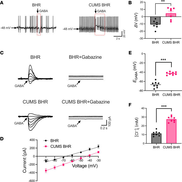
GABA commonly inhibits neurons, but its effect depends on ionic conditions when receptors open. CeA GABA and GABAA-receptor levels do not significantly differ across groups. Cellular responses do: GABA hyperpolarizes CRF cells in unstressed BHRs but depolarizes them after stress.
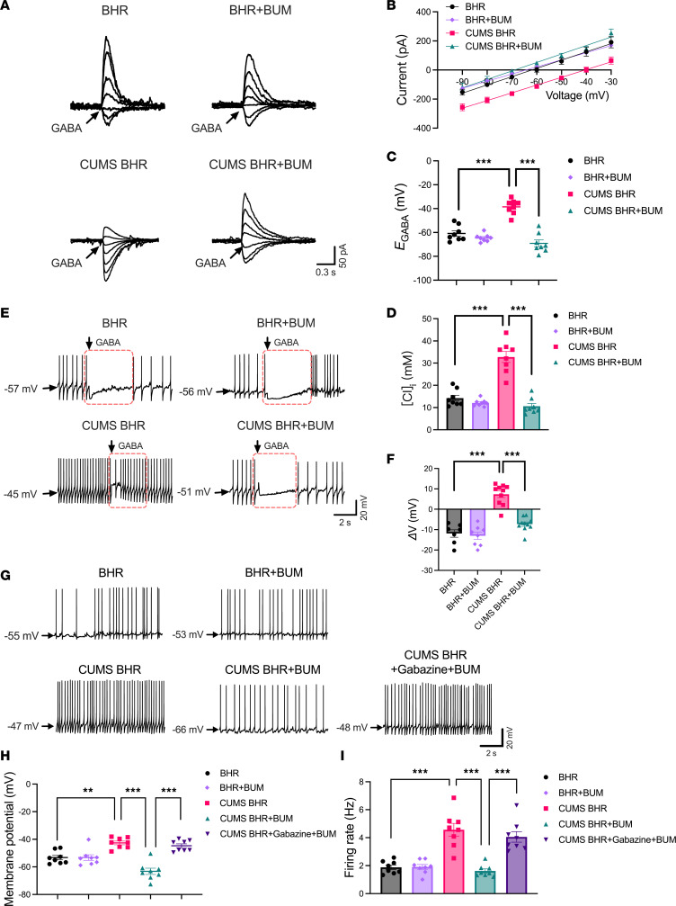
GABA reversal potential shifts approximately 21.5 mV toward depolarization in stressed BHRs, with increased intracellular chloride estimated from that potential; WKY shows no equivalent change. The reversal potential marks where GABA-associated current changes direction, revealing an altered ionic environment for the same receptor signal.
Source: Ma H et al. · The Journal of clinical investigation · CC BY
Original figure and English caption ↗
The change centers on NKCC1
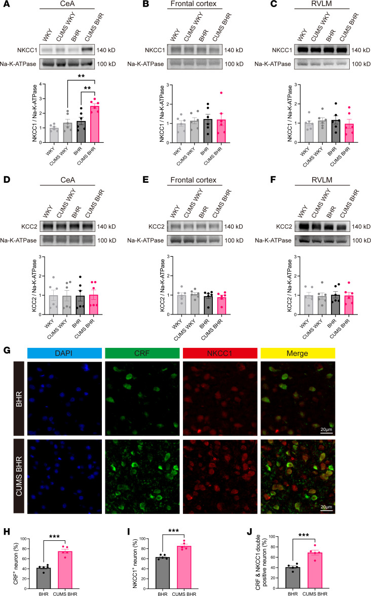
Chloride distribution depends on transport systems including NKCC1 and KCC2. Stressed BHR CeA shows increased membrane NKCC1 without significant KCC2 change; frontal cortex and rostral ventrolateral medulla lack equivalent changes in either protein, indicating regional and molecular selectivity.
CRF/NKCC1 colabeling increases in CeA, connecting transporter changes with the hyperexcitable population showing altered GABA responses. The next question is whether reducing NKCC1 activity can restore both ionic conditions and cellular activity.
Source: Ma H et al. · The Journal of clinical investigation · CC BY
Original figure and English caption ↗
NKCC1 blockade restores hyperpolarizing GABA responses
Bumetanide inhibition of NKCC1 restores GABA reversal potential and derived chloride in stressed BHR slices, reinstates hyperpolarizing responses and reduces spontaneous firing. Unstressed BHRs show no equivalent significant changes.
Adding the GABAA antagonist gabazine negates the firing reduction produced by bumetanide. Cellular inhibition after chloride-related restoration depends on GABAA signaling, linking transporter activity, reversal potential and firing functionally.
Source: Ma H et al. · The Journal of clinical investigation · CC BY
Original figure and English caption ↗
Central chloride regulation connects with sympathetic output
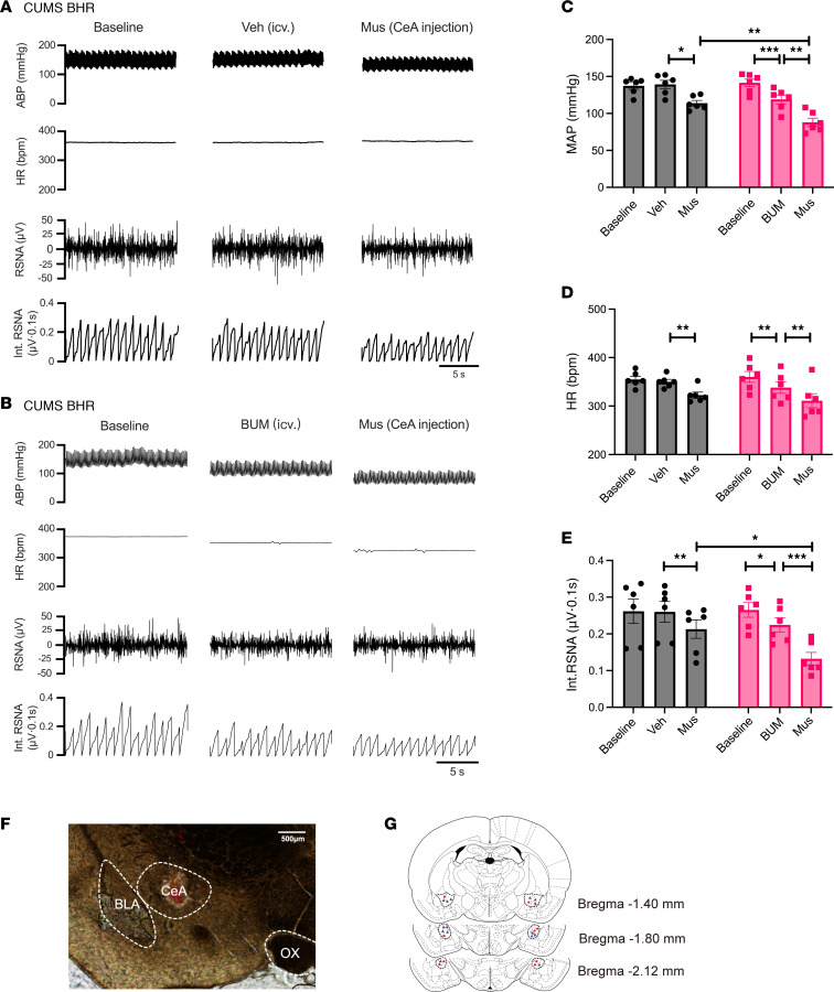
Intracerebroventricular bumetanide lowers pressure, heart rate and renal sympathetic activity in stressed BHRs, unlike vehicle. Subsequent CeA GABAA activation reduces outputs in both groups, with stronger pressure and sympathetic decreases after bumetanide pretreatment.
Bumetanide lacks equivalent circulatory effects in unstressed BHRs, and stressed WKY lacks the corresponding drug effects. Central NKCC1 inhibition has broader access than local CeA receptor activation; together they show dependence on susceptibility and stress state.
Source: Ma H et al. · The Journal of clinical investigation · CC BY
Original figure and English caption ↗
Stress changes the conditions for receiving inhibition
This male-rat study links sustained stress and cardiovascular changes to a cellular mechanism: inhibition depends on the receiving cell’s chloride conditions. In susceptible animals, NKCC1-associated changes weaken GABA inhibition of CRF cells alongside increased excitability and sympathetic output. Persistent hypertension involves how cells receive signals as well as signal strength.