When the pancreas inflames, does the brain participate? An oxytocin-cell–vagal output route
Starting with enzyme-producing acinar cells, a male-mouse study links hypothalamic and brainstem output with inflammation, showing how cholinergic effects depend on their targets.
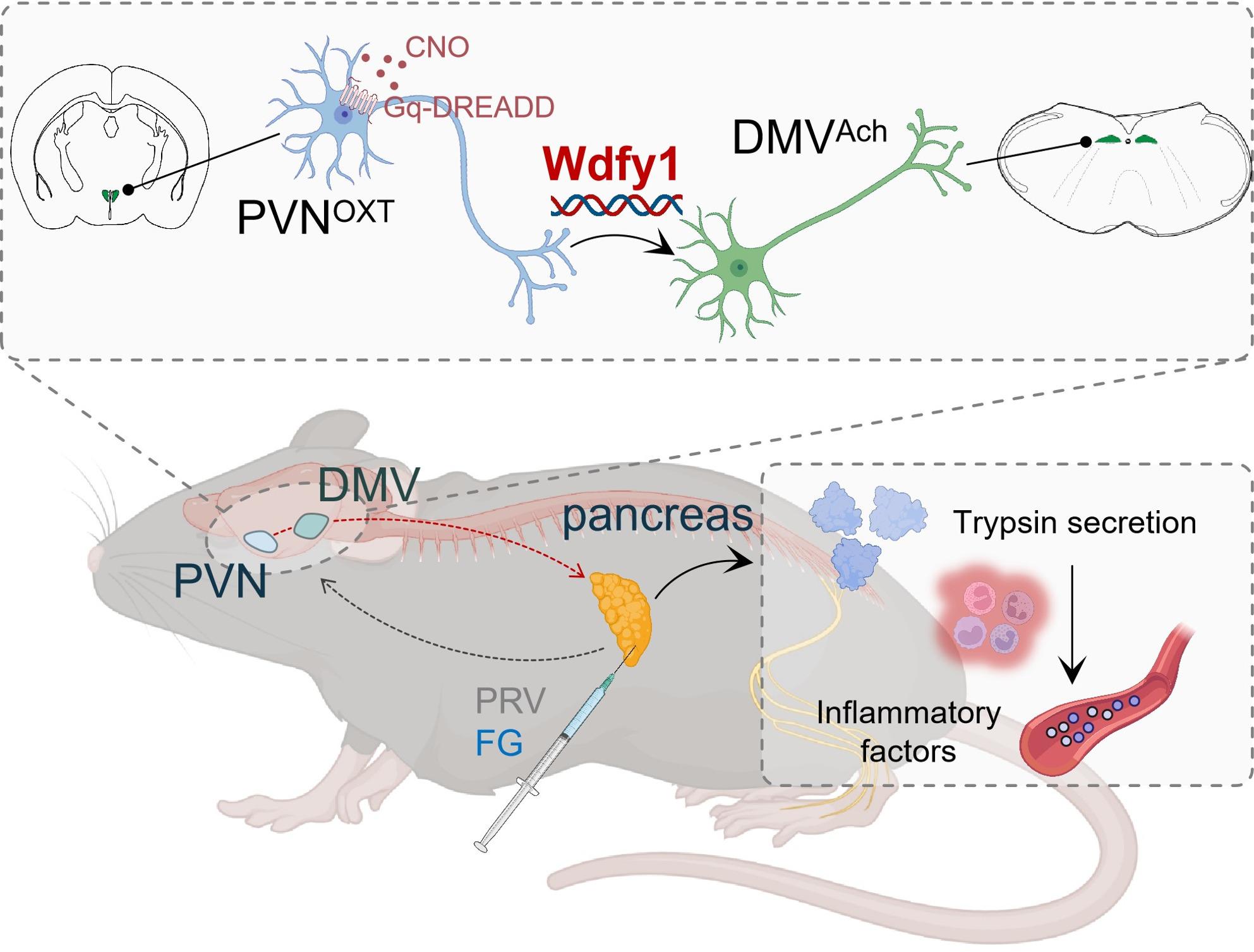
The pancreas is often associated with blood-glucose control through its islets. It also has another job: acinar cells produce digestive enzymes. During inflammation, secretion, cellular injury and immune responses become intertwined, potentially involving autonomic output from the brain. A 2026 Journal of Neuroinflammation study followed acinar-associated connections in male mice to a route through hypothalamus and a brainstem vagal motor nucleus. The finding brings a different perspective to cholinergic signaling: under particular target and disease conditions, it can contribute to worsening inflammation.
PVN oxytocin cells and DMV cholinergic output contribute to acinar and inflammatory changes in the model. Lower activity attenuates some abnormalities, while Wdfy1 links local neuronal state with downstream responses.
Contents · 7 sections
Enzyme-producing cells reveal another side of the pancreas
Acinar cells and islets have distinct roles. In a model combining a high-fat background with inflammatory induction, the study observed altered serum enzymes, tissue inflammation and pathology. This focused attention on exocrine function: which central outputs might change when enzyme-producing cells are injured?
Serum enzymes, inflammatory proteins and cellular structure describe different sides of the process. Relating them to neural activity explores how autonomic output might influence secretion and immune responses during injury, alongside organ function.
From pancreas to brainstem and hypothalamus
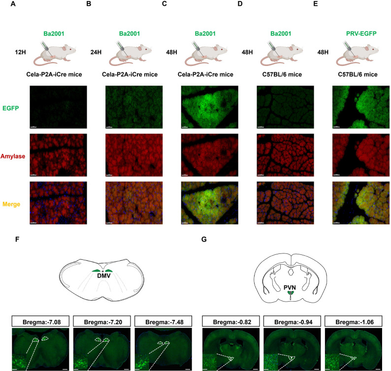
Tracing acinar-associated networks revealed earlier central labeling mainly in dorsal motor nucleus of the vagus (DMV), followed by more labeling in hypothalamic paraventricular nucleus (PVN). Pancreas, brainstem and hypothalamus emerged within a multilevel connection.
Cholinergic ganglia within the pancreas form another link. Interrupting the vagal connection removed labeling previously traced from pancreas to DMV, consistent with a vagal route. This is a relayed cross-organ network, with peripheral ganglia helping convey central output to acinar cells.
Source: Fang H et al. · Journal of neuroinflammation · CC BY-NC-ND
Original figure and English caption ↗
Oxytocin cells connect with vagal motor output
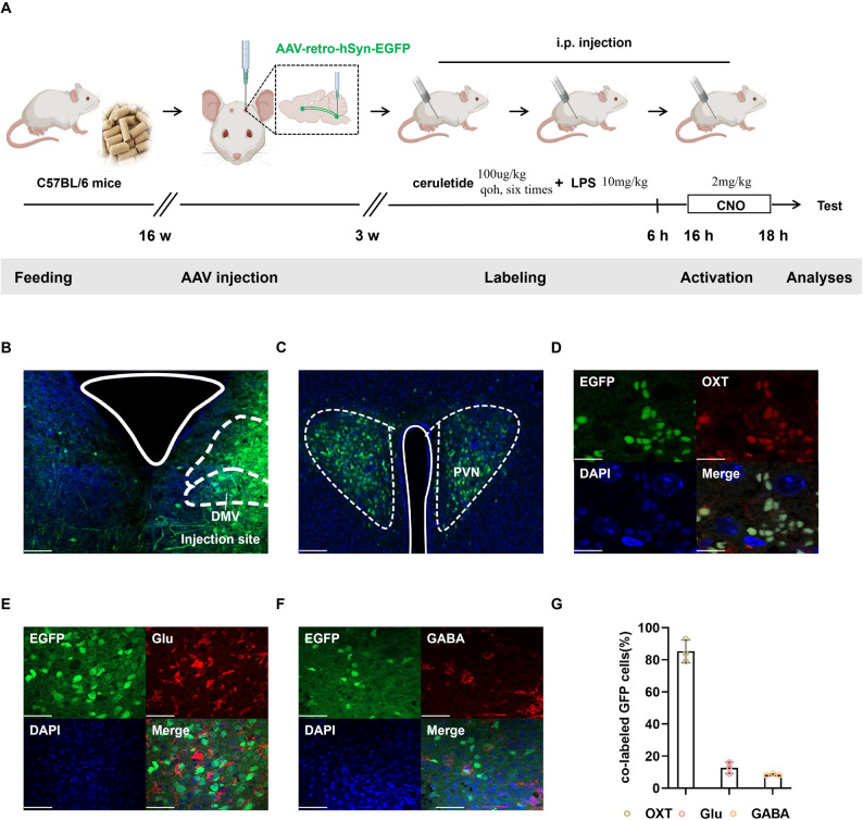
The inflammatory model showed stronger activity-related labeling in PVN and DMV, and pancreas-associated DMV cholinergic cells fired more readily. Following DMV input revealed predominantly oxytocin-associated cells in PVN, focusing the relation between organ injury and neuronal state on a specific combination.
Oxytocin here identifies a neuronal population. The question concerns how these cells influence pancreas through brainstem output, rather than a uniform change in circulating hormone. A familiar molecule can belong to different physiological stories depending on its cells and connections.
Source: Fang H et al. · Journal of neuroinflammation · CC BY-NC-ND
Original figure and English caption ↗
Normal and inflammatory states differ
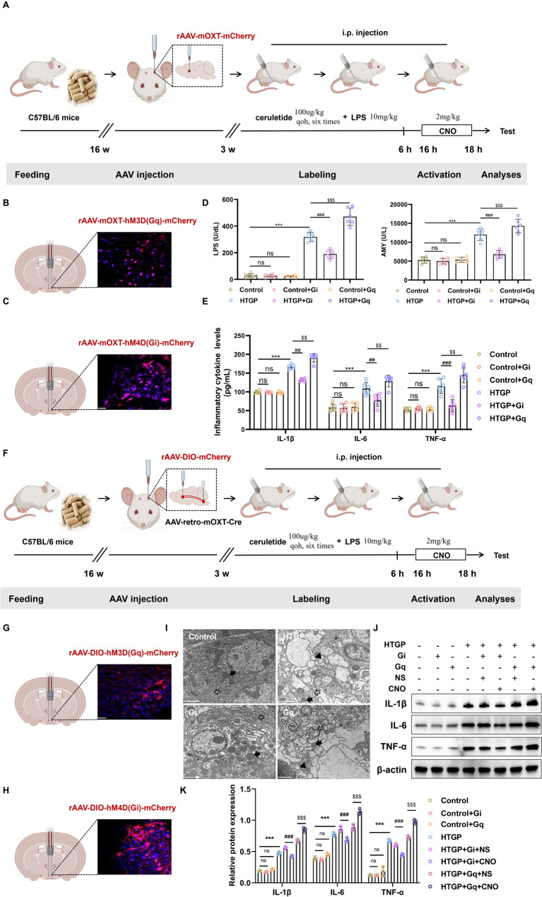
Reducing DMV cholinergic or PVN oxytocin-cell activity attenuated some enzyme and inflammatory measures in the model; increasing activity raised corresponding measures and worsened some tissue injury. DMV-directed PVN populations showed consistent effects. Comparable interventions under normal conditions produced no clear enzyme or inflammatory abnormalities, highlighting disease-state dependence.
Cells carried corresponding changes: expanded endoplasmic reticulum, altered zymogen granules and vacuolization improved after reduced neural activity, while some abnormalities remained. Neuronal output contributed alongside established cellular injury and immune responses.
Source: Fang H et al. · Journal of neuroinflammation · CC BY-NC-ND
Original figure and English caption ↗
Why can cholinergic signaling have different effects?
Vagal cholinergic signals are often associated with anti-inflammatory effects, but acinar secretion and immune responses can involve different receptors. In this model, muscarinic—particularly M3-associated—blockade reduced route-related inflammatory changes; nicotinic and α7 blockade did not have the same effect. Targets and receptors shape the direction of a signal’s effects.
Cellular involvement changed over time as well. Inflammatory labeling overlapped more with acinar cells early and with macrophages later. Local injury and immune recruitment connected within a changing inflammatory landscape.
Wdfy1 links molecular changes with neuronal output
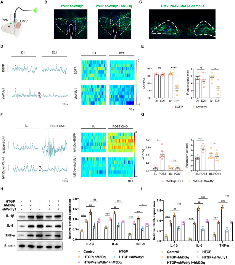
Single-cell transcriptomes brought Wdfy1 to the team’s attention. Reducing this candidate’s expression in PVN lowered DMV cholinergic calcium activity, attenuated downstream increases following PVN activation and reduced pancreatic inflammatory measures. Expression differences and functional effects converged at this node.
The change affected baseline activity as well as an evoked response. Wdfy1 was thus linked with local neuronal state; the paper’s proposed TLR-related interpretation offers a candidate framework for its molecular connections.
Source: Fang H et al. · Journal of neuroinflammation · CC BY-NC-ND
Original figure and English caption ↗
Brain–pancreas effects depend on cells and state
The study places exocrine pancreatic cells within brain–organ regulation and shows why a signal’s name alone does not determine its effect. Oxytocin cells, vagal output and cholinergic receptors influence tissue responses through a particular cellular combination and inflammatory state.
The findings concern a combined model in male mice. The central insight is that autonomic output depends on disease state and the cells it reaches. Here, acinar cells form an important endpoint connecting central activity with inflammatory change.